Báo cáo y học: "Giantin is the major Golgi autoantigen in human anti-Golgi complex sera" pptx
Bạn đang xem bản rút gọn của tài liệu. Xem và tải ngay bản đầy đủ của tài liệu tại đây (491.21 KB, 8 trang )
Introduction
The Golgi complex is an elaborate cytoplasmic organelle
that has a prominent function in the processing, transport-
ing, and sorting of intracellular proteins subsequent to
their synthesis in the rough endoplasmic reticulum. Struc-
turally, the Golgi complex is localized in the perinuclear
region of most mammalian cells and is characterized by
stacks of membrane-bound cisternae, as well as by func-
tionally distinct trans-Golgi and cis-Golgi networks [1].
Interestingly, several Golgi proteins have been reported to
be targets of the autoimmune response, even though they
are localized to the cytoplasmic face of Golgi membranes,
a site that is presumed to be privileged in that it is pro-
tected from immune surveillance. Autoantibodies directed
against the Golgi complex were first identified in the
serum of a Sjögren’s syndrome patient with lymphoma [2].
Several isolated reports have described anti-Golgi
complex antibodies (AGAs) in other systemic autoimmune
diseases such as systemic lupus erythematosus (SLE) [3],
rheumatoid arthritis [4], mixed connective tissue disease
[5], and Wegener’s granulomatosis [6]. AGAs were also
found in 10% of patients with HIV infection [7] and 35.7%
of HIV carriers [8]; however, in the more recent report by
Massabki and coworkers [9], AGAs were not found in
100 HIV-infected patients.
Within the past several years, our laboratories and others
have cloned and identified several novel Golgi autoanti-
AGA = anti-Golgi complex antibody; ELISA = enzyme-linked immunosorbent assay; IIF = indirect immunofluorescence; OD = optical density; SD =
standard deviation; SLE = systemic lupus erythematosus.
Available online />Research article
Giantin is the major Golgi autoantigen in human anti-Golgi
complex sera
Kazuhisa Nozawa
1
, Marvin J Fritzler
2
, Carlos A von Mühlen
3
and Edward K L Chan
1
1
Department of Oral Biology, University of Florida College of Dentistry, Gainesville, Florida, USA
2
Department of Biochemistry and Molecular Biology, University of Calgary, Calgary, Alberta, Canada
3
Department of Internal Medicine, Hospital São Lucas, Pontifícia Universidade Católica do Rio Grande do Sul, Porto Alegre, Brazil
Corresponding author: Edward K Chan (e-mail: )
Received: 29 Sep 2003 Revisions requested: 24 Oct 2003 Revisions received: 19 Nov 2003 Accepted: 27 Nov 2003 Published: 15 Dec 2003
Arthritis Res Ther 2004, 6:R95-R102 (DOI 10.1186/ar1035)
© 2004 Nozawa et al., licensee BioMed Central Ltd (Print ISSN 1478-6354; Online ISSN 1478-6362). This is an Open Access article: verbatim
copying and redistribution of this article are permitted in all media for any purpose, provided this notice is preserved along with the article's original
URL.
Abstract
Anti-Golgi complex antibodies (AGAs) are primarily associated
with systemic lupus erythematosus and Sjögren’s syndrome.
Here we report on the immunoreactivity of AGAs against five
Golgi autoantigens (giantin, golgin-245, golgin-160, golgin-
95/GM130, and golgin-97) and provide data from epitope
mapping on the most common Golgi autoantigen, namely giantin.
A total of 80 human sera containing AGAs, as defined by indirect
immunofluorescence on HEp-2 cells, were analyzed by ELISA
using recombinant autoantigens and immunoprecipitation. The
proportion of AGA sera that reacted with the five Golgi
autoantigens was correlated with the molecular mass of the
Golgi antigens. Autoantibodies to giantin, the largest Golgi
autoantigen, were the predominant AGAs, being found in 50% of
the AGA sera. Epitope mapping of giantin was performed using
six recombinant fragments spanning the entire protein.
Antigiantin-positive sera with low titer autoantibodies recognized
epitopes in the carboxyl-terminal fragments that are proximal to
the Golgi membrane, whereas higher titer sera exhibited strong
reactivity to amino-terminal and central domains that are likely to
extend from the Golgi membrane into the cytoplasm. Our
working hypothesis is that aberrantly expressed Golgi complex
autoantigens may be released into the immune system when
cells undergo lysis. By virtue of a carboxyl-terminal
transmembrane domain, giantin is likely to be more stably
associated with the cytoplasmic face of the Golgi complex than
are other golgins, which are peripheral proteins. The stable
association of giantin with the putative released Golgi complex
may contribute to its preferential autoantigenicity.
Keywords: anti-Golgi complex antibody, autoantibody, autoimmunity, cell death, epitope mapping
Open Access
R95
R96
Arthritis Research & Therapy Vol 6 No 2 Nozawa et al.
gens. This has been achieved primarily by expression
cloning using human autoantibody probes. These Golgi
autoantigens are referred to as giantin/macrogolgin/
GCP372, golgin-245/p230, golgin-160/GCP170,
golgin-95/GM130, golgin-97, and golgin-67, with their
names based in part on their molecular weights as esti-
mated from SDS-PAGE under denaturing conditions
[7,10–13]. A common feature of this family of Golgi
autoantigens is that they all have coiled-coil domains
throughout the entire protein except for short nonhelical
regions at the amino-terminus and carboxyl-terminus [1].
Golgin-245 was localized to the trans-Golgi compartment
[14], whereas GM130 has been reported to be localized
to the the cis-Golgi compartment [15]. It has been also
reported that several golgins, such as golgin-245 and
golgin-97, are attached to Golgi membranes through a
GRIP domain in the carboxyl-termini [16]. In contrast to
other Golgi autoantigens, giantin has a single transmem-
brane domain in the carboxyl-terminus [17]. A second
common feature among the Golgi autoantigens is that bio-
chemical evidence and immunoelectron microscopy data
show that they are peripheral or transmembrane (giantin)
proteins on the cytoplasmic face of the Golgi complex.
The implication is that these Golgi autoantigens may have
common biochemical characteristics and functions that
make them preferred autoimmune targets among the
approximately 100 Golgi complex proteins described to
date [18]. A third common feature among the Golgi
autoantigens is that none of these macromolecules are
localized to apoptotic blebs [19]; in fact, immunofluores-
cence analysis showed that the Golgi complex was
altered and developed distinctive characteristics during
apoptosis and necrosis [19].
It is striking that human autoimmune responses are selec-
tive for these proteins that are rich in coiled-coil motifs and
that reside on the cytoplasmic face of the Golgi complex.
How this family of coiled-coil proteins becomes auto-
immune targets remains to be determined. One possible
explanation is that these Golgi proteins may be recognized
as surface structures on the organelle that is exposed to
the immune system in aberrant disease states associated
with unregulated cell death (apoptosis and necrosis)
resulting from injury or infection, and defective clearance
of dying cells.
Although it is known that AGAs are associated with some
autoimmune diseases or viral infections, the prevalence of
AGAs and their fine specificity have not been reported.
Immunoblotting and immunoprecipitation studies have
shown that AGAs reacted with a number of cellular pro-
teins [20]. AGAs are generally considered to be rare
autoantibodies; however, Bizzaro and coworkers [21] sug-
gested that the presence of AGAs in high titer in the
absence of a clear clinical manifestation may constitute an
early sign of systemic autoimmune diseases. Here, we
present data on the reactivity of AGAs against known
Golgi autoantigens by ELISA using five recombinant pro-
teins. Because antigiantin autoantibodies were found to
be the most common reactivity in AGAs, epitope mapping
was performed using six overlapping recombinant frag-
ments of giantin.
Materials and method
Human sera and monitoring of anti-Golgi complex
antibody reactivity
Human putative AGA sera and normal control sera were
obtained from the laboratory serum bank and Advanced
Diagnostics Laboratory at the University of Calgary,
Canada. Some AGA sera were also provided by Drs R L
Humbel (Luxembourg), Kiyomitsu Miyachi (Keigu Clinic,
Yokohama, Japan), and Carlos A von Mühlen (Pontifícia
Universidade Católica do Rio Grande do Sul, Porto
Alegre, Brazil). All sera were provided as anonymous
samples and were stored at –80°C until use. The reactivity
to Golgi complex in all AGA sera was confirmed by indi-
rect immunofluorescence (IIF) microscopy on HEp-2 cells
(Immuno Concepts Inc., Sacramento, CA, USA). Double
staining was performed using the human sera (1 : 100 dilu-
tion) and rabbit antigiantin antiserum (1 : 500 dilution) as a
marker of the Golgi complex [19]. The secondary antibod-
ies were Alexa Fluor
®
488 conjugated goat antihuman IgG
reagents and Alexa Fluor
®
568 conjugated goat antirabbit
IgG reagents (Molecular Probes, Eugene, OR, USA) used
at a dilution of 1 : 400. Nuclei were counterstained with
4′,6-diamidino-2-phenylindole. By using this approach, a
total of 80 sera exhibited specific staining of the Golgi
complex.
Recombinant Golgi proteins
Recombinant human Golgi autoantigens were produced
using the expression plasmid pET28 system in
Escherichia coli BL21 (DE3; Novagen, Madison, WI,
USA) as previously described [19]. Recombinant proteins
of golgin-245 (amino acids 811–2083) [11], golgin-160
(amino acids 787–1348) [10], golgin-95/GM130 (amino
acids 370–990) [10], and golgin-97 (amino acids 1–767)
[12] were subcloned into pET28 vectors for the expres-
sion of recombinant bacterial proteins. Six overlapping
fragments P1–P6 representing the full-length giantin
cDNA (GenBank accession number NM_004487) [7]
were generated for epitope mapping analysis. Two frag-
ments (P1 and P2) were obtained by expression cloning
from a random-primed lambda phage cDNA library gener-
ated from human T24 cells using an antigiantin-specific
human serum. Three fragments were obtained from an
available expression sequence tag clone (P3, GenBank
accession number N_76853; P4, GenBank accession
number BG_567238; P5, GenBank accession number
AI_458639). One fragment (P6) was cloned from reverse
transcription polymerase chain reaction synthesis using
total RNA purified from HeLa cells. All six fragments of
R97
overlapping recombinant proteins cDNAs were inserted
into pET28 expression vector and introduced into
Escherichia coli BL21 (DE3). Sequencing was conducted
in both directions using custom primers. Bacterial pellets
were suspended in 6M guanidinium hydrochloride con-
taining buffer, and the recombinant proteins were purified
by nickel column chromatography according to manufac-
turer’s instructions (Qiagen, Valencia, CA, USA). The con-
centration of the purified recombinant proteins was
measured by a Protein DC Assay Kit (Bio-Rad, Hercules,
CA, USA) and these samples were stored at –80°C until
they were required for subsequent experiments.
Enzyme-linked immunosorbent assay
The ELISA protocol described by Rubin [22] was used
with some modifications. In brief, Ni column affinity purified
recombinant proteins were diluted in phosphate-buffered
saline to a final concentration of 1 µg/ml and then coated
on Immulon 2 microtiter plates (Dynatech Laboratories,
Alexandria, VA, USA). Human sera were diluted 1 : 1000
and then incubated in the antigen-coated wells. Horserad-
ish peroxidase-conjugated goat antihuman IgG (CALTAG
Laboratories, San Francisco, CA, USA) was used at
1 : 5000 dilution and the substrate 2,2′-azinobis (3-ethyl-
benzthiazoline) sulfonic acid was added as the detection
reagent. Each sample was analyzed in duplicate and the
average optical density (OD) at 405 nm with a substrate
development time of 15–45 min was used for data analy-
sis. The cutoff value designating a positive reaction was
the mean OD of 12 normal sera +3 standard deviations
(SDs).
Immunoprecipitation
HeLa cells (ATCC, Rockville, MD, USA) were metaboli-
cally labeled overnight with [
35
S]-methionine (Trans
35
S-label; ICN), as described previously [11,12]. Cell
extracts were harvested in a lysis buffer containing 1%
NP-40, 50 mmol/l Tris.HCl, pH 7.5 and 150 mmol/l NaCl,
and supplemented with Complete™ protease inhibitor
cocktail (Boehringer Mannheim, Indianapolis, IN, USA).
Soluble fractions were used as substrate for immunopre-
cipitation reactions by combining 100 µl 10% protein
A-Sepharose beads (Sigma, St. Louis, MO, USA), 10 µl
human serum, 500 µl NET2 buffer (50 mmol/l Tris-HCl,
150 mmol/l NaCl, 5 mmol/l EDTA, 0.5% Nonidet P-40,
0.5% deoxycholic acid, 0.1% SDS, 0.02% sodium azide,
pH 7.4), and 50–100 µl labeled cell extract. After 1 hour of
incubation at 8°C, the Sepharose beads were washed five
times in NET2. Proteins were eluted in 20 µl sample buffer
and analyzed by 10% gel SDS-PAGE [23], followed by
autoradiography.
Immunoblotting
Affinity purified recombinant proteins were loaded on
12.5% SDS-PAGE gels (4 µg/lane), separated by elec-
trophoresis, and transferred to nitrocellulose membranes
using a Semi-Dry Trans-Blot apparatus (Bio-Rad), as
described previously [19]. Human sera containing anti-
giantin antibodies were used at dilutions of 1 : 100 to
1 : 500. Detection of bound antibodies was achieved
using horseradish peroxidase-conjugated goat antihuman
IgG antibody (CALTAG Laboratories), used at 1 : 5000
dilution, in combination with enhanced chemilumines-
cence (Super Signal; PIERCE Products, Rockford, IL,
USA).
Results
Giantin is the most common autoantigen detected in
anti-Golgi complex antibody sera
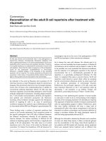
Reactivity to Golgi complex antigens in all sera was con-
firmed by IIF, and all sera exhibited a specific staining
pattern against Golgi complex structures as determined
by colocalization with rabbit antibodies to giantin (Fig. 1).
This approach yielded 80 human AGA sera that were
used to investigate the prevalence of autoantibodies to
five individual Golgi autoantigens represented by purified
recombinant proteins in an ELISA and by immunoprecipi-
tation using extracts from [
35
S]-methionine labeled HeLa
cells (Fig. 2). The number of positive sera and frequency of
reactivity of the 80 human AGA sera are summarized in
Table 1. The most common Golgi complex autoantigen
target was giantin (40/80 [50%]) and the second most
common target was golgin-245 (19/80 [24%]). The
lowest frequency reactivity (3.8%) was to golgin-97, and
25 AGA sera (31.3%) did not react with any of the five
Golgi autoantigens used in the present study. Interest-
Available online />Figure 1
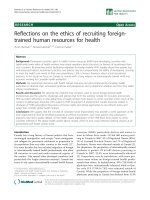
Immunofluorescence colocalization of putative human anti-Golgi
complex antibody (AGA) sera with rabbit antigiantin antibody to
confirm specificity to the Golgi complex. (a) A representative human
serum exhibiting staining specific to the Golgi complex in HEp-2 cells
colocalizes with (b) characteristic staining by rabbit antigiantin
antibody (arrows). (c) A serum with diffuse nuclear and cytoplasmic
staining unrelated to the Golgi complex as demonstrated by (d) lack of
costaining with rabbit antigiantin antibody.
ingly, the frequency of AGA sera reactive with the five
Golgi autoantigens was numerically correlated with the
molecular masses of the native Golgi autoantigens
(Table 1), and the number of positive sera that reacted
with giantin was remarkably higher than those for other
golgins. The confirmation by immunoprecipitation was
important because some of the Golgi autoantigens used as
substrate in the ELISA did not represent full-length proteins
and we were concerned that reactivity to these five Golgi
autoantigens may be underestimated by ELISA alone.
Among the 25 AGA sera that were negative for the five
Golgi autoantigens, there were no predominant reactivities
other than the five Golgi autoantigens described, even
though the immunoprecipitation assay showed unidentified
bands that were recognized by many of these sera.
Anti-Golgi complex antibody correlations
We then determined whether there were specific correla-
tions between any of the five specific AGAs with another
AGA. None of the sera had AGAs to four or more of these
five Golgi autoantigens. There were 6, 15, and 34 sera
with antibodies to three, two, and one of the five Golgi
autoantigens, respectively. Among the six sera with anti-
bodies to three of the five antigens, four sera had anti-
giantin, antigolgin-245 and antigolgin-160, which were the
three most common antibodies detected; one serum had
antigiantin, antigolgin-245 and antigolgin-95/GM130
(Fig. 2, lane 5); and the remaining serum had antigiantin,
antigolgin-245, and antigolgin-97. Among the 15 sera with
antibodies to two of the five antigens, five had antigiantin
and antigolgin-245, two had antigiantin and antigolgin-
160, two had antigiantin and anti-GM130, two had anti-
giantin and antigolgin-97, two had antigolgin-245 and
antigolgin-160, and two had antigolgin-245 and anti-
GM130 (Table 2). No specific correlations were observed
between the two most abundant antibodies, namely anti-
giantin and antigolgin-245. For example, among the
19 AGA sera positive for antibody to golgin-245, 11
(57.9%) were positive and 8 (42.1%) were negative for
antigiantin antibody. Among the 40 AGA sera positive for
antibody to giantin, 11 (27.5%) were positive and 29
(72.5%) were negative for antigolgin-245. Although the
number of sera that bound golgin-160, GM130, and
golgin-97 were relatively small, it was interesting that sera
with these three autoantibodies did not overlap. In other
words, sera positive for antigolgin-160 were negative for
antibody to GM130 and golgin-97, and sera positive for
anti-GM130 were negative for antigolgin-97 (Table 2).
Characterization of major epitopes in giantin
To examine the relative distribution of epitopes in giantin,
we performed mapping using six overlapping partial length
constructs of recombinant giantin. Expression vectors for
the recombinant proteins P1–P6 were constructed to
cover the full-length of giantin (amino acids 1–3259), with
the exception of the 38 amino acids at the amino-terminus
(Fig. 3). Forty sera containing antigiantin antibody were
analyzed using ELISA (Fig. 4, Table 3). Based on reactivity
to the P1–P6 peptides, we provisionally divided the posi-
tive sera into low-positive and high-positive groups. Low-
positive was defined as OD values between the mean OD
of normal sera plus 3–15 SDs, whereas high-positive was
defined as the OD greater than the mean OD of normal
sera plus 15 SDs. Giantin is known to be bound on the
cytoplasmic face of the Golgi complex via its single car-
boxyl-terminal transmembrane domain, and the amino-ter-
minal and the central domains extend into the cytoplasm
[24,25].
Arthritis Research & Therapy Vol 6 No 2 Nozawa et al.
R98
Table 1
Frequency of autoantibodies to specific Golgi complex
autoantigens in 80 human anti-Golgi complex antibody sera
Golgi autoantigen (molecular weight [kDa]) Positive sera (n [%])
Giantin (370) 40 (50.0)
Golgin-245 (245) 19 (23.8)
Golgin-160 (160) 11 (13.6)
Golgin-95/GM130 (130) 6 (7.5)
Golgin-97 (97) 3 (3.8)
Undefined anti-Golgi reactivity 25 (31.3)
A total of 80 sera were studied.
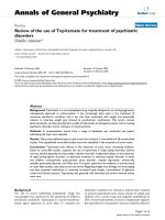
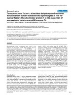
Figure 2
Representative data from the immunoprecipitation analysis of anti-Golgi
complex antibody (AGA) using extracts from HeLa cells metabolically
labeled with [
35
S]-methionine for 16 hours. Lane 1, normal human
serum; lanes 2–5, AGA sera. Lanes 2 and 3 show sera with primarily
antibody to golgin-160 (g160) and giantin, respectively. Lane 4 shows a
serum with antibodies to giantin and golgin-97 (g97). Lane 5 shows a
serum with antibodies to giantin, golgin-245 (g245), gm130, and an
unknown protein (arrowhead) migrated at approximately 90 kDa. Lane 6
shows a serum with strong reactivity to golgin-245 and weaker
reactivity to several unidentified lower molecular weight proteins (*).
None of antigiantin positive sera showed high positive
reactivity to P5 or P6 peptides; however, the highest pro-
portion of antibody reactivity (22/40 [55%]) was to P6,
which includes the carboxyl-terminus and the transmem-
brane signal sequence. P5, which is proximal to the trans-
membrane domain and the cytoplasmic face of the Golgi
membrane, also exhibited higher antibody frequency than
those for other fragments more distal to the transmem-
brane domain (P1–P4). In contrast to anti-P5 and anti-P6,
some AGA sera had antibodies to the distal fragments
P1–P4 exhibiting high-positive reactivity, but the overall
frequency of antibody to these fragments was relatively
low (Fig. 4 and Table 3). All of the antigiantin sera reacted
with one or more of the giantin subfragments used for
epitope mapping. There were no specific correlations
Available online />R99
Table 2
Correlation of the five antibodies detected in the current study among the 80 anti-Golgi complex antibody sera analyzed
Antigiantin Antigolgin-245 Antigolgin-160 Anti-GM130 Antigolgin-97
Positive Negative Positive Negative Positive Negative Positive Negative Positive Negative
Antigiantin 11 (27.5%) 29 (72.5%) 5 (12.5%) 35 (87.5%) 3 (7.5%) 37 (92.5%) 3 (7.5%) 37 (92.5%)
(n = 40)
Antigolgin-245 11 (57.9%) 8 (42.1%) 6 (31.5%) 13 (68.5%) 3 (15.9%) 16 (84.1%) 1 (5.2%) 18 (94.8%)
(n = 19)
Antigolgin-160 5 (45.5%) 6 (54.5%) 6 (54.5%) 5 (45.5%) 0 11 (100%) 0 11 (100%)
(n = 11)
Anti-GM130 3 (50%) 3 (50%) 3 (50%) 3 (50%) 0 6 (100%) 0 6 (100%)
(n = 6)
Antigolgin-97 3 (100%) 0 1 (33.3%) 2 (66.7%) 0 3 (100%) 0 3 (100%)
(n = 3)
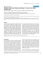
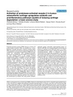
Figure 3
(a) Physical map of giantin cDNA fragments used in epitope analysis.
The open box denotes the open reading frame. P1–P6 represent
overlapping segments expressed as recombinant proteins, together
spanning almost the full length of giantin. The P1 fragment differs from
the published sequence and represents an alternatively mRNA spliced
product. (b) Coiled-coil domains of giantin, golgin-245, golgin-160,
golgin-95/GM130, and golgin-97. Each macromolecule is depicted
showing the coiled-coil regions, as predicted by the COILS program
[43]. A transmembrane (TM) hydrophobic region of 20–22 amino
acids at the carboxyl-terminus is postulated to be responsible for
anchoring giantin to Golgi membrane, with the molecule extending to
the cytoplasm. The cytoplasmic domains of giantin are responsible for
interaction with other Golgi proteins GM130 and p115.
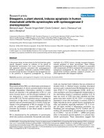
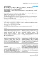
Figure 4
Reactivity of human antigiantin positive sera against six overlapping
giantin fragments. A total of 40 antigiantin positive sera were analyzed
by ELISA. Black squares represent optical density (OD) values for
each individual serum. Dotted lines represent cutoff values for the low-
positive group (between the mean OD of normal sera plus
3–15 standard deviations [SDs]), and dashed lines represent cutoff
values for the high-positive group (greater than mean OD of normal
sera +15 SDs).
between P1–P4 high-positive sera (Fig. 4) and another
coexisting AGA; among the P1–P4 high-positive sera,
only one serum had coexisting antigolgin-160 and a
second serum had coexisting anti-GM130. Taken
together, these data suggest that the major epitopes of
giantin are located in the carboxyl-terminal domain, includ-
ing the transmembrane signal sequence. However, the
epitopes localized in the distal amino-terminus or central
domains of giantin can generate stronger autoimmune
responses than can the epitopes in the transmembrane
region.
Discussion
In the present study we investigated the frequency of
autoantibodies to specific Golgi complex autoantigens in
a cohort of human sera containing AGAs as defined by IIF.
The most frequent target autoantigen was giantin.
Autoepitopes of giantin span across the entire protein, but
the most frequent reactivity was located in the carboxyl-
terminal fragments P5 and P6. These data are consistent
with the earlier report by Seelig and coworkers [7]
describing a diverse spectrum of AGAs that recognized
different recombinant fragments in a smaller cohort of
AGA sera. In contrast to antibodies to giantin, the least
common AGAs were those directed at golgin-97, which
also has the lowest molecular mass among the group of
Golgi complex autoantigens included in the present study.
The proportion of antibody to giantin was more than
10-fold greater than that to golgin-97.
To understand the mechanism of Golgi autoantibody pro-
duction, it is important to consider why giantin has a
greater frequency of reactivity than do other golgins. Dif-
ferences between giantin and other golgins include the
following: giantin is the highest molecular weight Golgi
protein and contains a greater number of coiled-coil
domain units than other golgins (Fig. 3b); and only giantin
possesses a transmembrane domain, which may ensure
its tight association with the Golgi complex.
Giantin is the most common target autoantigen in anti-
Golgi complex antibody sera
Although we showed that the majority of sera that react
with the Golgi complex in an IIF assay react with known
Golgi autoantigens, 25 out of 80 (31.3%) AGA sera did
not recognize any of the five Golgi autoantigens examined
in this study. The data suggest that these sera react with
other Golgi autoantigens. A candidate Golgi autoantigen is
GMAP-210, a reported cis-Golgi network associated
protein that also contains characteristic coiled-coil domains
[26]. Among the 80 AGA sera, our immunoprecipitation
data revealed three sera with a common band at approxi-
mately 210 kDa that might represent GMAP-210; however,
because we did not have the cDNA for GMAP-210 and the
frequency of this putative anti-GMAP-210 antibody was
low, we did not confirm these data using independent
methods. Two other Golgi proteins that may be candidate
autoantigens include golgin-84, an 84 kDa transmembrane
Golgi protein [27]; and βI Sigma spectrin, a 220 kDa
protein that is associated with Golgi complex and vesicles
[28]. However, our immunoprecipitation data did not yield
any bands consistent with these Golgi complex candidate
autoantigens. We did not include other known Golgi
autoantigens such as golgin-67 [13] and p115 [29]
because the frequencies of these autoantibodies are
known to be low. Thus, our data support the notions that
the five selected Golgi autoantigens are the most prevalent
in AGA sera and that giantin is the most common Golgi
autoantigen recognized in AGA sera.
Coiled-coil domain units may enhance selection as
autoantibody targets?
The Golgi autoantigens identified to date are related
because they have similar overall secondary structures, as
evidenced by extensive coiled-coil rod domains in the
central region and small non-coiled-coil or globular
domains at both the carboxyl-terminus and amino-terminus
[1]. The cumulative length of coiled-coil domains are thus
directly proportional to the molecular mass of the Golgi
Arthritis Research & Therapy Vol 6 No 2 Nozawa et al.
R100
Table 3
Epitope mapping of giantin
Recombinant fragments Total positive sera (n [%]) Low-positive sera (%) High-positive sera (%)
P1, aa 39–1040 (1001 aa) 12 (30.0) 7/12 (58.3) 5/12 (41.7)
P2, aa 851–1450 (599 aa) 11 (27.5) 8/11 (72.7) 3/11 (27.3)
P3, aa 1435–2204 (769 aa) 11 (27.5) 7/11 (63.6) 4/11 (36.4)
P4, aa 2019–2568 (549 aa) 7 (17.5) 3/7 (42.9) 4/7 (47.1)
P5, aa 2550–2843 (293 aa) 15 (37.5) 15/15 (100) 0
P6, aa 2818–3259 (441 aa) 22 (55.0) 22/22 (100) 0
A total of 40 antigiantin positive human sera were analyzed for reactivity in ELISA. Cutoff value for a positive reaction: the mean optical density
(OD) of normal human sera +3 standard deviations (SDs). Low-positive: the OD between the mean of normal sera +3 SDs to +15 SDs. High-
positive: OD greater than the mean of normal sera +15 SDs. aa, amino acids.
autoantigens (Fig. 3b). For example, giantin clearly has
more coiled-coil units than does golgin-97.
The human autoimmune response to Golgi autoantigens
appears to be highly specific because many AGA sera
react with only one (34/80 [42.5%]) or two (15/80
[18.8%]) of the five autoantigens. The specificity of the
autoimmune response is demonstrated in the present
study. For example, 23 of the 40 antigiantin positive sera
reacted with giantin without coexisting autoantibodies to
other five golgins. The lack of correlation with the fre-
quency of antibody, as shown in Table 2, is consistent
with the conclusion that it is unlikely that the immune
response is merely directed at cross-reactive coiled-coils
in these self-proteins. It is interesting to note that large
(approximately 100 kDa or greater) coiled-coil rich pro-
teins were noted in many non-Golgi cytoplasmic
organelles, including endosomal protein EEA1 [30] and
CLIP-170 [31], and the centrosomal proteins pericentrin
[32], ninein [33], and Cep250 and Cep110 [34]. The
mitotic organelles are also known to be associated with
large coiled-coil rich autoantigens, including the mitotic
apparatus proteins NuMA [35,36] and centromere-associ-
ated protein CENP-E [37] and CENP-F [38]. It is notewor-
thy that we did not observe coexisting autoantibodies to
these other coiled-coil rich organelles in our study of these
AGA sera. These endosome, centrosome, and mitotic
apparatus associated autoantigens are, like the golgins,
proteins with high molecular masses and high content of
coiled-coil domains. The combination of these two physi-
cal features in autoantigen may promote the induction and
production of autoimmune antibody in certain disease
states. As discussed above, this may have general signifi-
cance in other autoantigens other than those associated
with the Golgi complex.
Golgi autoantigens as surface structures on organelles
released to the immune system
Another possible reason why giantin has a high frequency
of reactivity among the Golgi autoantigens is that giantin is
a somewhat unique Golgi complex autoantigen in that it
possesses a transmembrane domain. It is not clear why
and how the immune system is able to recognize or target
these proteins because it is generally thought that the
immune system is not exposed to intact intracellular self-
antigens. One possible explanation is that they may be
surface structures represented on cytoplasmic organelles
that are recognized as foreign by the immune system in
aberrant disease states associated with unregulated cell
death (apoptosis or necrosis) resulting from injury or infec-
tion. A variety of autoantigens are cleaved into signature
fragments during apoptosis and necrosis [39]. The emerg-
ing view is that the modified forms of autoantigens gener-
ated during cell death might stimulate autoantibody
responses if presented to the immune system in a proin-
flammatory context [40]. We and others previously
reported that distinct cleavage fragments of Golgi
autoantigens were generated during apoptosis and necro-
sis, and we also observed that, compared with other
golgins, giantin is readily cleaved into multiple fragments
during apoptosis [19,41]. Furthermore, we observed that
the Golgi complex itself was also fragmented during apop-
tosis and necrosis [19]. It is interesting to note that, unlike
60 kDa SS-A/Ro and some other autoantigens targeted by
autoantibodies from Sjogren’s syndrome and SLE sera
[42], golgins do not appear to be expressed on membra-
nous apoptotic blebs [19]. One explanation for this appar-
ent paradox may be the unique nature of the
trans-membrane domain of giantin and the GRIP domain
of other golgins that allow the presentation of these Golgi
membrane-stabilized antigens to the immune system inde-
pendently of apoptotic blebs. It is possible that giantin is
more stably associated with the remaining Golgi surface
membrane than other golgins by virtue of its transmem-
brane domain when cells undergo cell death. Because the
cleaved Golgi autoantigens are antigenic [19,41], they
may play a role in sustaining autoantibody production in
certain autoimmune disease states.
Conclusion
Our work and that of other investigators have shown that
coiled-coil rich Golgi proteins are the predominant targets
of human anti-Golgi autoantibodies. Here we showed that
the most common Golgi autoantigen was giantin. Our data
suggest at least two possible explanations for the produc-
tion of human AGAs. One is that high molecular mass pro-
teins with high content of coiled-coils induce heightened
autoimmune responses. The other is that Golgi autoanti-
gens may be recognized as surface structures on cyto-
plasmic organelles that are released to the immune system
when cells undergo cell lysis. Giantin is likely to be more
stably associated with remnants of Golgi fragments than
other Golgi peripheral proteins, because only giantin has a
transmembrane domain.
Competing interests
None declared
Acknowledgments
This work was supported in part by National Institutes of Health Grants
AI39645 and AI47859 (EKLC), and Canadian Institutes for Health
Research Grant MOP-38034 (MJF).
References
1. Chan EKL, Fritzler MJ: Golgins: coiled-coil proteins associated
with the Golgi complex. Electronic J Biotechnol 1998, 1:1-10.
2. Rodriguez JL, Gelpi C, Thomson TM, Real FJ, Fernandez J: Anti-
golgi complex autoantibodies in a patient with Sjögren syn-
drome and lymphoma. Clin Exp Immunol 1982, 49:579-586.
3. Fritzler MJ, Etherington J, Sokoluk C, Kinsella TD, Valencia DW:
Antibodies from patients with autoimmune disease react with
a cytoplasmic antigen in the Golgi apparatus. J Immunol 1984,
132:2904-2908.
4. Hong HS, Morshed SA, Tanaka S, Fujiwara T, Ikehara Y, Nishioka
M: Anti-Golgi antibody in rheumatoid arthritis patients recog-
nizes a novel antigen of 79 kDa (doublet) by western blot.
Scand J Immunol 1992, 36:785-792.
Available online />R101
5. Rossie KM, Piesco NP, Charley MR, Oddis CV, Steen VD, Fratto
J, Deng JS: A monoclonal antibody recognizing golgi appara-
tus produced using affinity purified material from a patient
with connective tissue disease. Scand J Rheumatol 1992, 21:
109-115.
6. Mayet WJ, Hermann E, Csernok E, Knuth A, Poralla T, Gross WL,
Meyer zum Buschenfelde KH: A human renal cancer line as a
new antigen source for the detection of antibodies to cyto-
plasmic and nuclear antigens in sera of patients with Wegen-
er’s granulomatosis. J Immunol Methods 1991, 143:57-68.
7. Seelig HP, Schranz P, Schroter H, Wiemann C, Renz M: Macro-
golgin: a new 376 kD Golgi complex outer membrane protein
as target of antibodies in patients with rheumatic diseases
and HIV infections. J Autoimmun 1994, 7:67-91.
8. Gentric A, Blaschek M, Julien C, Jouquan J, Pennec Y, Berthelot
JM, Mottier D, Casburn-Budd R, Youinou P: Nonorgan-specific
autoantibodies in individuals infected with type 1 human
immunodeficiency virus. Clin Immunol Immunopathol 1991, 59:
487-494.
9. Massabki PS, Accetturi C, Nishie IA, da Silva NP, Sato EI,
Andrade LE: Clinical implications of autoantibodies in HIV
infection. AIDS 1997, 11:1845-1850.
10. Fritzler MJ, Hamel JC, Ochs RL, Chan EKL: Molecular characteri-
zation of two human autoantigens: unique cDNAs encoding
95- and 160-kD proteins of a putative family in the Golgi
complex. J Exp Med 1993, 178:49-62.
11. Fritzler MJ, Lung CC, Hamel JC, Griffith K, Chan EKL: Molecular
characterization of golgin-245: a novel Golgi complex protein
containing a granin signature. J Biol Chem 1995, 270:31262-
31268.
12. Griffith KJ, Chan EKL, Lung CC, Hamel JC, Guo X, Miyachi K, Frit-
zler MJ: Molecular cloning of a novel 97-kd Golgi complex
autoantigen associated with Sjögren’s syndrome. Arthritis
Rheum 1997, 40:1693-1702.
13. Eystathioy T, Jakymiw A, Fujita DJ, Fritzler MJ, Chan EKL: Human
autoantibodies to a novel Golgi protein golgin-67: high simi-
larity with golgin-95/gm 130 autoantigen. J Autoimmun 2000,
14:179-187.
14. Erlich R, Gleeson PA, Campbell P, Dietzsch E, Toh BH: Molecu-
lar characterization of trans-Golgi p230. A human peripheral
membrane protein encoded by a gene on chromosome 6p12-
22 contains extensive coiled-coil alpha-helical domains and a
granin motif. J Biol Chem 1996, 271:8328-8337.
15. Nakamura N, Rabouille C, Watson R, Nilsson T, Hui N,
Slusarewicz P, Kreis TE, Warren G: Characterization of a cis-
Golgi matrix protein, GM130. J Cell Biol 1995, 131:1715-1726.
16. Munro S, Nichols BJ: The GRIP domain: a novel Golgi-targeting
domain found in several coiled-coil proteins. Curr Biol 1999,
9:377-380.
17. Seelig HP, Schranz P, Schroter H, Wiemann C, Griffiths G, Renz
M: Molecular genetic analyses of a 376-kilodalton Golgi
complex membrane protein (giantin). Mol Cell Biol 1994, 14:
2564-2576.
18. Taylor RS, Jones SM, Dahl RH, Nordeen MH, Howell KE: Charac-
terization of the Golgi complex cleared of proteins in transit
and examination of calcium uptake activities. Mol Biol Cell
1997, 8:1911-1931.
19. Nozawa K, Casiano CA, Hamel JC, Molinaro C, Fritzler MJ, Chan
EKL: Fragmentation of Golgi complex and Golgi autoantigens
during apoptosis and necrosis. Arthritis Res 2002, 4:R3.
20. Kooy J, Toh BH, Gleeson PA: Heterogeneity of human anti-
Golgi auto-antibodies: reactivity with components from 35 to
260 kDa. Immunol Cell Biol 1994, 72:123-127.
21. Bizzaro N, Pasini P, Ghirardello A, Finco B: High anti-golgi
autoantibody levels: an early sign of autoimmune disease?
Clin Rheumatol 1999, 18:346-348.
22. Rubin RL: Enzyme-linked immunosorbent assay for antibod-
ies to native DNA, histones and (H2A-H2B)-DNA. In Manual of
Clinical Laboratory Immunology. Edited by Rose NR, de Macario
EC, Folds JD, Lane HC, Nakamura RM. Washington, DC: Ameri-
can Society for Microbiology, 1997:935-941.
23. Laemmli UK: Cleavage of structural protein during the assem-
bly of the head of bacteriophage T4. Nature 1970, 227:680-
685.
24. Linstedt AD, Hauri HP: Giantin, a novel conserved Golgi mem-
brane protein containing a cytoplasmic domain of at least 350
kDa. Mol Biol Cell 1993, 4:679-693.
25. Sonnichsen B, Lowe M, Levine T, Jamsa E, Dirac-Svejstrup B,
Warren G: A role for giantin in docking COPI vesicles to Golgi
membranes. J Cell Biol 1998, 140:1013-1021.
26. Infante C, Ramos-Morales F, Fedriani C, Bornens M, Rios RM:
GMAP-210, A cis-Golgi network-associated protein, is a
minus end microtubule-binding protein. J Cell Biol 1999, 145:
83-98.
27. Bascom RA, Srinivasan S, Nussbaum RL: Identification and
characterization of Golgin-84, a novel Golgi integral mem-
brane protein with a cytoplasmic coiled-coil domain. J Biol
Chem 1999, 274:2953-2962.
28. Godi A, Santone I, Pertile P, Devarajan P, Stabach PR, Morrow
JS, Di Tullio G, Polishchuk R, Petrucci TC, Luini A, De Matteis
MA: ADP ribosylation factor regulates spectrin binding to the
Golgi complex. Proc Natl Acad Sci USA 1998, 95:8607-8612.
29. Fritzler MJ, Chan EKL: Autoantibodies to endosomes and the
golgi complex. In Autoantigens and Autoantibodies: Diagnostic
Tools and Clues to Understanding Autoimmunity. Lengerich,
Germany: Pabst Scientific Publishers, 2000:339-361.
30. Selak S, Chan EKL, Schoenroth L, Senécal JL, Fritzler MJ: Early
endosome antigen. 1: An autoantigen associated with neuro-
logical diseases. J Investig Med 1999, 47:311-318.
31. Griffith KJ, Ryan JP, Senécal JL, Fritzler MJ: The cytoplasmic
linker protein CLIP-170 is a human autoantigen. Clin Exp
Immunol 2002, 127:533-538.
32. Doxsey SJ, Stein P, Evans L, Calarco PD, Kirschner M: Pericen-
trin, a highly conserved centrosome protein involved in micro-
tubule organization. Cell 1994, 76:639-650.
33. Bouckson-Castaing V, Moudjou M, Ferguson DJ, Mucklow S,
Belkaid Y, Milon G, Crocker PR: Molecular characterisation of
ninein, a new coiled-coil protein of the centrosome. J Cell Sci
1996, 109:179-190.
34. Mack GJ, Rees J, Sandblom O, Balczon R, Fritzler MJ, Rattner JB:
Autoantibodies to a group of centrosomal proteins in human
autoimmune sera reactive with the centrosome. Arthritis
Rheum 1998, 41:551-558.
35. Price CM, McCarty GA, Pettijohn DE: NuMA protein is a human
autoantigen. Arthritis Rheum 1984, 27:774-779.
36. Andrade LEC, Chan EKL, Peebles CL, Tan EM: Two major
autoantigen-antibody systems of the mitotic spindle appara-
tus. Arthritis Rheum 1996, 39:1643-1653.
37. Rattner JB, Rees J, Arnett FC, Reveille JD, Goldstein R, Fritzler
MJ: The centromere kinesin-like protein, CENP-E. An autoanti-
gen in systemic sclerosis. Arthritis Rheum 1996, 39:1355-
1361.
38. Rattner JB, Rees J, Whitehead CM, Casiano CA, Tan EM, Humbel
RL, Conrad K, Fritzler MJ: High frequency of neoplasia in
patients with autoantibodies to centromere protein CENP-F.
Clin Invest Med 1997, 20:308-319.
39. Casiano CA, Ochs RL, Tan EM: Distinct cleavage products of
nuclear proteins in apoptosis and necrosis revealed by
autoantibody probes. Cell Death Differ 1998, 5:183-190.
40. Casciola-Rosen L, Andrade F, Ulanet D, Wong WB, Rosen A:
Cleavage by granzyme B is strongly predictive of autoantigen
status: implications for initiation of autoimmunity. J Exp Med
1999, 190:815-826.
41. Mancini M, Machamer CE, Roy S, Nicholson DW, Thornberry NA,
Casciola-Rosen LA, Rosen A: Caspase-2 is localized at the
Golgi complex and cleaves golgin-160 during apoptosis. J
Cell Biol 2000, 149:603-612.
42. Casciola-Rosen LA, Anhalt G, Rosen A: Autoantigens targeted
in systemic lupus erythematosus are clustered in two popula-
tions of surface structures on apoptotic keratinocytes. J Exp
Med 1994, 179:1317-1330.
43. Lupas A, Van Dyke M, Stock J: Predicting coiled coils from
protein sequences. Science 1991, 252:1162-1164.
Correspondence
Edward K L Chan, PhD, Department of Oral Biology, University of
Florida College of Dentistry, PO Box 100424, Gainesville, FL 32610-
0424, USA. Tel: +1 352 392 6190; fax: +1 352 392 4620; e-mail:
Arthritis Research & Therapy Vol 6 No 2 Nozawa et al.
R102