Báo cáo y học: "Flow cytometric assessment of the signaling status of human B lymphocytes from normal and autoimmune individuals" doc
Bạn đang xem bản rút gọn của tài liệu. Xem và tải ngay bản đầy đủ của tài liệu tại đây (273.67 KB, 11 trang )
28
EMSA = electrophoretic mobility shift assays; ERK = extracellular regulated kinase; IFN = interferon; IκB = inhibitors of κB; JNK = jun N-terminal
kinase; mAB = monoclonal antibody; MAPK = mitogen activated protein kinase; MFI = mean fluorescence intensity; NF = nuclear factor; PBMC =
peripheral blood mononuclear cell; PE = phycoerythrin; PerCP = peridinin chlorophyll protein; pTyr = phosphorylation of tyrosine; SLE = systemic
lupus erythematosus; STATs = signal transducers and activators of transcription.
Arthritis Research & Therapy Vol 6 No 1 Grammer et al.
Introduction
Engagement of surface molecules on lymphocytes initi-
ates signaling cascades that change the quantity and bio-
chemical nature of transcription factors that interact with
DNA, thus altering gene expression and cellular function.
Numerous contributions from the scientific community
have yielded insights into the complex nature of the initia-
tion and control of these intracellular signaling pathways.
The vast majority of these studies were performed with
human cell lines or genetically manipulated mice, using
biochemical techniques to follow cytoplasmic events with
in vitro kinase assays or Western blotting experiments
with phosphospecific antibodies and nuclear events with
electrophoretic mobility shift assays (EMSA) or with trans-
Review
Flow cytometric assessment of the signaling status of human
B lymphocytes from normal and autoimmune individuals
Amrie C Grammer
1
, Randy Fischer
1
, Olivia Lee
1
, Xuan Zhang
1
and Peter E Lipsky
2
1
B Cell Biology Group in the
2
Autoimmunity Branch of the Intramural Research Program of the National Institute of Arthritis and Musculoskeletal
and Skin Diseases, National Institutes of Health, Department of Health and Human Services, Bethesda, Maryland, USA
Corresponding author: Amrie C Grammer (email: )
Received: 28 Jan 2004 Accepted: 2 Feb 2004 Published: 5 Feb 2004
Arthritis Res Ther 2004, 6:28-38 (DOI 10.1186/ar1155)
© 2004 BioMed Central Ltd (Print ISSN 1478-6354; Online ISSN 1478-6362)
Abstract
Abnormalities in lymphocyte signaling cascades are thought to play an important role in the development
of autoimmune disease. However, the large amount of cellular material needed for standard biochemical
assessment of signaling status has made it difficult to evaluate putative abnormalities completely using
primary lymphocytes. The development of technology to employ intracellular staining and flow cytometry
to assess the signaling status of individual cells has now made it possible to delineate the perturbations
that are present in lymphocytes from patients with autoimmune disease. As an example, human B cells
from the Ramos B cell line and the periphery of systemic lupus erythematosus (SLE) patients or normal
nonautoimmune controls were assessed for activation of the NF-κB and mitogen activated protein
kinase (MAPK) signaling cascades by intracellular multiparameter flow cytometric analysis and
biochemical Western blotting. In combination with fluorochrome conjugated antibodies specific for
surface proteins that define B cell subsets, antibodies that recognize activated, or phosphorylated
inhibitors of κB (IκB) as well as the extracellular regulated kinase (ERK), jun N-terminal kinase (JNK) or
p38 MAPKs were used to stain fixed and permeabilized human B cells and analyze them flow
cytometrically. Examination of the known signaling pathways following engagement of CD40 on human
B cells confirmed that intracellular flow cytometry and Western blotting equivalently assay CD154-
induced phosphorylation and degradation of IκB proteins as well as phosphorylation of the MAPKs ERK,
JNK and p38. In addition, B cells from the periphery of SLE patients had a more activated status
immediately ex vivo as assessed by intracellular flow cytometric analysis of phosphorylated ERK, JNK
and p38 when compared with B cells from the periphery of normal, nonautoimmune individuals.
Together, these results indicate that multiparameter intracellular flow cytometric analysis of signaling
pathways, such as the NF-κB and MAPK cascades, can be used routinely to assess the activation
status of a small number of cells and thus delineate abnormalities in signaling molecules expressed in
primary lymphocytes from patients with autoimmune disease.
Keywords: B lymphocytes, flow cytometry, human, IκB, intracellular staining, MAPK, SLE
29
Available online />fected reporter constructs that assay the induction of tran-
scription regulated by specific factors. While informative, it
has been difficult to adapt these biochemical approaches
to the study of primary human cells, especially those col-
lected from lymphopenic patients with autoimmune dis-
eases for which minimal amounts of cellular material are
available. Specifically, analysis of signal transduction in
primary cells, especially in primary systemic lupus erythe-
matosus (SLE) B cells that constitute a small percentage of
the peripheral blood cells, has been challenging because
of the large number of cells needed for biochemical
assessment of signaling status and the relatively poor effi-
ciency of transfection of primary cells. Recent advances in
the instrumentation and reagents commercially available for
multiparameter flow cytometry have encouraged the devel-
opment of intracellular staining techniques to assess the
status of signaling proteins that, when phosphorylated,
translocate to the nucleus, such as signal transducers and
activators of transcription (STATs), and kinases that are
phosphorylated when activated, such as mitogen activated
protein kinases (MAPKs).
Multiparameter intracellular flow cytometric
analysis of STAT proteins and MAPKs
Intracellular flow cytometric assays have been developed
to assay general phosphorylation of tyrosine (pTyr) as well
as to analyze specific amino acid phosphorylation of
STATs (tyrosines) of the JAK-STAT signaling cascade
(STAT-1, -4, -5 and -6) as well as of the MAPKs (threo-
nine/tyrosine), extracellular regulated kinase (ERK), jun N-
terminal kinase (JNK) and p38.
pTyr
The earliest experiments that utilized multiparameter intra-
cellular flow cytometry to follow kinase activation were
performed using activated human primary T cells and were
published 10 years ago [1]. In this 1994 study, human
peripheral blood mononuclear cells (PBMCs) were stimu-
lated with anti-CD3 monoclonal antibody (mAb), stained
for CD2 with a phycoerythrin (PE)-conjugated mAb, fixed
with 1% paraformaldehyde, permeabilized with 0.2%
saponin and analyzed for tyrosine phosphorylation using
fluoroscein (FITC)-conjugated anti-pTyr mIgG1 antibody
(clone PT-66; Sigma, St Louis, MO, USA). A later paper
from this laboratory also showed pTyr-FITC staining in
activated primary human peripheral T cell subsets with the
addition of PE-conjugated antibody to CD4 or CD8 [2].
Similar results were obtained by biochemical Western
blotting as well as by multiparameter flow cytometric
analysis. A 1995 study demonstrated analysis of pTyr in
activated human PBMCs that had been stained with PE-
conjugated anti-CD3 or anti-CD4 following fixation with
3% paraformaldehyde and permeabilization with 0.15%
Triton X-100 with a rabbit anti-pTyr antiserum followed by
an FITC-conjugated donkey F(ab)′
2
anti-rabbit Ig secondary
[3]. As a control, phosphorylated tyrosine, but not serine,
competitively inhibited staining of pTyr detected by intracel-
lular flow cytometry.
A subsequent study demonstrated that biotinylated anti-
pTyr mIgG2b mAb (clone 4G10; Euromedex, Souffelwey-
ersheim, France) developed with FITC-conjugated strepavidin
could be used in combination with peridinin chlorophyll
protein (PerCP)-conjugated mAb specific for CD4 or CD8
in activated whole blood samples from normal or lym-
phopenic individuals to identify pTyr staining in human T
cell subsets [4]. In this study, surface proteins were
stained followed by red blood cell lysis with the proprietary
‘FACSlyse’ reagent from Becton Dickinson (San Jose, CA,
USA), with fixation in 4% paraformaldehyde and permeabi-
lization with 0.1% saponin.
Another study demonstrated that pTyr staining with the ini-
tially published [1] FITC-conjugated PT-66 anti-pTyr mAb
(Sigma) could be performed in activated whole blood fol-
lowing red blood cell lysis and lymphocyte fixation using
the proprietary ‘FACSlyse’ reagent from Becton Dickinson
or a combination of ammonium chloride and 1% para-
formaldehyde, with either treatment followed by permeabi-
lization with 0.05% saponin. Simultaneous staining was
performed with PerCP or PE-Cy5 conjugated mAb to
surface markers such as CD45. Importantly, this study
demonstrated that fresh or cryopreserved cells from
sources such as normal or malignant (acute myelogenous
leukemia, chronic lymphocyte leukemia) peripheral blood,
cord blood or bone marrow yielded equivalent results.
Recently, an additional study demonstrated staining of
pTyr (clone pTyr-100; Cell Signaling Technologies,
Beverly, MA, USA) with fluorochrome conjugated mAb in
the activated human Jurkat T cells using a 1%
paraformaldehyde fixation step followed by permeabiliza-
tion with 0.1% saponin [5].
These five studies suggest that pTyr can be analyzed by
multiparameter flow cytometry in bone marrow as well as
in PBMCs or whole blood from the periphery or umbilical
cord using 1–4% fixation with paraformaldehyde following
by permeabilization with 0.05–0.2% saponin or 0.15%
Triton X-100. Equivalent results were obtained from
Western blotting and multiparameter intracellular flow
cytometric analysis of pTyr. Importantly, cryopreservation
did not affect intracellular flow cytometric analysis of pTyr
when staining was directly compared with pTyr staining in
parallel fresh samples. Finally, surface staining could be
performed before or after staining for pTyr depending
upon the surface marker analyzed.
Phosphorylated STAT (pSTAT)
Initially, pTyr [1–6] was analyzed using antibodies that rec-
ognize phosphorylated tyrosine but did not specifically
recognize the identity of the kinase phosphorylated. Later
studies examined specific kinases phosphorylated at
30
Arthritis Research & Therapy Vol 6 No 1 Grammer et al.
unique amino acids. For example, activation of STAT-1, -4,
-5 and -6 that are phosphorylated at specific tyrosine
residues were examined under a variety of circumstances.
The first study demonstrating phosphorylation of the
STATs by intracellular flow cytometry was published in
1999 and showed detection of pSTAT1 following in vitro
activation. PBMCs from normal individuals or patients with
a genetic deficiency in IFNγR1 were activated with IFNγ,
fixed and permeabilized using a two-step Caltag
(Burlingame, CA, USA) proprietary reagent (‘Fix and
Perm’) with the addition of methanol, and stained with a
mAb specific for pSTAT1 (Transduction Laboratories, Los
Angeles, CA, USA) followed by an FITC-conjugated
F(ab)′
2
specific for mouse mAb [7]. A later study showed
staining of the U937 monocytic cell line with Alexa 488-
conjugated anti-pSTAT1 mAb (pY701, Clone 14; BD
Pharmingen, San Jose, CA, USA/Transduction Laborato-
ries) [8] following fixation with 1.5% paraformaldehyde
and permeabilization with methanol. This study also
demonstrated staining of pSTAT5 (pY694, Alexa 488-
conjugated clone 47; BD Pharmingen/Transduction Labo-
ratories) and pSTAT6 (pY641, Alexa 647-conjugated
clone 18; BD Pharmingen/Transduction Laboratories) in
the human U937 monocytic cell line. An important aspect
of this study showing phosphostaining of STAT-1, -5 and -
6 was the demonstration that cells could be stored at
–20°C for several weeks before staining. An additional
study at this same time demonstrated staining of phospho-
rylated STAT5 (Y694, mAb clone ST5P-4A9; Zymed, San
Francisco, CA, USA) followed by FITC-conjugated rabbit
anti-mouse Ig secondary (DAKO, Carpenteria, CA, USA)
in CD45
+
gated blasts (PerCP anti-CD45 mAb) from fresh
or cyropreserved blood or bone marrow samples from
untreated acute myelogenous leukemia patients that were
fixed and permeabilized with the ‘Cell Permeabilization Kit’
from Harlan Seralab (Loughborough, UK) followed by
methanol [9]. Of note, this study demonstrated that liquid
nitrogen cyropreserved samples could be used for
pSTAT5 analysis and that surface staining for CD45 could
be performed after the pSTAT5 staining. Finally, an inde-
pendent study detected phosphorylated STAT4 in acti-
vated human PBMCs or mouse splenic cells following the
two-step Caltag ‘Fix and Perm’ proprietary reagent and
methanol with rabbit anti-pSTAT4 (Zymed) followed by
FITC-conjugated goat anti-rabbit secondary (Caltag) [10].
Splenocytes from STAT4-deficient mice were used as a
control. This study demonstrated that flow cytometric
analysis of phosphorylated STAT4 was much more sensi-
tive than Western blotting and that total STAT4, in addi-
tion to pSTAT4, could also be detected by intracellular
flow cytometry. These four studies [7–10] suggest that
pSTATs can be analyzed by multiparameter flow cytometry
in PBMCs or murine splenic cells using fixation with the
proprietary Caltag system or 1.5% paraformaldehyde, with
either treatment followed by exposure to methanol. Addi-
tionally, it was confirmed that cells can be cryopreserved
at –20°C or with liquid nitrogen for several weeks before
staining. Moreover, similar to the results with pTyr, surface
staining can be performed before or after staining for
pSTATs depending upon the surface marker analyzed.
Finally, multiparameter intracellular flow cytometric analy-
sis of phosphorylated proteins not only used many fewer
cells than biochemical techniques such as Western blot-
ting, but the flow cytometric technique, at least for analysis
of pSTAT4, appeared to be tenfold more sensitive when
compared directly to the Western blotting technique.
Phosphorylated MAPKs (pMAPKs)
The latest studies examining multiparameter intracellular
flow cytometric analysis of signaling molecules in lympho-
cytes have assessed activation status of MAPKs such as
ERK, JNK and p38 with mAb specific for dually phospho-
rylated threonine and tyrosine residues (ERK: pThr202/
pTyr204; JNK: pThr183/pTyr185; p38: pThr180/pTyr182).
A study in 2001 examined activation of ERK in activated
whole blood and PBMCs [11]. Following lysis of red blood
cells in whole blood by hypotonic shock, lymphocytes in
blood or PBMCs were fixed with 2% paraformaldehyde
and permeabilized with methanol. Phosphorylated ERK
(pERK) was assessed in T cells with rabbit anti-pERK1/2
(Cell Signaling Technology, Beverly, MA, USA) followed
by FITC-conjugated goat F(ab)′
2
anti-rabbit IgG and PE-
conjugated anti-CD3 mAb. Demonstration of specificity
was provided by the finding that pERK staining was inhib-
ited in the presence of chemical inhibitors of upstream
kinases such as MKK1 (PD98059) or RAF (BAY37-
9751). An independent laboratory subsequently published
two studies that assessed pERK, phosphorylated JNK
(pJNK) and phosphorylated p38 (p-p38) in activated lym-
phocyte subpopulations [5,8]. The first paper [5] analyzed
activation of MAPKs in phorbol ester-activated Jurkat T
cells or negatively selected peripheral blood T cells follow-
ing fixation with 1% paraformaldehyde and permeabiliza-
tion with 0.1% saponin. The second paper [8] tested a
range of fixation and permeabilization conditions and came
to the conclusion that fixation with 0.5–3% paraformalde-
hyde followed by permeabilization with methanol, Triton X-
100 or saponin, gave similar staining of MAPKs in
activated Jurkat T cells or peripheral blood T cells,
although the authors concluded that 1.5% paraformalde-
hyde followed by saponin was the optimal combination for
intracellular flow cytometric analysis of phosphorylated
proteins. MAPKs were detected with directly conjugated
mAb (pERK-Alexa 488, pJNK-Alexa 647 and p-p38 Alexa
647; Cell Signaling Technology). Ethidium monoazide, but
not propidium iodide, was successfully used for live/dead
cell discrimination. Surface staining for lymphoctye subset
markers was performed before fixation. These studies con-
firmed previous results that cryopreserved or fresh
samples gave similar staining patterns for pMAPKs. In
addition, these studies demonstrated that the exact
method of fixation and permeabilization is not crucial for a
31
successful analysis of many activated kinases (pTyr,
pMAPKs, pSTATs, pAKT) by multiparameter intracellular
flow cytometry.
The role of NF-
κκ
B and MAPKs in B cell
differentiation leading to humoral immunity
and autoimmunity
The signaling cascades involving MAPKs (ERK, JNK, p38)
and proteins involved in regulation of the nuclear translo-
cation of NF-κB (IκBs) have been shown to be critically
involved in B cell differentiation leading to humoral immu-
nity. Abberations in these signaling pathways have been
shown to lead to, and be associated with, autoimmune
diseases such as SLE [12–14]. SLE is an autoimmune
disease characterized by the differentiation of plasma cells
that secrete pathogenic autoantibodies [15]. Examination
of the role of signaling molecules in the differentiation of
Ig-secreting plasma cells has been performed in vitro as
well as in vivo. For example, mice deficient in the NFATc1
and NFATc2 transcription factors, that are located down-
stream of the ERK and JNK MAPK pathways, have
increased numbers of polyclonal, spontaneously gener-
ated, long-lived plasma cells producing IgG1 and IgE [16].
Moreover, (NFAT) c2
–/–
c3
–/–
double knockout mice have
increased numbers of all types of Ig-secreting cells [17].
The relationship of these results to autoimmunity is
emphasized by the observation that NFATc1 (18q23),
NFATc2 (20q13.2-3) and NFAT-c3 (16q13-14) are
located in genetic regions [18–21] of previously
described human SLE susceptibility loci (AC Grammer,
unpublished observation).
In addition to being negatively regulated by NFAT, the
plasma cell differentiation program is negatively regulated
by ERK, which has been shown to inhibit expression of the
BLIMP1 transcription factor that is required for the gene
expression program leading to plasma cell differentiation
[22]. By contrast, NF-κB activation has been shown to
induce expression of IRF4 that is required for differentia-
tion of B cells to Ig-secreting plasma cells [22] and is
located (IRF4, 6p24-25; AC Grammer et al., unpublished
observation) in a genetic region of a previously described
SLE susceptibility locus [18–21]. Notably, the p38 MAPK
regulates the production of a number of cytokines, includ-
ing IL6 [23] that promotes differentiation and survival of
plasma cells [24–27].
Using mice generated in the classic HEL/anti-HEL double
transgenic murine system, in which the mouse expresses
both the autoAg (HEL) and the surface Ig specific for the
HEL autoAg, differential signaling through surface
immunoglobulin has been shown to lead to tolerance or a
positive immune response [28]. Whereas activation of
JNK and NF-κB leads to an immunogenic response, NFAT
has been shown to mediate B cell tolerance. Interestingly,
NFAT activation that leads to tolerance in this system has
been shown to induce a E2 ubiquitin ligase family member
called E2-20K [28] that is upstream of the GRAIL E3
ubiquitin ligase molecule reported to be expressed in
anergic T cells [29].
Prolonged expression of CD154 on T cells in the circula-
tion of active SLE patients has been shown to be related
to a defect in the anergic pathway leading to enhanced
activation of ERK [30]. In addition, inducible CD154
expression on human B cells has been shown to be medi-
ated by the MKK1-ERK as well as the NF-κB signaling
cascades [31]. The importance of these observations is
emphasized by the finding that interrupting in vivo
CD154–CD40 interactions in active SLE patients leads to
a decrease in circulating plasma cells, anti-dsDNA anti-
body levels and disease activity [32].
As an example of the use of multiparameter intracellular
flow cytometry to assess signaling status of lymphocytes
in patients with autoimmune disease, methodololgy was
developed to analyze activated kinases in primary B cells
using antibodies that recognize pMAPKs such as ERK,
JNK and p38. In addition, novel techniques were devel-
oped that assess the status of NF-κB activation in primary
B cells by analysis of the phosphorylation and degradation
of IκB proteins that release active NF-κB dimers for
translocation to the nucleus and control of gene expres-
sion. These examples clearly demonstrate the potential of
multiparameter intracellular flow cytometric analysis to
assess signaling abnormalities in primary lymphocytes
from nonautoimmune normal individuals or those with
autoimmune diseases such as SLE.
Methodology of the analysis of MAPK and
NF-
κκ
B activation by multiparameter
intracellular flow cytometry
Analysis of MAPK phosphorylation
To validate flow cytometric analysis of MAPKs in human B
cells, 300,000 cells from the Epstein-Barr virus-negative
Ramos B cell line were preincubated at 37°C for 1 hour to
normalize baseline kinase activation before a 15 min stim-
ulation in the presence or absence of recombinant CD154
(hCD154-mCD8 fusion protein, Ancell, Bayport, MN,
USA), a stimulus that has been previously been shown to
induce phosphorylation of ERK, JNK and p38 [33]. Kinase
activation was frozen in time before intracellular flow cyto-
metric analysis by fixation and permeabilization for 10 min
at room temperature with the proprietary ‘FACSjuice’
reagent from Becton Dickinson. Cells were washed with a
1% BSA/PBS solution before nonspecific staining was
blocked at 4°C for 15 min with a 10% solution of rat and
donkey serum (Jackson ImmunoResearch, West Grove,
PA, USA) in PBS. Cells were stained for the presence of
activated MAPKs for 30 min at 4°C with 3 µg mouse
IgG2a anti-pERK (Santa Cruz Biotechnologies, Santa
Cruz, CA, USA) or the isotype-matched control P1.17
Available online />32
(ATCC, Gaithersburg, MD, USA) to analyze activated
ERK, 3 µg mouse IgG1 anti-pJNK (Santa Cruz Biotech-
nologies) or the isotype-matched control MOPC (ATCC)
to analyze activated JNK or 3 µg mouse IgM anti-p-p38
(Santa Cruz Biotechnologies) or the isotype-matched
control mouse anti-pan Pig (BD Pharmingen) to analyze
activated p38. Cells were washed with a 1% BSA/PBS
solution before additional nonspecific staining was
blocked at 4°C for 15 min with a 10% solution of rat and
donkey serum (Jackson ImmunoResearch) in PBS. Speci-
ficity of staining was demonstrated by incubation in the
presence or absence of the peptide used to immunize
animals for the preparation of the mAb (Santa Cruz
Biotechnologies). Activated kinase staining was devel-
oped by an incubation for 30 min at 4°C in the dark with
6 µl rat anti-mouse IgG2a/2b-PE, 6 µl rat anti-mouse
IgG1-PE (Becton Dickinson) or biotinylated donkey anti-
mouse IgM (Jackson Immunoreasearch) and 6 µl strepa-
vidin PE (Becton Dickinson). Cells were washed and
resuspended in 1% paraformaldehyde before analysis
using the FACS Calibur (Becton Dickinson). The condi-
tions used for the Western blots that confirmed results
obtained by intracellular flow cytometry have been
described previously [34].
Analysis of NF-
κκ
B activation
A novel technique was developed to assess NF-κB activa-
tion in single cells by intracellular flow cytometry. Three
hundred thousand cells from the Epstein-Barr virus-nega-
tive Ramos B cell line were preincubated at 37°C for
1 hour in the presence of 10 µg/ml cycloheximide to
inhibit new protein synthesis and thus normalize baseline
IκB levels. Ramos B cells were then activated in the pres-
ence or absence of recombinant CD154 (hCD154-mCD8
fusion protein) for 15 min, a stimulus that has been previ-
ously shown to induce IκB phosphorylation and degrada-
tion that lead to NF-κB activation [33]. IκB
phosphorylation and degradation were frozen in time
before intracellular flow cytometric analysis by fixation and
permeabilization for 10 min at room temperature with the
proprietary ‘FACSjuice’ reagent (Becton Dickinson). Cells
were washed with a 1% BSA/PBS solution before non-
specific staining was blocked at 4°C for 15 min with a
10% solution of rat and donkey serum (Jackson
ImmunoResearch) in PBS. Cells were analyzed for phos-
phorylated and degraded IκB proteins by staining for
30 min at 4°C with 3 µg mouse IgG1 anti-IκBα mAb
(Becton Dickinson/Transduction Laboratories), phosphos-
pecific IκBα mAb (Ser 32/ser 36; Cell Signaling Tech-
nologies) or the isotype-matched control MOPC (ATCC).
Cells were washed and blocked as above before incuba-
tion for 30 min at 4°C in the dark with 6 µl rat anti-mouse
IgG1-PE (Becton Dickinson). Alternatively, cells were
stained with 3 µg rabbit anti-IκBβ or -ε antibody or 3 µg
control rabbit Ig (Santa Cruz Biotechnologies), washed,
and blocked before development with a 1: 50 dilution of
donkey anti-rabbit-FITC (Jackson ImmunoResearch). Cells
were washed and resuspended in 1% paraformaldehyde
before analysis using FACS Calibur (Becton Dickinson).
The conditions used for the Western blots that confirmed
results obtained by intracellular flow cytometry have been
described previously [34].
Assessment of MAPK and NF-
κκ
B activation in peripheral
B cells from normal individuals or SLE patients
To compare MAPK and NF-κB activation in human B cells
from the periphery of SLE patients or normal, nonauto-
immune controls, blood was collected in cell preparation
tubes (CPT; Becton Dickinson) and PBMCs isolated
following the manufacturer’s instructions. PBMCs were
preincubated at 37°C for 1 hour to normalize baseline
conditions before staining for CD19 with APC-conjugated
anti-CD19 mAb (Becton Dickinson) followed by staining
for MAPKs and IκB proteins as described above.
Discussion
Intracellular assessment of MAPK and NF-
κκ
B activation
in human B cells
Human B cells from the Ramos B cell line R2G6 were
used to develop a method to follow the status of MAPKs
and the NF-κB cascade by multiparameter intracellular
flow cytometry (Figs 1 and 2). To assess phosphorylated
and thus activated MAPKs, R2G6 cells were fixed, perme-
abilized and stained with phosphospecific antibodies to
ERK, JNK and p38 in the presence or absence of peptide
immunogens used to generate the pERK, pJNK and p-p38
antibodies. pERK, pJNK and p-p38 were identified in
B cells from the Ramos cell line by Western Blotting
(Fig. 1c) or by intracellular flow cytometry (Fig. 1a,b).
Importantly, intracellular flow cytometric staining of Ramos
B cells with pERK, pJNK or p-p38 was reversed by
peptide preincubation (Fig. 1a). CD40 ligation on Ramos
B cells further increased pERK, pJNK and p-p38 in a
manner that was equivalently detectable by Western Blot-
ting or intracellular flow cytometry (Fig. 1b,c).
The experiments presented in Fig. 1 conclusively demon-
strate that activation of the MAPKs ERK, JNK and p38 can
be detected in human B cells in a specific manner that is
blocked with the peptide immunogen used to make the
phosphospecific antibodies. Monoclonal antibodies to
pERK, pJNK and p-p38 have cleaner staining with less
nonspecific fluorescence background detectable than
polyclonal goat or rabbit antibodies to pERK, pJNK or p-
p38 tested (data not shown). In addition, the staining
obtained with the IgM anti-p-p38 mAb was much brighter
than that obtained with IgG monoclonal antibodies to p-
p38 (data not shown), presumably because of the pen-
tavalency of the IgM molecule. The results presented in
Fig. 1 in B cells are consistent with previously published
reports [5,8,11] that demonstrated staining of phosphory-
lated MAPKs in activated peripheral T cells or T cell lines.
Arthritis Research & Therapy Vol 6 No 1 Grammer et al.
33
The status of the NF-κB pathway was assessed in Ramos
B cells following preincubation with an inhibitor of protein
synthesis, cycloheximide. Inhibition of new protein synthe-
sis is necessary, since IκB proteins are degraded as part
of the signaling pathway and NF-κB activity itself induces
new protein synthesis of IκB proteins. Following engage-
ment of CD40 on Ramos B cells with rCD154, the phos-
phorylation of IκBα and the degradation of IκBα, -β and -ε
were detected by Western Blotting (Fig. 2c) and intracel-
lular flow cytometry (Fig. 2a,b). CD154-induced degrada-
tion of all IκB isoforms was reversed by an inhbitor of
proteosome activity, lactacystein (Fig. 2a–c).
The experiments presented in Fig. 2 have described a
novel method to assess NF-κB activation in human cells
and have conclusively demonstrated that NF-κB activation
can be detected in human B cells in a specific manner that
is blocked by inhibiting activity of the proteosome, with
lactacystein, that degrades phosphorylated IκB proteins.
Importantly, cells must be preincubated with cyclohex-
imide that blocks new protein synthesis so that changes
only in existing IκB proteins can be assessed.
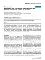
Figure 3 demonstrates the practical usefulness of multi-
parameter intracellular flow cytometry to compare and
Available online />Figure 1
Assessment of extracellular regulated kinase (ERK), jun N-terminal kinase (JNK) and p38 mitogen activated protein kinase (MAPK) activity in human
B lymphocytes by intracellular flow cytometric analysis. R2G6 cells (3 × 10
5
) were fixed, permeabilized, incubated in the presence or absence of a
blocking peptide and stained with isotype matched control antibodies or phosphospecific antibodies that recognize pERK, pJNK and p-p38
(a) immediately or (b) following incubation with increasing amounts of rCD154 for 15 min at 37°C. Dot plots of cell size on the x-axis versus
fluorescence intensity in the channel that detects the fluorochrome conjugated secondary antibody on the y-axis are shown in (a). Histograms of
fluorescence intensity of the channel that detects the fluorochrome conjugated secondary antibody are shown in (a) and (b). The percentage of
positive cells and the mean fluorescence intensity (MFI) of positive staining are indicated. The solid line indicates the division between negative
background staining of isotype-matched control antibody and positive staining with the pMAPK antibody. (c) Western blot analysis of pERK, pJNK
or p-p38 expression in R2G6 cells following incubation with rCD154 for 15 min at 37°C.
Control Ab
Cell size
Fl
uor
escence
i
nt
ensi
t
y
p-ERK
p-JNK
p-p38
M1
M1
M1
M1
M1
M1
M1
M1
M1
pMAPK Ab
pMAPK Ab
+ peptide
9%
MFI=100
17%
MFI=71
30%
MFI=56
p-ERK p-JNK p-p38
Unstim,
Control Ab
Unstim,
p-Specific Ab
CD154 Stim,
p-Specific Ab
85%
MFI=1280
95%
MFI=2797
36%, MFI=239
8%
MFI=96
85%, MFI=
330
83%
MFI=259
CD154
unstim
(a)
(b) (c)
34
Arthritis Research & Therapy Vol 6 No 1 Grammer et al.
Figure 2
Assessment of NF-κB activation in human B lymphocytes by multiparameter intracellular flow cytometric analysis of IκB isoforms (-α, -β, -ε) and the
phosphorylation status of IκBα. R2G6 cells (3 × 10
5
) were incubated with rCD154 in the presence or absence of 30 µM of the proteosome
inhibitor lactacystein (LAC) or the inhibitor of IκB phosphorylation BAY11-7082 (BAY; Calbiochem) for 15 min at 37°C. Cells were fixed,
permeabilized and stained with isotype-matched control antibody or antibody that recognize (a) IκBα, pIκBα, (b) IκBβ or IκBε. Two independent
experiments are presented in both (a) and (b). Experiment 1 in (a) depicts IκBα staining following CD154 stimulation in the presence or absence of
LAC. Experiment 2 in (b) depicts IκBα and pIκBα staining in the presence or absence of LAC. It is important to note that R2G6 cells are known to
have a high level of constitutive NF-κB activation, accounting for the large percentage of cells expressing p-IκBα. Experiment 1 in (b) depicts
staining for IκBβ following stimulation with CD154 in the presence or absence of LAC. Experiment 2 in (b) depicts staining for IκBε following
stimulation with CD154 in the presence or absence of LAC. Dot plots of cellular size or complexity on the x-axis versus fluorescence intensity in the
channel that detects the fluorochrome conjugated secondary antibody on the y-axis are shown in (a) and (b). Insets of histograms of fluorescence
intensity of the channel that detects the fluorochrome conjugated secondary antibody are shown in (a) and (b). The percentage of positive cells and
the mean fluorescence intensity (MFI) of positive staining are indicated. The solid line indicates the division between negative background staining
of isotype-matched control antibody and positive staining with the antibody that recognizes a specific IκB. (c) Western blot analysis of pIκBα as
well as total IκBα, -β and -ε following CD154 stimulation in the presence or absence of LAC or BAY11-7082 (BAY).
M1
M1
M1
M1
M1
M1
M1
IκBα
12%
MFI=21
5%
MFI=14
58%
MFI=9
4%
MFI=1
41%
MFI=5
61%
MFI=6
14%
MFI=17
Cell complexity (SSC)
Control
IκBα
Control
p-IκBα
p-IκBα
IκBα
Unstim
CD154
CD154 +
LAC
Expt. 1
Expt. 2
Cell size (FSC)
IκBβ
Unstim
CD154
CD154 +
LAC
M1
45%
MFI=36
M1
37%
MFI=23
M1
49%
MFI=31
IκBε
M1
M1
M1
23%
MFI=101
9%
MFI=66
31%
MFI=11
4
- IκBα
- IκBβ
- IκBε
- actin
CD154
unstim
- pIκBβ
- actin
CD154
unstim
–
BAY
LAC
(a)
(b) (c)
35
contrast signaling status of lymphocytes in patients with
autoimmune diseases, such as SLE, with lymphocytes
from normal, nonautoimmune individuals. PBMCs from
active SLE patients or normal individuals were stained for
CD19 and then for phosphorylated IκBα, ERK, JNK or
p38. Although many groups have demonstrated that
surface proteins can be stained before or after the
phosphospecific antibodies, it should be noted that
surface proteins should always be stained after kinase
staining when using indirect methods with a fluorochrome-
conjugated secondary to eliminate cross-reactivity of the
secondary with the antibody to the surface protein. Seven
active SLE patients and seven normal control individuals
were examined. The percentage of pIκBα
+
cells (Fig. 3)
and the level, or mean fluorescence intensity (MFI), of
pIκBα staining (data not shown) was not significantly
different in CD19
+
-gated B cells from active SLE patients
or normal control individuals. By contrast, there was a
significantly higher percentage of CD19
+
B cells from
active SLE patients that were positive for pERK, pJNK and
p-p38 compared with CD19
+
B cells from normal control
individuals (Fig. 3). The level, or MFI, of staining for pERK,
pJNK and p-p38 was not significantly different in SLE or
normal B cells (data not shown). These experiments have
demonstrated that assessment of signaling status in
lymphocytes from lymphopenic patients with autoimmune
disease is possible and informative. The importance of the
development of a sensitive assay of kinase activation in a
small number of cells is particularly important for analysis
of peripheral B cells from human patients. The specific
experiments in Figs 1–3 show that a novel multiparameter
intracellular flow cytometric method has been developed
to analyze both the MAPK and NF-κB signaling cascades
in a small number of primary B cells from human patients.
This technique will allow assessment of kinase activation
in situations where cell numbers are limiting and/or the
number of samples is great, such as in a clinical trial.
Advantages and disadvantages of multiparameter
intracellular flow cytometric analysis of lymphocyte
signaling status
Multiparameter intracellular flow cytometric analysis of sig-
naling status of human lymphocytes was pioneered
10 years ago by Bernard Rossi’s laboratory at the
INSERM in Nice, France [1]. Many groups have followed
up the initial report of Rossi and colleagues that cells
containing proteins with pTyr residues could be individu-
ally identified immediately ex vivo or following in vitro
stimulation by surface staining in combination with intra-
cellular staining with a phosphospecific antibody.
Presently, there are now published reports of the utiliza-
tion of multiparameter intracellular flow cytometry to iden-
tify specifically not only phosphorylated residues, but also
individual phosphorylated proteins such as STATs,
MAPKs, AKT [5,8,35], BTK [36] and VASP [37] in cells
from many sources. Flow cytometric analysis of kinase
activation utilizing phosphospecific antibodies to the
kinases listed above has been utilized to detect these acti-
vated signaling proteins in peripheral blood (platelets, lym-
phocytes, malignant cells), cord blood (lymphocytes),
G-CSF mobilized CD34
+
stem cells and bone marrow
(lymphocytes precursors, malignant cells).
There are numerous advantages to multiparameter intra-
cellular flow cytometric analysis of signaling status in cells
compared to traditional biochemical techniques that
measure cytoplasmic events such as in vitro kinase assays
or Western blotting with phosphospecific antibodies and
nuclear events with EMSA or with transfected reporter
constructs that assay the presence of specific activated
transcription factors. The primary advantage is that the
flow cytometric technique requires many fewer cells and is
at least 10-fold more sensitive when parallel samples are
compared to Western blotting with the same phosphos-
pecific antibodies. The ability to examine signaling status
in a small number of cells expands the source of cells that
can be examined to include such populations as the rare
CD34
+
stem cells that may number less than one in a
hundred in a G-CSFmobilized sample at one extreme to
rare B cell populations in lymphopenic patients such as
plasma cells which are often 0.1% of the circulating B cell
pool, which in lymphopenic SLE patients may be less than
0.05% of the PBMCs. In addition, rare subpopulations of
circulating lymphocytes can be examined for signaling
status in a multiparameter fashion using many fluo-
rochrome conjugated antibodies to surface proteins to
identify multiple subsets and/or many different phosphos-
pecific antibodies to unique cellular proteins. Moreover,
signaling status in many cellular subsets can be examined
Available online />Figure 3
Multiparameter intracellular flow cytometry reveals elevated
percentages of B lymphocytes in the periphery of systemic lupus
erythematosus (SLE) patients with spontaneous activation of
extracellular regulated kinase (ERK), jun N-terminal kinase (JNK) and
p38 mitogen activated protein kinases (MAPKs). Peripheral B
lymphocytes isolated from SLE patients (n = 7) or nonautoimmune
normal individuals (n = 7) were fixed, permeabilized and stained with
phosphospecific antibody for pIκBα, pERK, pJNK or p-p38. The
mean ± SEM percentages of CD19
+
B cells positive for each
phospho-Ab are shown graphically. *P < 0.05 by Student’s t test.
0 50 100
p-p38
pJNK
pERK
pIκBα
% B cells positive for phosphoprotein
normal
SLE
*
*
*
*
*
*
36
simultaneously with dyes that discriminate live and dead
cells such as ethidium monoazide. The second major
advantage to the multiparameter flow cytometric tech-
nique to measure signaling status in cellular subsets is
that many studies have demonstrated that cryopreserved
cells are not much different to fresh cells in terms of sig-
naling status if preserved and stored correctly. This allows
serial samples, as may occur during a clinical trial, to be
stored as they arrive and then stained at one time.
Although theoretically this serial analysis of signaling
status would be possible with biochemical techniques, the
large number of purified cells required at each time point
often makes this approach unfeasible. The third advantage
is that samples can be rapidly fixed and permeabilized at
the whole blood stage, thus preserving the immediate ex
vivo examination of signaling status that may be altered by
the cellular subset purification that is required for bio-
chemical techniques.
There are several important points to be highlighted when
performing multiparameter intracellular flow cytometric
analysis of cellular signaling status. Importantly, temperature
control of samples from the harvest to the time of the experi-
ment must be monitored carefully. Often, the temperature of
the samples and the time period that elapses until the
researcher receives the sample after harvest are not easily
controlled. For this reason, a brief preincubation period at a
constant temperature should be employed to normalize
baseline cellular activity. Furthermore, if samples are ana-
lyzed following stimulation with an exogeneous agent, the
time of activation should be as brief as possible to be sure
that direct signaling events following stimulation are being
analyzed.
During the development of a new phosphospecific experi-
mental techniques, there are several controls that are
useful to demonstrate specific staining of the phospho-
protein. One approach is to use cells from subjects or mice
genetically deficient in the kinase itself or an upstream
kinase [7,10]. Another approach is to demonstrate
reversibility of staining in the presence of the immunogen
peptide as shown in Fig. 1 or in the presence of a known
inhibitor of the signaling pathway as shown in Fig. 2.
Data analysis is an important aspect of multiparameter
flow cytometric analysis of signaling status of cells. The
flow cytometric technique allows simultaneous gating of
multiple cellular subsets within a sample. In addition, the
percentage of cells positive for a given phosphoprotein as
well as the level of the phosphoprotein that is directly pro-
portional to the MFI of staining for the phosphoprotein can
be assessed. Each of these parameters alone as well as
an index can be used to assess signaling status of a given
phosphoprotein. For example, biochemical techniques
such as Western blotting give a measure of total phospho-
protein present in a particular sample that takes into
account both the percentage of the population that is pos-
itive as well as the level of expression in that population. In
some instances, an index of phosphoprotein staining that
is the percentage of the population that is positive for a
phosphoprotein multiplied by the MFI that is directly pro-
portional to the expression level of the phosphoprotein
can be used to compare signaling status results obtained
by Western blotting directly versus multiparameter flow
cytometric staining.
There are a few disadvantages to multiparameter flow
cytometric analysis of cellular signaling status when com-
pared with biochemical techniques. These include the
necessity for an antibody that recognizes the phosphory-
lated form of the protein. Biochemically, the signaling state
of a protein can be examined in the absence of such
reagents directly by antibody immunoprecipitation of the
protein followed by an in vitro kinase assay with substrate
and radioactively labeled ATP or indirectly by transfection
with a reporter construct that measures a downstream
outcome of kinase activation such as ERK activation of
AP-1. Secondly, multiparameter flow cytometric analysis
of cellular signaling status cannot easily discriminate
between events that occur cytoplasmically versus in the
nucleus. By contrast, biochemical purification of lysates
from either cytoplasm or nucleus can be used for in vitro
kinase assays or EMSAs described above. Finally, multipa-
rameter flow cytometric analysis of signaling status with
phosphospecific antibodies cannot reliably tell the differ-
ence between kinase isoforms, such as ERK1 (p42) and
ERK2 (p44) that can be easily detected by Western blot-
ting (Fig. 1c). As a corollary, one can only state the total
phosphorylated state of a particular kinase. For example, if
cells expresss both ERK1 and ERK2, following a given
stimulation one cannot tell if one or both ERK isoforms is
phosphorylated. By contrast, this question can be easily
answered by biochemical immunoprecipitation of each
isoform individually followed by in vitro kinase assays. In
summary, the advantages of multiparameter intracellular
flow cytometric analysis of cellular signaling status are
numerous and, if one has enough cells from a given sample
and would like to ask the questions brought up as disadvan-
tages to the flow cytometric technique, one can do the bio-
chemical experiments in parallel.
Conclusions
Multiparameter intracellular flow cytometry is a valuable
technique that has been developed over the last 10 years
from the primitive analysis of total pTyr-containing proteins
in activated primary human T cells to the current ability to
follow specific phosphorylated proteins, such as those in
the MAPK and NF-κB signaling cascades, in rare lympho-
cyte populations as might be found in lymphopenic SLE
patients. Fixation of the cells to be analyzed is important to
cross-link and stabilize the cellular structure in preparation
for permeabilization and access of the phosphospecific
Arthritis Research & Therapy Vol 6 No 1 Grammer et al.
37
antibody to their targets, although the exact percentage of
paraformaldehyde (range 1–4%) used for this procedure
does not seem to be of great importance. Effective perme-
abilization of fixed cells has been achieved with methanol,
Triton X-100 or saponin. Importantly, a recent study has
compared these various fixation and permeabilization con-
ditions and concluded that they all work, but that 1.5%
paraformaldehyde followed by methanol treatment gives
optimal results [8]. Surface staining can be performed
before or after intracellular staining but conditions should
be tested as new surface stains are incorporated in the
assay. In addition, various permeabilization conditions may
individually affect particular surface stains. Furthermore,
cryopreserved and fresh samples gave equivalent results
for phosphoproteins tested, but this variable should be
confirmed for each phosphoprotein analyzed.
In summary, multiparameter intracellular flow cytometry is
a valuable tool to assess signaling status quantitatively in a
variety of lymphoctye populations. This technique will
allow assessment of kinase activation in situations where
cell numbers are limiting and/or the number of samples is
great, such as in clinical trials of patients with autoimmune
diseases.
Competing interests
None declared.
References
1. Far DF, Peyron JF, Imbert V, Rossi B: Immunofluorescent quan-
tification of tyrosine phosphorylation of cellular proteins in
whole cells by flow cytometry. Cytometry 1994, 15:327-334.
2. Far DF, Rossi B: Measurement of protein tyrosine phosphory-
lation in T-cell subsets by flow cytometry. Methods Mol Biol
2000, 134:301-306.
3. Vuillier F, Scott-Algara D, Cayota A, Siciliano J, Nugeyre MT,
Dighiero G: Flow cytometric analysis of protein-tyrosine phos-
phorylation in peripheral T cell subsets. Application to healthy
and HIV-seropositive subjects. J Immunol Methods 1995, 85:
43-56.
4. Hubert P, Grenot P, Autran B, Debre P: Analysis by flow cytom-
etry of tyrosine-phosphorylated proteins in activated T-cell
subsets on whole blood samples. Cytometry 1997, 29:83-91.
5. Perez OD, Nolan GP: Simultaneous measurement of multiple
active kinase states using polychromatic flow cytometry. Nat
Biotechnol 2002, 20:155-162.
6. Tsao PW, Mills GB, Diaz RJ, Radde IC, Parkinson D, Waddell J,
Wilson GJ, Coles JG: Evidence that increases in lymphocyte
tyrosine phosphorylation precede cardiac allograft rejection.
Effects of cyclosporine and potential use in clinical manage-
ment. Transplantation 1994, 58:451-457.
7. Fleisher TA, Dorman SE, Anderson JA, Vail M, Brown MR, Holland
SM: Detection of intracellular phosphorylated STAT-1 by flow
cytometry. Clin Immunol 1999, 90:425-430.
8. Krutzik PO, Nolan GP: Intracellular phospho-protein staining
techniques for flow cytometry: monitoring single cell signal-
ing events. Cytometry 2003 55A:61-70.
9. Pallis M, Seedhouse C, Grundy M, Russell N: Flow cytometric
measurement of phosphorylated STAT5 in AML: lack of spe-
cific association with FLT3 internal tandem duplications. Leuk
Res 2003, 27:803-805.
10. Uzel G, Frucht DM, Fleisher TA, Holland SM: Detection of intra-
cellular phosphorylated STAT-4 by flow cytometry. Clin
Immunol 2001, 100:270-276.
11. Chow S, Patel H, Hedley DW. Measurement of MAP kinase
activation by flow cytometry using phospho-specific antibod-
ies to MEK and ERK: potential for pharmacodynamic monitor-
ing of signal transduction inhibitors. Cytometry 2001, 46:72-
78.
12. Criscione LG, Pisetsky DS: B lymphocytes and systemic lupus
erythematosus. Curr Rheumatol Rep 2003, 5:264-269.
13. Tsokos GC, Nambiar MP, Tenbrock K, Juang YT: Rewiring the T-
cell: signaling defects and novel prospects for the treatment
of SLE. Trends Immunol 2003, 24:259-263.
14. Khan IU, Tsokos GC, Kammer GM: Abnormal B cell signal
transduction in systemic lupus erythematosus. Curr Dir
Autoimmun 2003, 6:89-104.
15. Grammer AC, Lipsky PE: CD154-CD40 interactions mediate
differentiation to plasma cells in healthy individuals and
persons with systemic lupus erythematosus. Arthritis Rheum
2002, 46:1417-1429.
16. Peng SL, Gerth AJ, Ranger AM, Glimcher LH: NFATc1 and
NFATc2 together control both T and B cell activation and dif-
ferentiation. Immunity 2001, 14:13-20.
17. Ranger AM, Oukka M, Rengarajan J, Glimcher LH: Inhibitory
function of two NFAT family members in lymphoid homeosta-
sis and Th2 development. Immunity 1998, 9:627-635.
18. Shai R, Quismorio Jr FP, Li L, Kwon OJ, Morrison J, Wallace DJ,
Neuwelt CM, Brautbar C, Gauderman WJ, Jacob CO: Genome-
wide screen for systemic lupus erythematosus susceptibility
genes in multiplex families. Hum Mol Genet 1999, 8:639-644.
19. Gray-McGuire C, Moser KL, Gaffney PM, Kelly J, Yu H, Olson JM,
Jedrey CM, Jacobs KB, Kimberly RP, Neas BR, Rich SS, Behrens
TW, Harley JB: Genome scan of human systemic lupus ery-
thematosus by regression modeling: evidence of linkage and
at 4p16-15.2. Am J Hum Genet 2000, 67:1460-1469.
20. Gaffney PM, Kearns GM, Shark KB, Ortmann WA, Selby SA,
Malmgren ML, Rohlf KE, Ockenden TC, Messner RP, King RA,
Rich SS, Behrens TW: A genome-wide search for susceptibil-
ity genes in human systemic lupus erythematosus sib-pair.
Proc Natl Acad Sci U S A 1998, 95:14875-14879.
21. Moser KL, Neas BR, Salmon JE, Yu H, Gray-McGuire C, Asundi
N, Bruner GR, Fox J, Kelly J, Henshall S, Bacino D, Dietz M,
Hogue R, Koelsch G, Nightingale L, Shaver T, Abdou NI, Albert
DA, Carson C, Petri M, Treadwell EL, James JA, Harley JB:
Genome scan of human systemic lupus erythematosus: evi-
dence for linkage on chromosome 1q in African-American
pedigrees. Proc Natl Acad Sci U S A 1998, 95:14869-14874.
22. Lin KI, Tunyaplin C, Calame K: Transcriptional regulatory cas-
cades controlling plasma cell differentiation. Immunol Rev
2003, 194:19-28.
23. Mainiero F, Colombara M, Antonini V, Strippoli R, Merola M, Poffe
O, Tridente G, Ramarli D: p38 MAPK is a critical regulator of
the constitutive and the beta4 integrin-regulated expression
of IL-6 in human normal thymic epithelial cells. Eur J Immunol
2003, 33:3038-3048.
24. Minges Wols HA, Underhill GH, Kansas GS, Witte PL: The role
of bone marrow-derived stromal cells in the maintenance of
plasma cell longevity. J Immunol 2002, 169:4213-4221.
25. Poudrier J, Weng X, Kay DG, Pare G, Calvo EL, Hanna Z, Kosco-
Vilbois MH, Jolicoeur P: The AIDS disease of CD4C/HIV trans-
genic mice shows impaired germinal centers and
autoantibodies and develops in the absence of IFN-gamma
and IL-6. Immunity 2001, 15:173-185.
26. Kitani A, Hara M, Hirose T, Harugai M, Suzuki K, Kawakami M,
Kawaguchi Y, Hidaka T, Kawagoe M, Nakamura H: Autostimula-
tory effects of IL-6 on excessive B cell differentiation in
patients with systemic lupus erythematosus: analysis of IL-6
production and IL-6R expression. Clin Exp Immunol 1992, 88:
75-83.
27. Linker-Israeli M, Deans RJ, Wallace DJ, Prehn J, Ozeri-Chen T, Kli-
nenberg JR: Elevated levels of endogenous IL-6 in systemic
lupus erythematosus. J Immunol 1991, 147:117-123.
28. Jun JE, Goodnow CC: Scaffolding of antigen receptors for
immunogenic versus tolerogenic signaling. Nat Immunol
2003, 4:1057-1064.
29. Anandasabapathy N, Ford GS, Bloom D, Holness C, Paragas V,
Seroogy C, Skrenta H, Hollenhorst M, Fathman CG, Soares L:
GRAIL: an E3 ubiquitin ligase that inhibits cytokine gene tran-
scription is expressed in anergic CD4+ T cells. Immunity 2003,
18:535-547.
30. Yi Y, McNerney M, Datta SK: Regulatory defects in Cbl and
mitogen-activated protein kinase (extracellular signal-related
Available online />38
kinase) pathways cause persistent hyperexpression of CD40
ligand in human lupus T cells. J Immunol 2000, 165:6627-
6634.
31. Grammer AC, McFarland RD, Heaney J, Darnell BF, Lipsky PE:
Expression, regulation, and function of B cell-expressed
CD154 in germinal centers. J Immunol 1999, 163:4150-4159.
32. Grammer AC, Slota R, Fischer R, Gur H, Girschick H, Yarboro C,
Illei GG, Lipsky PE: Abnormal germinal center reactions in sys-
temic lupus erythematosus demonstrated by blockade of
CD154-CD40 interactions. J Clin Invest 2003, 112:1506-1520.
33. Grammer AC, Lipsky PE: CD40-mediated regulation of
immune responses by TRAF-dependent and TRAF-indepen-
dent signaling mechanisms. Adv Immunol 2000, 76:61-178.
34. Grammer AC, Swantek JL, McFarland RD, Miura Y, Geppert T,
Lipsky PE: TNF receptor-associated factor-3 signaling medi-
ates activation of p38 and Jun N-terminal kinase, cytokine
secretion, and Ig production following ligation of CD40 on
human B cells. J Immunol 1998, 161:1183-1193.
35. Tazzari PL, Cappellini A, Bortul R, Ricci F, Billi AM, Tabellini G,
Conte R, Martelli AM: Flow cytometric detection of total and
serine 473 phosphorylated Akt. J Cell Biochem 2002, 86:704-
715.
36. Nisitani S, Kato RM, Rawlings DJ, Witte ON, Wahl MI: In situ
detection of activated Bruton’s tyrosine kinase in the Ig sig-
naling complex by phosphopeptide-specific monoclonal anti-
bodies. Proc Natl Acad Sci U S A 1999, 96:2221-2226.
37. Schwarz UR, Geiger J, Walter U, Eigenthaler M: Flow cytometry
analysis of intracellular VASP phosphorylation for the assess-
ment of activating and inhibitory signal transduction path-
ways in human platelets: definition and detection of
ticlopidine/clopidogrel effects. Thromb Haemost 1999, 82:
1145-1152.
Correspondence
Amrie C Grammer, B cell Biology Group Leader, Autoimmunity Branch,
NIAMS, NIH, 9000 Rockville Pike, Bldg 10, Rm 6D47A, Bethesda, MD
20892, USA. Tel: +1 301 594 3493; fax: +1 301 402 2209; email:
Arthritis Research & Therapy Vol 6 No 1 Grammer et al.