Báo cáo y học: "CD14 mediates the innate immune responses to arthritopathogenic peptidoglycan–polysaccharide complexes of Gram-positive bacterial cell walls" docx
Bạn đang xem bản rút gọn của tài liệu. Xem và tải ngay bản đầy đủ của tài liệu tại đây (423.6 KB, 9 trang )
Open Access
Available online />R273
Vol 6 No 3
Research article
CD14 mediates the innate immune responses to
arthritopathogenic peptidoglycan–polysaccharide complexes of
Gram-positive bacterial cell walls
Xiangli Li
1,2
, BlairUBradford
3
, Frederick Dalldorf
4
, Sanna M Goyert
5
, Stephen A Stimpson
6
,
Ronald G Thurman
3
and Sergei S Makarov
2,7,8
1
Department of Nutrition, University of North Carolina at Chapel Hill, Chapel Hill, North Carolina, USA
2
Thurston Arthritis Center, University of North Carolina at Chapel Hill, Chapel Hill, North Carolina, USA
3
Department of Pharmacology, University of North Carolina at Chapel Hill, Chapel Hill, North Carolina, USA
4
Department of Pathology, University of North Carolina at Chapel Hill, Chapel Hill, North Carolina, USA
5
Division of Molecular Medicine, North Shore University Hospital, Cornell University Medical College, Manhasset, New York, USA
6
GlaxoSmithKline, Research Triangle Park, North Carolina, USA
7
Department of Endodontics, University of North Carolina at Chapel Hill, Chapel Hill, North Carolina, USA
8
Comprehensive Center for Inflammatory Disorders, University of North Carolina at Chapel Hill, Chapel Hill, North Carolina, USA
Corresponding author: Sergei S Makarov,
Received: 30 Dec 2003 Revisions requested: 16 Jan 2004 Revisions received: 27 Feb 2004 Accepted: 15 Mar 2004 Published: 27 Apr 2004
Arthritis Res Ther 2004, 6:R273-R281 (DOI 10.1186/ar1175)
http://arthr itis-research.com/conte nt/6/3/R273
© 2004 Li et al.; licensee BioMed Central Ltd. This is an Open Access article: verbatim copying and redistribution of this article are permitted in all
media for any purpose, provided this notice is preserved along with the article's original URL.
Abstract
Bacterial infections play an important role in the multifactorial
etiology of rheumatoid arthritis. The arthropathic properties of
Gram-positive bacteria have been associated with
peptidoglycan–polysaccharide complexes (PG-PS), which are
major structural components of bacterial cell walls. There is little
agreement as to the identity of cellular receptors that mediate
innate immune responses to PG-PS. A
glycosylphosphatidylinositol-linked cell surface protein, CD14,
the lipopolysaccharide receptor, has been proposed as a PG-
PS receptor, but contradictory data have been reported. Here,
we examined the inflammatory and pathogenic responses to
PG-PS in CD14 knockout mice in order to examine the role for
CD14 in PG-PS-induced signaling. We found that PG-PS-
induced responses in vitro, including transient increase in
intracellular calcium, activation of nuclear factor-κB, and
secretion of the cytokines tumor necrosis factor-α and
interleukin-6, were all strongly inhibited in CD14 knockout
macrophages. In vivo, the incidence and severity of PG-PS
induced acute polyarthritis were significantly reduced in CD14
knockout mice as compared with their wild-type counterparts.
Consistent with these findings, CD14 knockout mice had
significantly inhibited inflammatory cell infiltration and synovial
hyperplasia, and reduced expression of inflammatory cytokines
in PG-PS arthritic joints. These results support an essential role
for CD14 in the innate immune responses to PG-PS and
indicate an important role for CD14 in PG-PS induced
arthropathy.
Keywords: bacterial, cell surface molecules, rheumatoid arthritis, transcription factors
Introduction
Rheumatoid arthritis (RA) is characterized by a chronic,
erosive inflammation of peripheral joints. The etiology of RA
is not known, but both genetic and environmental factors
contribute to the disease. Gram-positive bacterial infection
is one environmental factor that has been linked with RA
pathology [1]. The link between systemic bacterial infection
and RA is based on observations that bowel-related dis-
eases such as Crohn's disease and ulcerative colitis are
frequently complicated by arthritis, that RA patients have
significantly elevated levels of antibodies to bacterial prod-
ucts (e.g. antipeptidoglycan antibodies), and that bacterial
products can frequently be found in RA synovial fluids and
tissues [2-5]. The arthropathogenic properties have been
attributed to poorly degradable peptidoglycan–polysac-
charide complexes (PG-PS) – covalently bound polymers
[Ca
2+
]
i
= intracellular calcium concentration; DMEM = Dulbecco's modified Eagle's medium; GAPDH = glyceraldehyde-3-phosphate dehydrogenase;
IL = interleukin; LPS = lipopolysaccharide; NF-κB = nuclear factor-κB; PAMP = pathogen-associated molecular patterns; PG-PS = peptidoglycan–
polysaccharide complexes; RA = rheumatoid arthritis; RT-PCR = reverse transcription polymerase chain reaction; TLR = Toll-like receptor; TNF =
tumor necrosis factor.
Arthritis Research & Therapy Vol 6 No 3 Li et al.
R274
that comprise cell walls of Gram-positive bacteria [2,6].
When injected in susceptible strains of animals, crude
preparations of bacterial cell walls or purified PG-PS pro-
duce disease with clinical features resembling those of
human RA [6].
Injected PG-PS accumulate in macrophages of the spleen,
liver, and mesenteric lymph nodes [7], resulting in persist-
ent cell activation and secretion of inflammatory cytokines.
The activation of macrophages by PG-PS represents an
example of innate immune response, a defensive reaction
based on recognition of conserved pathogen-associated
molecular patterns (PAMPs) shared by large groups of
micro-organisms (for review [8]). PAMP recognition in
mammals is mediated by the Toll-like receptor (TLR) family
of pattern recognition receptors. Engagement of TLRs trig-
gers signal transduction pathways that culminate in the
activation of transcription of defensive genes. Individual
TLRs produce overlapping but distinct patterns of gene
expression, permitting tailoring of host responses to differ-
ent pathogens. The precise molecular mechanisms
whereby distinct PAMPs activate individual TLRs are not
well defined. Recognition of lipopolysaccharide (LPS), a
major component of the outer membrane of Gram-negative
bacteria, is the best studied model of innate immunity [9].
In serum, LPS molecules are opsonized by LPS-binding
proteins that recruit LPS to CD14 – a glycosylphosphati-
dylinositol-anchored cell surface protein that is expressed
by phagocytic cells. The tertiary LPS–LPS binding protein–
CD14 complex generates the transmembrane signal of cell
activation through an association with the TLR-4 receptor.
A principal target and mediator of the TLR-4-induced sign-
aling is the transcription factor nuclear factor-κB (NF-κB),
which controls the expression of a variety of proinflamma-
tory cytokines, including IL-1, IL-6, and tumor necrosis fac-
tor (TNF)-α [8,10].
It is less clear which primary events and cellular receptors
mediate the innate immune responses to and the arthri-
topathogenic properties of PG-PS. The intracellular signal-
ing that occurs in response to stimulation with
peptidoglycans of Gram-positive bacteria is triggered by
TLR-2 [11]. A number of in vitro studies have suggested
that, similar to the involvement of CD14 in LPS signaling,
CD14 plays an essential role in the activation of PG-PS
induced innate immune responses [12-16]. However, there
are reports that peptidoglycans can directly bind TLR-2
[17] and activate cells independently of CD14 [18]. Stud-
ies conducted in vivo have also yielded contradictory
results. Whereas CD14 knockout mice were resistant to
Gram-negative bacteria-induced and LPS-induced shock
[19], CD14-deficient mice, when challenged with live or
killed Gram-positive bacteria, had survival rates similar to
those in wild-type animals [20].
In order to elucidate the role played by CD14 in the innate
immune response to PG-PS in vitro and in vivo, we
assessed the inflammatory responses to PG-PS in CD14
knockout primary macrophages and examined the develop-
ment of PG-PS induced arthritis in CD14 knockout mice.
Our findings demonstrate that, in CD14 knockout macro-
phages, PG-PS failed to activate inflammatory responses
in vitro, and that the development of PG-PS induced
arthropathy was significantly inhibited, albeit not abolished,
in CD14 knockout mice.
Materials and methods
Purified PG-PS (fraction 100P) from group A Streptococ-
cus pyogenes was purchased from Lee Labs (Garrison,
GA, USA). Endotoxin contamination was under 0.35 pg
endotoxin/µg PG-PS, as determined using a Limulus ame-
bocyte lysate assay system (Pyrogent
®
plus; Biowhittaker,
Walkersville, MD, USA). Therefore, the concentration of
endotoxin in in vitro experiments was under 35 pg/ml, even
at the highest (100 µg/ml) concentrations of PG-PS. All
reagents, unless otherwise noted, were from Sigma (St.
Louis, MO, USA) and the culture medium was from GIBCO
(Grand Island, NY, USA).
Animals and arthritis model
Female CD14 knockout mice [19] were backcrossed 10
times onto the BALB/c background, as described previ-
ously [20,21]. The expression of CD14 mRNA was evalu-
ated by using RNase protection assay, as described
previously [21]. Age-matched wild-type female BALB/c
mice were used as controls. To induce acute polyarthritis,
animals were injected intravenously with PG-PS (3 mg
rhamnose/kg). At this dose, the maximal amount of co-
injected contaminating endotoxin was less than 30 pg/
mouse. The development of arthritis was evaluated by
using a scoring system based on a scale from 0 to 4 for
each of four paws, as described elsewhere [22]: 0 = nor-
mal paw; 1 = swelling of individual digits; 2 = moderate
swelling and redness of ankle or wrist joints; 3 = swelling
and redness of at least two joints; and 4 = swelling of the
whole paw. According to this system, the total arthritis
score of a mouse varied in the range 0–16. To compare the
frequencies of arthritis in wild-type and CD14 knockout ani-
mals, each mouse that had any signs of arthritis (i.e. total
score ≥ 1) was considered positive. To minimize bias, ani-
mals were coded and assessed randomly.
Histologic analysis
Following the intravenous PG-PS injection on day 0, mice
were killed on the next day after the joint inflammation had
reached a peak (usually, on day 4). The hind ankle joint and
paws were fixed in 10% formalin, decalcified, paraffinized,
cut into 5 µm sections, deparaffinized, stained with hema-
toxylin–eosin, and evaluated histologically. Morphologic
assessments of arthritis were performed in a blinded
Available online />R275
manner using a scoring system with a scale from 0 (no
damage) to 4 (severe damage), based on the overall infiltra-
tion of inflammatory cells, synovial hyperplasia, and deposi-
tion of fibrinous exudates in the joints and surrounding
tissue [4].
Cell culture
Splenocytes were harvested as described elsewhere [23].
Cells were resuspended in Dulbecco's modified Eagle's
medium (DMEM) supplemented with 10% fetal bovine
serum, HEPES (10 mmol/l) and antibiotics (100 U/ml pen-
icillin G and 100 µg/ml streptomycin sulfate), and incu-
bated at 37°C in 5% carbon dioxide for 1 hour. The
nonadherent cells were washed out gently twice with phos-
phate-buffered saline and once with DMEM. Cell viability
was typically in excess of 95%, as assessed by trypan blue
exclusion. Peritoneal macrophages were collected as pre-
viously described [20]. Cells were recruited by an intraperi-
toneal injection of a sterile solution of 3% thioglycollate.
Three days after intraperitoneal injection, the peritoneal
exudates were collected and washed thee times, and cells
were allowed to adhere to plastic for 30 min, followed by
removal of nonadherent cells. Unless otherwise indicated,
cells were maintained for 24 hours before use in a DMEM
media at 37°C in 5% carbon dioxide.
Assessments of intracellular calcium
The intracellular calcium concentration ([Ca
2+
]
i
) in macro-
phages was measured using fura-2, a fluorescent [Ca
2+
]
i
indicator, as described elsewhere [23]. Macrophages were
plated on coverslips in a DMEM media supplemented with
10% fetal bovine serum at 24 hours before measurement.
Growth media was replaced with a modified Hanks' bal-
anced buffer (115 mmol/l NaCl, 5 mmol/l KCl, 0.3 mmol/l
Na
2
HPO
4
, 0.4 mmol/l KH
2
PO
4
, 5.6 mmol/l glucose, 0.8
mmol/l MgSO
4
, 1.26 mmol/l CaCl
2
, and 15 mmol/l HEPES;
pH = 7.4) containing 5 µmol/l acetoxymethyl ester of fura-
2 (Molecular Probes, Eugene, OR, USA). Cells were incu-
bated at room temperature for 30 min and stimulated with
indicated concentrations of PG-PS. The [Ca
2+
]
i
measure-
ments were performed in individual cells by using a micro-
spectrofluorometer (InCyt Im2™ Imaging, Cincinnati, OH,
USA) interfaced with an inverted microscope TMS-F
(Nikon, Kanagawa, Japan) [23]. The fluorescence intensity
was monitored at excitation wavelengths 340 and 380 nm,
and emission wavelength 510 nm. Each value was cor-
rected by subtracting the system dark noise and autofluo-
rescence. The [Ca
2+
]
i
was calculated as [Ca
2+
]
i
= Kd([R -
Rmin]/[Rmax - R]) × (F
0
/Fs) as previously described [23].
Cells were selected at random and analyzed using InCyt
Im2™ image acquisition and analysis software.
Assessments of nuclear factor-κB activation
Cellular localization of the RelA (p65) subunit of NF-κB was
detected by immunocytochemistry. Splenic macrophages
were stimulated with PG-PS for 30 min, fixed for 10 min
with ice cold methanol, and blocked for 30 min in a 10%
normal goat serum (Sigma). Cells were immunostained
using a primary polyclonal rabbit anti-RelA antibody (Rock-
land Immunochemicals, Gilbertsville, PA, USA) diluted at
1:200 in 10% normal goat serum, and visualized with the
secondary rhodamine isothiocyanate-conjugated antirabbit
antibody. Nuclei were counterstained with Hoechst 33342
(Molecular Probes).
RNA isolation and reverse transcription polymerase
chain reaction
Total RNA was extracted from homogenized tissues by
using Trizol reagent (Sigma). One nanogram of RNA was
subjected to reverse transcription by using MLV reverse
transcriptase (Promega, Madison, WI, USA). The resulting
cDNA was PCR amplified with the following primers: TNF-
α: 5'-TAC TGA ACT TCG GGG TGA TTG GTC C-3' (for-
ward) and 5'-CAG CCT TGT CCC TTG AAG AGA ACC-
3' (reverse); and glyceraldehyde-3-phosphate dehydroge-
nase (GAPDH): 5'-TGA AGG TCG GTG TCA ACG GAT
TTG-3' (forward) and 5'-GTA CAT CCG TAC TCC AGG
TGG TG-3' (reverse). In a typical PCR reaction, 50 ng
cDNA template was added to a mixture containing 75
µmol/l of primers, 2.5 mmol/l dNTPs, and 1 U Taq polymer-
ase. For detection of TNF-α message, the template was
amplified by 26 cycles of PCR (at the annealing tempera-
ture 56°C). GAPDH was amplified by using 24 cycles
(annealing temperature 63°C). The PCR products were vis-
ualized by agarose gel electrophoresis.
Statistical analysis
Unless otherwise indicated, all results are expressed as
means ± standard error of the mean. Statistical differences
between groups were determined using analysis of vari-
ance with Tukey's or Scheffe's post hoc test. Scores were
compared using the Mann–Whitney rank sum test. P <
0.05 was considered statistically significant.
Results
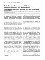
CD14 mediates the PG-PS-induced increase in
intracellular calcium concentration
PG-PS stimulation induces a rapid, transient increase in
[Ca
2+
]
i
, which is a prerequisite for induction of inflammatory
gene expression by PG-PS and LPS [23-25]. Here, we
compared the increase in [Ca
2+
]
i
in primary spleen macro-
phages from wild-type and CD14 knockout mice. Stimula-
tion of cells with PG-PS in a serum-free medium did not
induce an increase in [Ca
2+
]
i
(data not shown), which is
consistent with previously published data [14]. In the pres-
ence of serum, PG-PS caused a rapid and transient
increase in [Ca
2+
]
i
(Fig. 1a), which peaked at approximately
2 min after stimulation. This response was dose dependent
and reached a plateau at concentrations above 20 µg/ml
(Fig. 1b). The increase in [Ca
+2
]
i
was significantly reduced
Arthritis Research & Therapy Vol 6 No 3 Li et al.
R276
in CD14 knockout cells. At the saturating concentrations of
PG-PS (20 µg/ml), the maximal increase in [Ca
2+
]
i
in the
wild-type cells was 47 ± 5 nmol/l as compared with 12 ±
7 nmol/l in CD14 knockout cells (Fig. 1b), indicating that
the PG-PS-induced transient increase in [Ca
2+
]
i
was medi-
ated by CD14.
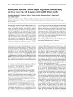
PG-PS-induced nuclear factor-κB activation is mediated
by CD14
The transcription factor NF-κB is a pivotal regulator of
genes that are involved in inflammation and immunity [26].
NF-κB is an essential component of TLR signaling [8]. The
activity of NF-κB is controlled by interaction an inhibitory
molecule known as IκB. Cell stimulation induces degrada-
tion of IκB, thereby allowing NF-κB to enter the nucleus
and initiate the transcription of target genes [27]. Thus, the
nuclear localization of NF-κB is indicative of NF-κB
activation.
To assess NF-κB activation, wild-type and CD14 knockout
spleen macrophages were stimulated with PG-PS and
immunostained with antibodies against the RelA (p65) sub-
unit of NF-κB. As shown in Fig. 2, NF-κB was largely cyto-
plasmic in resting cells (top panels) whereas PG-PS
stimulation induced nuclear translocation of NF-κB in the
majority of wild-type cells (middle panels). In contrast, only
few, if any, of the PG-PS-stimulated CD14 knockout cells
had nuclear NF-κB (lower panels), indicating that PG-PS-
induced NF-κB activation is CD14-dependent.
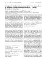
CD14 mediates PG-PS-induced production of
inflammatory cytokines
The signal transduction pathways activated by TLRs culmi-
nate in activation of transcription of defensive genes. The
expression of many inflammatory cytokines, including IL-1,
TNF-α, and IL-6, depends on NF-κB, because their promot-
ers contain NF-κB-binding sites, and specific suppression
of NF-κB blocks their production [28]. Here, we examined
the production of these cytokines in response to PG-PS.
Upon stimulation with PG-PS wild-type peritoneal macro-
phages secreted large quantities of IL-6 (Fig. 3a), whereas
the IL-6 production by CD14 knockout macrophages was
below the level of detection (<10 pg/ml). Similar to that
finding, PG-PS-stimulated CD14 knockout macrophages
produced much less TNF-α than did wild-type cells (Fig.
3b). The results in peritoneal macrophages were in good
agreement with those observed in spleen macrophages
(data not shown). Therefore, PG-PS-induced production of
inflammatory cytokines is CD14-dependent.
The severity of PG-PS-induced arthritis is attenuated in
CD14 knockout mice
To elucidate the role for CD14 in development of PG-PS-
induced arthropathy, CD14 knockout mice were back-
crossed on a BALB/c background, which is highly
Figure 1
Peptidoglycan–polysaccharide -induced increase in intracellular cal-cium concentration ([Ca
2+
]
i
) is inhibited in CD14 knockout cellsPeptidoglycan–polysaccharide (PG-PS)-induced increase in intracellu-
lar calcium concentration ([Ca
2+
]
i
) is inhibited in CD14 knockout cells.
The levels of [Ca
2+
]
i
in spleen macrophages were evaluated by using
fura-2, a fluorescent [Ca
2+
] indicator. (a) Kinetics of PG-PS-induced
[Ca
2+
]
i
(average from six to eight cells). Representative data from two
experiments are shown. (b) The average maximal increase in [Ca
2+
]
i
shown as a function of PG-PS concentration. Data represent means ±
standard error of the mean for 12–34 cells from two independent
experiments. The significance of the difference between the groups
was calculated using one-way analysis of variance with Scheffe's post-
hoc test. *P < 0.05 versus wild-type.
Available online />R277
susceptible to PG-PS-induced acute arthritis [22]. Intrave-
nous injection of PG-PS caused polyarthritis that reached
a peak at around 2 days after injection. At that time, each
animal in the wild-type group had developed arthritis (19/
19 mice [100%]), with an average arthritis index of 4.1 ±
0.5 (Fig. 4). In CD14 knockout mice, both the incidence of
arthritis (11/16 [68%]; P < 0.05) and the average arthritis
index (2.3 ± 0.5; P < 0.05) were significantly inhibited.
The pathomorphologic changes within arthritic joints were
consistent with those previously described in this model
[22] (i.e. the infiltration of inflammatory cells into the syn-
ovium, synovial hyperplasia, and fibrinous deposits; Fig.
5a). Infiltration of inflammatory cells was apparent in the
tibio-tarsal joint space, synovium, tendon, toes, and sur-
rounding soft tissues. Extensive deposition of fibrinous exu-
date in the tibio-tarsal joint and in the bursa of the Achilles'
tendon was also observed. There was neither pannus for-
mation nor destruction of cartilage and subchondral bone.
All of these changes were observed in some CD14 knock-
out mice as well, but the magnitude of these changes was
Figure 2
Peptidoglycan–polysaccharide -induced activation of nuclear factor-κB (NF-κB) is inhibited in CD14 knockout spleen macrophagesPeptidoglycan–polysaccharide (PG-PS)-induced activation of nuclear
factor-κB (NF-κB) is inhibited in CD14 knockout spleen macrophages.
Spleen macrophages were stimulated for 30 min with PG-PS at a con-
centration of 20 µg/ml. The left column shows immunostaining of the
p65 (RelA) subunit of NF-κB, and the right column shows counterstain-
ing of nuclei with Hoechst 33342. The rows show the following: top –
unstimulated cells from wild-type mice; middle – PG-PS stimulated
cells from wild-type mice; and bottom row – PG-PS-stimulated cells
from CD14 knockout mice. Representative data from three independ-
ent experiments are shown.
Figure 3
CD14 mediates peptidoglycan–polysaccharide complex -induced secretion of IL-6 and tumor necrosis factor (TNF)-αCD14 mediates peptidoglycan–polysaccharide complex (PG-PS)-
induced secretion of IL-6 and tumor necrosis factor (TNF)-α. Peritoneal
macrophages were stimulated for 4 hours with the indicated concentra-
tions of PG-PS. Concentrations of TNF-α and IL-6 were determined in
supernatants by ELISA. Each determination was done in duplicates. (a)
TNF-α production by peritoneal macrophages. Data represent means ±
standard error of the mean from three independent experiments. (b)
IL-6 production by peritoneal macrophages. Data represent mean ±
SEM from two independent experiments.
Arthritis Research & Therapy Vol 6 No 3 Li et al.
R278
much lower. On average, the morphologic arthritis score
was 3.3 ± 0.4 in wild-type mice as compared with 1.0 ± 0.5
in CD14 knockout animals (n = 8–10 mice/group; P <
0.05; Fig. 5b). The morphologic changes correlated well
with the gross observation data.
To assess inflammation at the molecular level, we examined
the expression of inflammatory cytokines TNF-α, IL-1β, and
IL-6 in arthritic joints by RT-PCR. In normal joints the mes-
sages were below the level of detection, but at the peak of
arthritis these cytokines were readily detectable in the wild-
type mice. The expression of TNF-α mRNA was substan-
tially lower in the arthritic joints of CD14 knockout mice
(Fig. 6). Similar to that finding, IL-6 message was inhibited
in the arthritic joints of CD14 knockout mice (data not
shown). Combined, these data indicate that the PG-PS-
induced arthropathy is significantly reduced in CD14
knockout animals.
Discussion
The etiologic agents of RA remain to be identified. How-
ever, in spite of the inability to isolate a specific infectious
organism from the joints of RA patients, there is evidence
suggesting that transient exposure to indigenous bacterial
products may provoke an initial response that could even-
tually perpetuate and amplify itself. This notion has been
supported by numerous animal studies. Single intraperito-
neal injection of crude bacterial cell walls or water-soluble
PG-PS from Gram-positive bacteria into susceptible
strains of rats induces polyarthritis that closely resembles
human RA. The joint lesions have a biphasic course with an
initial acute inflammation of the ankle, wrist, and small joints
of extremities, followed by chronic erosive arthritis (for
review [6]). The initial, acute, phase has features of the
innate immune response in that inflammation is driven by
neutrophils and macrophages and is T-cell independent.
The chronic stage of arthritis is T-cell dependent and thus
has features of the adaptive immune response [7,29]. Sys-
temic administration of Gram-positive bacterial cell walls or
PG-PS in susceptible strains of mice also produces arthri-
tis, although, for unknown reasons, mice develop only the
acute phase [22,30]. The minimal essential arthritogenic
structures have been identified as PG-PS, the major struc-
tural components of Gram-positive bacterial cell walls [31].
The peptidoglycan moiety is responsible for the patho-
genicity of PG-PS, because enzymatic digestion of the
peptidoglycan moiety eliminates the proinflammatory and
arthritopathogenic properties of PG-PS [32]. The pepti-
doglycan can substitute for PG-PS in the induction of the
acute [22] but not chronic phase of arthritis [2,33]; it has
been proposed that the polysaccharide moiety protects the
peptidoglycan moiety from degradation in vivo, thereby
facilitating chronic persistence of PG-PS in the host [2].
The primary events and the identity of cellular receptors
that mediate the innate immune response to and the
arthritogenic properties of peptidoglycan and PG-PS are
incompletely characterized. There is substantial evidence
that the transmembrane signal leading to cell activation in
response to stimulation with Gram-positive peptidoglycans
is triggered by TLR-2 [11]. CD14, has been proposed as
an intermediate, connecting peptidoglycan with TLR-2-
induced cell activation, but the role of CD14 is controver-
sial. On the one hand, neutralizing antibody to CD14 pre-
vented peptidoglycan-induced cell activation, and
transfection of CD14-negative cells with exogenous CD14
conferred responsiveness to peptidoglycan [14], suggest-
ing that CD14 plays an essential role. On the other hand,
peptidoglycan was shown to bind TLR-2 directly [17] and
to induce the activation of TLR-2-transfected cells regard-
less of CD14, although CD14 increased the affinity of PG–
Figure 4
Peptidoglycan–polysaccharide -induced polyarthritis is attenuated in CD14 knockout animals: gross observation scorePeptidoglycan–polysaccharide (PG-PS)-induced polyarthritis is attenu-
ated in CD14 knockout animals: gross observation score. Acute PG-
PS polyarthritis was induced by intravenous injection of PG-PS (3 mg/
kg). The arthritis index was scored as described in the Materials and
methods section. Each point represents a mean ± standard error of the
mean in 16–19 animals. The significance of the difference between the
groups was evaluated by Mann–Whitney rank sum test. *P < 0.05.
Available online />R279
Figure 5
Peptidoglycan–polysaccharide -induced polyarthritis is attenuated in CD14 knockout animals: morphological assessmentPeptidoglycan–polysaccharide (PG-PS)-induced polyarthritis is attenuated in CD14 knockout animals: morphological assessment. (a) Ankle joints
at day 3 after intravenous injection of PG-PS. The upper row shows the joint of a wild-type mouse and the lower row the joint of a CD14 knockout
mouse. Synovium (S), tibia (T), tarsal (Ta), joint space (J), cartilage (C), and fibrinous exudates (F) are indicated. Hematoxylin–eosin counterstaining
was employed and the original magnifications are as follows: left column 12.5× and right column 40×. Representative data from 10 joints in each
group are shown. (b) Histological score. The severity of arthritis was assessed as described in the Materials and methods section. Data are shown
as means ± standard error of the mean (n = 10). The significance of difference between groups was calculated by using the Mann–Whitney rank
sum test. *P < 0.05.
Arthritis Research & Therapy Vol 6 No 3 Li et al.
R280
TLR-2 interactions [17] and potentiated cell activation [18],
suggesting a facultative role for CD14. It is possible that
the discrepancies between the studies can be attributed to
differences in experimental conditions. In this regard, using
primary cells with genetically inactivated CD14 allowed for
clearer interpretation. We demonstrate that, in CD14
knockout macrophages, each step of PG-PS induced sig-
nal transduction, including the transient increase in [Ca
2+
]
i
,
nuclear translocation of NF-κB, and secretion of TNF-α and
IL-6, were almost completely suppressed. Thus, our data
strongly support an essential role for CD14 in the innate
immune responses to PG-PS.
To examine the role for CD14 in PG-PS induced arthropa-
thy, CD14 knockout mice were backcrossed to a suscepti-
ble BALB/c genetic background [22]. Because mice do
not develop chronic arthritis, we were restricted to exami-
nation of acute arthritis, which is driven by the innate
immune response to PG-PS. The CD14 knockout mice
developed arthritis significantly less frequently (68% versus
100% in wild-type group), and the severity of arthritis was
significantly reduced (average arthritis score of 2.5 versus
4.8 in the wild-type group). The gross observation data
largely correlated with morphologic assessments, the most
pronounced difference being in the degree of infiltration of
inflammatory cells. Because CD14 knockout macrophages
were largely unresponsive to PG-PS stimulation, these in
vivo results were not unexpected. It was rather surprising to
find that PG-PS was able to induce arthritis in a significant
proportion of CD14 knockout mice. The reason for that is
not clear. Our analysis of mRNA expression has shown that
inflammatory gene expression in arthritic joints of CD14
knockout mice was strongly inhibited but not abolished
(Fig. 6). It is possible that resident nonphagocytic cells
within joints can be activated by PG-PS via CD14-inde-
pendent mechanisms. In this regard, Kyburz and coworkers
[34] showed that stimulation of synovial fibroblasts with
staphylococcal peptidoglycan caused cell activation and
elevated expression of matrix metalloproteinases and
cytokines IL-6 and IL-8; it appeared that cell activation was
mediated by both TLR-2-dependent and -independent
pathways. It is not known whether CD14 is the major
receptor for PG-PS in each cell type; alternative receptors
have been proposed, including the peptidoglycan
recognition proteins and nucleotide-binding oligomeriza-
tion proteins (NODs) [35-37]. Nonetheless, our data indi-
cate that CD14 is an essential receptor for activation of the
innate immune response in macrophages by the arthritio-
genic PG-PS, and that CD14-dependent mechanisms sig-
nificantly contribute to PG-PS-induced arthropathy.
Conclusion
Bacterial infection has frequently been associated with RA
pathology. The arthropathic properties have been attrib-
uted to PG-PS, which are major structural components of
bacterial cell walls. The identity of receptors that mediate
the arthropathic properties of PG-PS is controversial. Here,
we examined the role played by CD14 in PG-PS-induced
arthritis in mice. To do so, we used CD14-deficient knock-
out mice. We found that CD14 knockout macrophages
were almost completely deficient in inflammatory
responses to PG-PS stimulation in vitro. In vivo, the inci-
dence and severity of PG-PS-induced polyarthritis in CD14
knockout mice were significantly reduced but not abol-
ished, as compared with their wild-type counterparts.
These results support an essential role for CD14 in the
innate immune responses to PG-PS and indicate an impor-
tant role for CD14 in PG-PS-induced arthropathy.
Competing interests
None declared.
Acknowledgments
The authors gratefully acknowledge the technical expertise of Mrs Julie
V Mitchell and Charlotte Walters at the Center for Gastrointestinal Biol-
ogy and Disease (P30 KD34987). This work was supported by NIH
public health grants AR/AI-44564, 5-P60 AR-30701-14, and AR/AI-
44030. SM is the recipient of an Investigator Award from The Arthritis
Foundation.
References
1. Firestein GS: Evolving concepts of rheumatoid arthritis. Nature
2003, 423:356-361.
2. Schwab JH: Phlogistic properties of peptidoglycan-polysac-
charide polymers from cell walls of pathogenic and normal-
flora bacteria which colonize humans. Infect Immun 1993,
61:4535-4539.
Figure 6
The expression of inflammatory cytokines in arthritic joints of mice with peptidoglycan–polysaccharide (PG-PS)-induced arthritisThe expression of inflammatory cytokines in arthritic joints of mice with
peptidoglycan–polysaccharide (PG-PS)-induced arthritis. Total RNA
was extracted from ankle joints, reversely transcribed, amplified by
PCR, and resolved on gel electrophoresis. The upper panel shows 24
cycles of amplification with tumor necrosis factor (TNF)-α specific prim-
ers, and the lower panel shows 22 cycles of amplification with glyceral-
dehyde-3-phosphate dehydrogenase (GAPDH)-specific primers. The
first two lanes on the left represent DNA molecular weight markers.
Lane (-) represents a negative control (RT-PCR amplification in the
absence of RNA sample). Lanes N1 and N2 represent RT-PCR ampli-
fied RNA from nonarthritic joints of normal (i.e. not injected with PG-PS)
wild-type and CD14 knockout animals, respectively. Lanes a–d repre-
sent RT-PCR amplified RNA from four different arthritic joints of PG-
PS-injected wild-type animals. Lanes e–g represent RT-PCR amplified
RNA from three arthritic joints of PG-PS injected CD14 knockout ani-
mals. Representative data from two independent experiments are
shown.
Available online />R281
3. van der Heijden IM, Wilbrink B, Tchetverikov I, Schouls LM, Hazen-
berg MP, Breedveld FC, Tak PP: Presence of bacterial DNA and
bacterial peptidoglycans in joints of patients with rheumatioid
arthritis and other arthritides. Arthritis Rheum 2000,
43:593-598.
4. Lichtman SN, Wang J, Sartor RB, Zhang C, Bender D, Dalldorf FG,
Schwab JH: Reactivation of arthritis induced by small bowel
bacterial overgrowth in rats: role of cytokines, bacteria, and
bacterial polymers. Infect Immun 1995, 63:2295-2301.
5. Gravallese EM, Kantrowitz FG: Arthritic manifestations of
inflammatory bowel disease. Am J Gastroenterol 1988,
83:703-709.
6. Schwab JH: Bacterial cell-wall induced arthritis: models of
chronic recurrent polyarthritis and reactivation of monoarticu-
lar arthritis. In Mechanisms and Models in Rheumatoid Arthritis
Edited by: Henderson B, Edwards JCW, Pettipher ER. London:
Academic Press; 1995:431-446.
7. Allen JB, Malone DG, Wahl SM, Calandra GB, Wilder RL: Role of
the thymus in streptococcal cell wall-induced arthritis and
hepatic granuloma-formation: comparative studies of pathol-
ogy and cell-wall distribution in athymic and euthymic rats. J
Clin Invest 1985, 76:1042-1056.
8. Janeway CA Jr, Medzhitov R: Innate immune recognition. Annu
Rev Immunol 2002, 20:197-216.
9. Beutler B: TLR4 as the mammalian endotoxin sensor. Curr Top
Microbiol Immunol 2002, 270:109-120.
10. Zhang G, Ghosh S: Toll-like receptor-mediated NF-kappaB
activation: a phylogenetically conserved paradigm in innate
immunity. J Clin Invest 2001, 107:13-19.
11. Takeuchi O, Hoshino K, Kawai T, Sanjo H, Takada H, Ogawa T,
Takeda K, Akira S: Differential roles of TLR2 and TLR4 in recog-
nition of gram-negative and gram-positive bacterial cell wall
components. Immunity 1999, 11:443-451.
12. Pugin J, Heumann D, Tomasz A, Kravchenko VV, Akamatsu Y,
Nishijima M, Glauser MP, Tobias PS, Ulevitch RJ: Cd14 is a pat-
tern-recognition receptor. Immunity 1994, 1:509-516.
13. Dziarski R, Tapping RI, Tobias PS: Binding of bacterial peptidog-
lycan to CD14. J Biol Chem 1998, 273:8680-8690.
14. Gupta D, Kirkland TN, Viriyakosol S, Dziarski R: CD14 is a cell-
activating receptor for bacterial peptidoglycan. J Biol Chem
1996, 271:23310-23316.
15. Rietschel ET, Schletter J, Weidemann B, El-Samalouti V, Mattern
T, Zahringer U, Seydel U, Brade H, Flad H-D, Kusumoto S, Gupta
D, Dziarski R, Ulmer AJ: Lipopolysaccharide and peptidoglycan:
CD14-dependent bacterial inducers of inflammation. Microb
Drug Resist 1998, 4:37-44.
16. Gupta D, Wang Q, Vinson C, Dziarski R: Bacterial peptidoglycan
induces CD14-dependent activation of transcription factors
CREB/ATF and AP-1. J Biol Chem 1999, 274:14012-14020.
17. Iwaki D, Mitsuzawa H, Murakami S, Sano H, Konishi M, Akino T,
Kuroki Y: The extracellular toll-like receptor 2 domain directly
binds peptidoglycan derived from Staphylococcus aureus. J
Biol Chem 2002, 277:24315-24320.
18. Schwandner R, Dziarski R, Wesche H, Rothe M, Kirschning CJ:
Peptidoglycan- and lipoteichoic acid-induced cell activation is
mediated by toll-like receptor 2. J Biol Chem 1999,
274:17406-17409.
19. Haziot A, Ferrero E, Kontgen F, Hijiya N, Yamamoto S, Silver J,
Stewart CL, Goyert SM: Resistance to endotoxin shock and
reduced dissemination of gram-negative bacteria in CD14-
deficient mice. Immunity 1996, 4:407-414.
20. Haziot A, Hijiya N, Schultz K, Zhang F, Gangloff SC, Goyert SM:
CD14 plays no major role in shock induced by Staphylococcus
aureus but down-regulates TNF-α production. J Immunol 1999,
162:4801-4805.
21. Yin M, Bradford BU, Wheeler MD, Uesugi T, Froh M, Goyert SM,
Thurman RG: Reduced early alcohol-induced liver injury in
CD14-deficient mice. J Immunol 2001, 166:4737-4742.
22. Koga T, Kakimoto K, Hirofuji T, Kotani S, Ohkuni H, Watanabe K,
Okada N, Okada H, Sumiyoshi A, Saisho K: Acute joint inflamma-
tion in mice after systemic injection of the cell wall, its pepti-
doglycan, and chemically defined peptidoglycan subunits from
various bacteria. Infect Immun 1985, 50:27-34.
23. Li X, Bradford BU, Wheeler MD, Stimpson SA, Pink MH, Brodie
AT, Schwab JH, Thurman RG: Dietary glycine prevents pepti-
doglycan polysaccharide-induced reactive arthritis in the rat:
role for glycine-gated chloride channel. Infect Immun 2001,
69:5883-5891.
24. Watanabe N, Suzuki J, Kobayashi Y: Role of calcium in tumor
necrosis factor-α produced by activated macrophages. J
Biochem 1996, 120:1190-1195.
25. Ikejima K, Enomoto N, Seabra V, Ikejima A, Brenner DA, Thurman
RG: Pronase destroys the lipopolysaccharide receptor CD14
on Kupffer cells. Am J Physiol 1999, 276:G591-598.
26. Barnes PJ, Karin M: Nuclear factor-kappaB: a pivotal transcrip-
tion factor in chronic inflammatory diseases. N Engl J Med
1997, 336:1066-1071.
27. Baldwin AS: The NF-κB and IκB proteins: new discoveries and
insights. Annu Rev Immunol 1996, 14:649-681.
28. Makarov SS, Johnston WN, Olsen JC, Watson JM, Mondal K, Rine-
hart C, Haskill JS: NF-kappa B as a target for anti-inflammatory
gene therapy: suppression of inflammatory responses in
monocytic and stromal cells by stable gene transfer of I kappa
B alpha cDNA. Gene Ther 1997, 4:846-852.
29. Schrier DJ, Schimmer RC, Flory CM, Tung DKL, Ward PA: Role of
chemokines and cytokines in a reactivation model of arthritis
in rats induced by injection with Streptococcal cell walls. J Leu-
kocyte Biol 1998, 63:359-363.
30. Onta T, Sashida M, Noriyuki F, Sugawara S, Rikiishi H, Kumagai K:
Induction of acute arthritis in mice by peptidoglycan derived
from Gram-positive bacteria and its possible role in cytokine
producion. Microbiol Immunol 1993, 37:573-582.
31. Schwab JH, Brown RR, Anderle AK, Schlievert PM: Superantigen
can reactivate bacterial cell wall-induced arthritis. J Immunol
1993, 150:4151-4159.
32. Janusz MJ, Esser RE, Schwab JH: In vivo degradation of bacte-
rial cell wall by the muralytic enzyme mutanolysin. Infect
Immun 1986, 52:459-467.
33. Fox A, Brown RR, Anderle SK, Chetty C, Cromartie WJ, Gooder H,
Schwab JH: Arthropathic properties related to the molecular
weight of peptidoglycan-polysaccharide polymers of strepto-
coccal cell walls. Infect Immun 1982, 35:1003-1010.
34. Kyburz D, Rethage J, Seibl R, Lauener R, Gay RE, Carson DA, Gay
S: Bacterial peptidoglycans but not CpG oligodeoxynucle-
otides activate synovial fibroblasts by toll-like receptor
signaling. Arthritis Rheum 2003, 48:642-650.
35. Girardin SE, Boneca IG, Carneiro LA, Antignac A, Jehanno M, Viala
J, Tedin K, Taha MK, Labigne A, Zahringer U, Coyle AJ, DiStefano
PS, Bertin J, Sansonetti PJ, Philpott DJ: Nod1 detects a unique
muropeptide from gram-negative bacterial peptidoglycan. Sci-
ence 2003, 300:1584-1587.
36. Dziarski R: Peptidoglycan recognition proteins (PGRPs). Mol
Immunol 2004, 40:877-886.
37. Girardin SE, Hugot JP, Sansonetti PJ: Lessons from Nod2 stud-
ies: towards a link between Crohn's disease and bacterial
sensing. Trends Immunol 2003, 24:652-658.