Báo cáo y học: "Psychological stress and fibromyalgia: a review of the evidence suggesting a neuroendocrine link" pps
Bạn đang xem bản rút gọn của tài liệu. Xem và tải ngay bản đầy đủ của tài liệu tại đây (84.51 KB, 9 trang )
98
ACTH = adrenocorticotrophic hormone; CNS = central nervous system; CRH = corticotrophin-releasing hormone; CSF = cerebrospinal fluid;
DHEA = dehydroepiandrosterone; FSH = follicle-stimulating hormone; HPA = hypothalamic–pituitary–adrenal; IGF-I = insulin-like growth factor I;
LH = luteinising hormone.
Arthritis Research & Therapy Vol 6 No 3 Gupta and Silman
Introduction
Fibromyalgia is the second most common diagnosis made
in rheumatology clinics [1], yet its aetiology remains a
source of controversy. It has been suggested that
fibromyalgia is a functional/psychological disorder and
that the symptoms of fibromyalgia are simply due to
somatisation of distress [2]. In support of this construct,
there is definite evidence from population-based studies
that psychological distress, particularly early-life trauma
such as parental loss and abuse, can predict the future
development of chronic widespread pain and fibromyalgia
[3,4]. However, such observations leave unanswered the
question of exactly how psychological factors translate
into chronic physical pain.
The alternative hypothesis is that fibromyalgia has an
organic basis [5]. The possible neuroendocrine origins of
fibromyalgia have been extensively investigated, based on
the specific hypothesis that abnormalities of various
endocrine axes, and certain neurotransmitters, might be
responsible for the development of the fibromyalgia
syndrome [6–8].
The present review attempts to reconcile the conflict
between psychological factors and physiological factors
as a basis for fibromyalgia, by determining whether there
are cogent neuroendocrine pathways that explain how
psychological stress could lead to the symptoms of the
fibromyalgia syndrome. Although these systems are clearly
interconnected, the review will consider separately the
potential role of the hypothalamic–pituitary–adrenal (HPA)
axis, the role of the growth hormone axis, the role of sex
steroids (both androgens and oestrogens), and the role of
the neurotransmitters serotonin and substance P.
Schematic representations of these systems are shown in
Figs 1 and 2.
The HPA axis
Normal physiology and response to stress
The HPA axis, along with the sympatho-adrenal system,
is the principal stress-response system in the human
body. Acute stress causes the hypothalamus to release
corticotrophin-releasing hormone (CRH) into the
hypothalamic–hypophysial portal system. CRH releases
adrenocorticotrophic hormone (ACTH) from the anterior
Review
Psychological stress and fibromyalgia: a review of the evidence
suggesting a neuroendocrine link
Anindya Gupta and Alan J Silman
ARC Epidemiology Unit, School of Epidemiology and Health Sciences, Manchester, UK
Corresponding author: Anindya Gupta (e-mail: )
Received: 23 Dec 2003 Revisions requested: 27 Jan 2004 Revisions received: 3 Mar 2004 Accepted: 18 Mar 2004 Published: 7 Apr 2004
Arthritis Res Ther 2004, 6:98-106 (DOI 10.1186/ar1176)
© 2004 BioMed Central Ltd
Abstract
The present review attempts to reconcile the dichotomy that exists in the literature in relation to
fibromyalgia, in that it is considered either a somatic response to psychological stress or a distinct
organically based syndrome. Specifically, the hypothesis explored is that the link between chronic
stress and the subsequent development of fibromyalgia can be explained by one or more abnormalities
in neuroendocrine function. There are several such abnormalities recognised that both occur as a
result of chronic stress and are observed in fibromyalgia. Whether such abnormalities have an
aetiologic role remains uncertain but should be testable by well-designed prospective studies.
Keywords: fibromyalgia, hormone, neurotransmitter, psychological stress
99
Available online />pituitary, which leads to cortisol being secreted from the
adrenals. Elevated ACTH levels and elevated cortisol
levels can be detected in the serum, which return to
normal once the stressor has been dealt with.
Investigations of the HPA axis, like all endocrine axes, can
be ‘static’ or ‘dynamic’. Static tests include estimation of 24-
hour free cortisol excretion in urine, and estimation of serum
cortisol levels in the morning and the evening to detect the
loss of normal diurnal variation. The most common dynamic
test is the ‘dexamethasone suppression test’, which tests
the suppressiblity of the HPA axis in response to an
exogenous steroid. The ACTH response to exogenous
CRH is a good indicator of the existing CRH ‘tonus’. In
CRH deficiency states there is an exaggerated ACTH
response due to upregulation of CRH receptors in the
anterior pituitary, while in conditions of CRH excess, such
as classical depression, the ACTH response is muted.
Several studies, especially in animals, seem to suggest
that the HPA axis becomes permanently hyperactive
following exposures to early and severe stressors. Adult
rats subjected to maternal deprivation as pups exhibit
higher basal ACTH and higher ACTH response to stress
[9], as well as a higher plasma cortisol response to stress
[10]. This enhanced HPA responsivity to early life stress
persisted throughout life [11].
Data from humans are less clearcut. Parental loss before
the age of 17 years was only associated with higher basal
levels of plasma cortisol in subjects who had abnormal
psychiatric status as adults [12]. Even in children with a
history of sexual abuse or other abuse the data are
inconsistent, with both a decreased ACTH response to
exogenous CRH [13] and an increased ACTH response
to exogenous CRH [14] being reported. In the latter study,
the abused children who had been selected for
depression had a higher ACTH response compared with
depressed but nonabused controls [14].
Abnormalities in fibromyalgia
Given the, albeit not completely clear, influence of stress
on the HPA axis, there has been considerable research
into the latter’s role in fibromyalgia. In contrast to the
stress data, available evidence suggests that the HPA axis
is underactive in fibromyalgia. Several studies have shown
reduced basal plasma cortisol or decreased 24-hour
urinary free cortisol excretion [15–18]. Dynamic testing
shows an exaggerated ACTH response but a blunted
cortisol response to ovine CRH [7,18,19], and possibly
reflects a CRH deficiency state and secondary atrophy of
the adrenals due to chronic understimulation by reduced
ACTH levels. This is consistent with a central abnormality
of the HPA axis in fibromyalgia, resulting from the
undersecretion of CRH by the hypothalamus.
There is indirect evidence supporting fibromyalgia as a
low-cortisol state, in that it has several clinical features in
common with other hypocortisolic states (namely, fatigue,
somnolence, and muscle and joint pain). Fibromyalgia has
indeed been reported to develop after hypophysectomy
for Cushing’s disease [20].
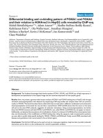
Figure 1
Scheme showing the hormonal pathways implicated in stress and
fibromyalgia. GHRH, growth hormone releasing hormone; CRH,
corticotrophin releasing hormone; GnRH, gonadotrophin releasing
hormone; GH, growth hormone; ACTH, adrenocorticotrophic hormone;
FSH, follicle-stimulating hormone; LH, luteinising hormone; DHEA,
dehydroepiandrosterone.
Hypothalamus
Anterior
Pi
tuitary
GHRH CRH GnRH
GH
ACTH FSH
LH
Muscle Adrenals Ovaries
Cortisol
DHEA
Oestrogens
Figure 2
Scheme showing the pain-modulating pathways in the dorsal horn of
the spinal cord. Nociceptive A-delta fibres and nociceptive C fibres
cause release of substance P in the dorsal horn. Serotonin released by
the dorsolateral inhibitory tracts inhibits the release of substance P.
Serotonin
–
A delta
C fibres
Substance P
+
Dorsal
Horn
Descending
Inhibitory
tracts
100
Arthritis Research & Therapy Vol 6 No 3 Gupta and Silman
Some investigators have, confusingly, pointed out that
fibromyalgia displays endocrine responses observed in the
hypercortisolic state of Cushing’s syndrome. These
responses include blunting of the diurnal variation of
serum cortisol [16,17] and a failure in approximately 35%
of fibromyalgia patients to suppress serum cortisol levels
with low-dose dexamethasone [21,22]. However, these
abnormalities have been reported in clinical depression
and alcohol abuse, and are therefore not specific to
fibromyalgia [23,24].
The confounding effect of depression, a relatively frequent
comorbidity associated with fibromyalgia, is an important
consideration in hormonal studies. In particular, HPA axis
abnormalities are often found in depression [25–27].
While there are certain similarities between depression
and fibromylagia, as already mentioned, there are
significant differences. Unlike fibromyalgia, plasma-free
cortisol levels are increased in classical depression
[24,28], and the ACTH response to exogenous CRH is
blunted rather than increased [24,25]. Only one of these
studies adjusted for coexistent depression, and none of
them adjusted for alcohol use.
It is also relevant to consider whether these
abnormalities are a feature of all chronic pain states or
are a particular feature of the otherwise unexplained pain
observed in fibromyalgia. Few studies have addressed
this. In one study, compared with patients with
rheumatoid arthritis, patients with fibromyalgia showed a
significant loss of diurnal variation and a lack of
suppression of serum cortisol with dexamethasone [16].
None of the subjects had clinical depression, and the
depression rating scale was similar in both groups. Griep
and colleagues [18] similarly reported that the
exaggerated ACTH response to CRH challenge was
significantly less in noninflammatory low back pain
patients compared with fibromyalgia patients. There was,
however, no difference between the two groups in 24-
hour free cortisol excretion, both groups being lower
than healthy controls.
In summary, animal studies would indicate that exposure
to stress during childhood has the effect of raising the
‘tonus’ of the HPA axis, with exaggerated ACTH response
and exaggerated cortisol response to stressors in later life.
These findings contrast with the observed central deficiency
of CRH in fibromyalgia, and thus the relationship between
changes in the HPA axis in stress and in fibromylagia
remain to be clarified.
Growth hormone — insulin-like growth factor I
Normal physiology and response to stress
Growth hormone is released from the anterior pituitary in
response to a releasing hormone from the hypothalamus.
Growth hormone causes release of insulin-like growth
factor I (IGF-I) from the liver, which exerts its effect on
target organs such as muscles.
The effects of acute and chronic stress on growth
hormone secretion are diametrically different. Several
authors have reported that acute stress has the effect of
raising growth hormone levels in the plasma manifold
[29–32].
Conversely, chronic psychosocial stress has the effect of
lowering growth hormone levels. This phenomenon has
been well studied in children and adolescents because of
its implications in terms of growth failure [33–35]. Powell
and colleagues [34] were the first to describe a syndrome
of emotional deprivation and growth retardation
associated with low growth hormone levels. Skuse and
colleagues [35] more recently described a condition of
growth failure and hyperphagia associated with low
growth hormone levels in children who came from
stressful homes. When the children were removed from
their stressful home circumstances, growth hormone
insufficiency resolved spontaneously.
Abnormalities in fibromyalgia
Given the aforementioned, it is appropriate to consider
evidence that fibromyalgia is associated with a growth
hormone deficiency state. In support, several authors have
demonstrated low serum growth hormone levels or low
IGF-I levels in patients with fibromyalgia compared with
controls [7,36–40].
A case control study of 500 patients with fibromyalgia and
152 controls (74 healthy subjects, 26 patients with
regional pain and 52 patients with other rheumatic
diseases) found significantly lower mean serum IGF-I
levels in those with fibromyalgia [36]. The low levels of
IGF-I in the fibromyalgia group were not explained by
depression, tricyclic antidepressants, nonsteroidal anti-
inflammatory medications, poor aerobic conditioning,
obesity or pain levels. Controls with regional pain had
normal IGF-I levels, as did most subjects with other
rheumatic disorders, unless they had concomitant
fibromyalgia.
The low growth hormone levels in fibromyalgia may be a
consequence rather than a cause: the hormone is largely
secreted during stage 3 and stage 4 of nonrapid eye
movement sleep, which are known to be disrupted in
fibromyalgia [41]. It was thus of interest that patients with
fibromyalgia with initially normal IGF-I levels followed-up
over 2 years often showed a rapid decline [36]. The
majority of patients with fibromyalgia who had low IGF-I
levels had markedly reduced stimulation of growth
hormone secretion with secretagogues. The authors
performed a similar study on a separate subset of patients,
controlling for concomitant therapy, weight and disease
101
severity, and again reported significantly lower levels of
IGF-I in fibromyalgia patients compared with controls.
Overall, one-third of subjects with fibromyalgia had low
IGF-I levels [37].
Support for a causal role for growth hormone deficiency,
however, comes from observations that such deficiency in
adults has been associated with many of the symptoms
described by fibromyalgia patients. These symptoms are
poor general health [42], low energy, reduced exercise
capacity, cold intolerance, dysthymia [43,44], muscle
weakness [45], impaired cognition [46] and reduced lean
body mass [47]. Growth hormone is important in
maintaining muscle homeostasis [48], and it has been
suggested that low levels of the hormone may be
responsible for delayed healing of muscle microtrauma in
fibromyalgia [36].
Further evidence that growth hormone deficiency may
have a role to play in fibromyalgia comes from a
randomised, double-blinded, placebo-controlled study in
which subjects with fibromyalgia, who gave themselves
daily subcutaneous injections of growth hormone over 9
months, showed significant improvement in overall
symptoms and tender points [49].
Androgens
Normal physiology and response to stress
Dehydroepiandrosterone (DHEA) is the major androgen
produced by the adrenal glands, both in women and men.
Up to 20% of DHEA in women is produced by the ovaries.
The adrenal gland is also the major source of testosterone
in women, where it is directly responsible for the
production of the hormone. Testosterone is also produced
peripherally in women by conversion from adrenal
steroids. DHEA is present in high concentrations in the
blood, lacks diurnal variation and has a long half-life [50].
Accordingly, serum DHEA levels are a good marker of
adrenocortical function and are probably a more sensitive
indicator of adrenocortical hypofunction than gluco-
corticoid secretion [51].
Serum DHEA levels are inversely related to perceived
stress [52]. DHEA production and cortisol production vary
inversely, and DHEA antagonises the physiological effects
of corticosteroids [53]. This is illustrated by the existence
of low levels of DHEA in depression [54], which, in its
classical form, is now known to be a high cortisol state.
Levels of DHEA have similarly been found to be low in
anorexia nervosa relative to cortisol [55]. Testosterone
levels also seem to be similarly lowered by stress. How-
ever, most studies on androgens (DHEA and testosterone)
have been of acute stress, such as that resulting from
military endurance courses [56–58]. Interestingly, many of
these studies involved sleep deprivation, which was
consistently associated with lower testosterone.
Abnormalities in fibromyalgia
There have been few studies on clinic patients. In one
study involving 56 women with fibromyalgia, serum DHEA
and testosterone levels were markedly decreased in
fibromyalgia patients compared with healthy controls [50].
Interestingly, low DHEA levels were significantly
correlated with pain. The authors adjusted for important
confounders such as age, menopausal status, body mass
index and oral contraceptive use, and they excluded those
who had recently taken glucocorticoids or other
medications. No adjustments were made for levels of
physical activity, however, which have been known to raise
androgen levels [59].
There is indirect evidence that fibromyalgia may be a
consequence of low androgens. Fibromyalgia has many
anti-anabolic features, such as muscle pain and fatigue,
typically seen in androgen-deficiency states. Androgens
exert anabolic effects, particularly on muscle. They
promote muscle growth and healing, and androgens have
been used for this purpose after trauma, after prolonged
immobilisation and in individuals with debilitating illness
[60]. However, no therapeutic trials of androgens have
been conducted in fibromyalgia.
Oestrogens
Normal physiology and response to stress
Oestrogens in women are produced by the ovaries in
response to the gonadotrophic hormones, namely follicle-
stimulating hormone (FSH) and luteinising hormone (LH).
FSH and LH are themselves released from the anterior
pituitary by the gonadotrophin-releasing hormone, a
product of the hypothalamus. Oestrogen levels vary
throughout the menstrual cycle in response to
fluctuations in LH levels, and the levels peak just before
ovulation.
It has been known for some time that stress has a
profound effect on the female reproductive system. The
development of functional amenorrhoea in response to
psychological stress is termed ‘hypothalamic’ amenor-
rhoea [61]. Animal studies have shown that socially
subordinate macaques have impaired ovarian function,
resulting in low oestrogen levels [62]. The effect is not
confined to premenopausal females. Ballinger [63] found
that stress lowered oestrogen levels in women in the early
postmenopausal phase.
Oestrogens have also been shown to ameliorate the
physiological response to stress. In perimenopausal
women exposed to time-restricted mental arithmetic as a
stressor, supplementation with oestradiol significantly
blunted the increases in both systolic blood pressure and
diastolic blood pressure, and in levels of plasma cortisol,
of ACTH, of epinephrine and of norepinephrine in
response to the challenge [64]. Oestradiol has also been
Available online />102
shown to lower the cardiovascular response to stress in
young women [65].
Abnormalities in fibromyalgia
Given the effect of stress on oestrogens, it is logical to
consider whether oestrogen levels are lower in women
with fibromyalgia, and whether this might contribute to the
pathogenesis of this condition. There are indeed several
lines of evidence suggesting that oestrogen deficiency
may be relevant. Women with fibromyalgia report more
pain perimenstrually compared with the ovulatory phase,
consistent with the fact that oestrogen levels peak during
the ovulatory phase and then nadir around menstruation
[66,67]. Female fibromyalgia patients have significantly
lower oestrogen levels than controls during the follicular
phase despite elevated FSH levels [7]. None of these
subjects were on hormones such as oral contraceptives at
the time of the study nor had coexistent rheumatic
conditions.
In another study, 65% of female patients with fibromyalgia
experienced menopause prior to the onset of the condition
[68]. In this study, 30% of fibromyalgia patients between
the ages of 24 and 45 years were found to be prematurely
menopausal. Despite these findings, however, Macfarlane
and colleagues [69] failed to find an association between
sex hormonal factors, including oestrogen levels, and
chronic widespread pain in a large unselected population.
The relationship of oestrogen deficiency with pain is not
confined to fibromyalgia. Rheumatoid arthritis tends to
improve during pregnancy, during oestrogen replacement
therapy and during treatment with oestrogen-containing
oral contraceptives [70].
If lower oestrogen levels do predispose to pain, what are
the pathways involved? Changes in oestrogen levels in
the plasma are accompanied by changes in a variety of
neurotransmitters, particularly serotonin and substance P
[71,72]. Both of these neurotransmitters are closely involved
in the pathogenesis of nociception. Increased serotonin
levels suppress the production of substance P within the
central nervous system (CNS) [8]. It has also been shown
that oestrogens upregulate serotonin [73]. It is therefore
possible that serotonin production in the CNS is
decreased in low oestrogen states, thereby leading to
increased substance P levels and to more pain.
Serotonin
Normal physiology and response to stress
Serotonin (5-hydroxytryptamine) acts as an antinociceptive
transmitter in the descending tracts located in the
dorsolateral funiculus of the spinal cord [74] (Fig. 2).
These descending tracts inhibit input from pain receptors
in deep tissues in preference to input from cutaneous
nociceptors. Serotonin is thought to exert its anti-
nociceptive effect by suppressing the production of
substance P, a nociceptive neurotransmitter. In the spinal
cord, substance P acts on the neurokinin-1 receptors
located in the dorsal horn [75]. Loss of pain modulation by
the descending inhibitory tracts subserved by serotonin
may result in spontaneous pain and tenderness, mainly in
the deep tissues. As the terminations of the descending
neurones have a widespread distribution in the spinal
cord, a dysfunction of the descending system due to a
lack of serotonin is likely to cause widespread pain [74].
Serotonin has been implicated in various psychiatric
disorders such as depression and anxiety. Its association
with stress has also been studied, as part of the paradigm
of stress-induced depression. Several studies indicate
that acute stress results in activation of the brain
serotonergic system. Various forms of stressors (namely,
physical stressors, metabolic stressors, psychological
stressors or immunological stressors) cause a rise in
extracellular serotonin in most regions of the brain [76],
and increase serotonin synthesis and turnover [77]. For
example, levels of brain tryptophan, the amino acid
precursor of serotonin, is markedly increased by exposure
to insulin injection in fasted rats (metabolic stressor), by
running (physical stressor) and by immobilisation (psycho-
logical stressor) [77].
However, chronic stress affects the brain serotonergic
system quite differently from acute stress. Sustained
stress is thus accompanied by diminution of serotonin
turnover [78,79]. An inverse relation has been found
between the plasma corticosterone level in rats and
serotonin turnover in the CNS [80]. In subordinate
(chronically stressed) rats, serotonin receptor binding
throughout the entire hippocampus was decreased [81].
Abnormalities in fibromyalgia
Consistent with the data on the influence of chronic
stress, most studies of serotonin in serum of fibromyalgia
patients reveal lower levels than in controls [82–85]. The
association between pain and low serum serotonin levels
is not limited to fibromyalgia. Both patients with fibro-
myalgia and those with rheumatoid arthritis have thus
been shown to have low serum levels of serotonin compared
with healthy controls [84,86]. However, serum concentra-
tions of serotonin were significantly lower in patients with
fibromyalgia compared with arthritis sufferers [86].
Serum serotonin levels do not simply reflect CNS
serotonin levels, however, since serotonin does not cross
the blood–brain barrier and since CNS serotonin makes
up less than 2% of total body serotonin. Moreover, serum
serotonin is obtained from platelets, and therefore can
vary with the platelet count [87]. Serotonin levels have not
been measured in the cerebrospinal fluid (CSF), but levels
of its immediate precursor (5-hydroxytryptophan) and its
Arthritis Research & Therapy Vol 6 No 3 Gupta and Silman
103
metabolite (5-hydroxy indoleacetic acid) were found to be
lower than normal concentrations in the CSF of
fibromyalgia patients [83,88].
Serotonin levels are, however, altered in psychiatric
disorders, particularly in depression and in those patients
receiving antidepressant therapy [89,90]. These factors
are of relevance in interpreting most of the aforementioned
studies, which were based on fibromyalgia patients from
rheumatology clinics who are more likely to have
associated depression and anxiety, resulting in healthcare-
seeking behaviour [91,92]. The only population-based
study found that serotonin levels were significantly lower
in subjects with fibromyalgia compared with a composite
group with no pain, regional pain or nonfibromyalgia
chronic widespread pain [85]. However, serotonin levels
were not significantly different between fibromyalgia
subjects and the pain-free group, considered alone.
Unexpectedly, serum serotonin levels rose corres-
pondingly with depression scores, contrary to what has
been reported in clinic patients [83,84]. Concurrent anti-
depressant therapy did not alter the relationship between
fibromyalgia and serotonin levels, or that between
depression and serotonin levels.
Substance P
Normal physiology and response to stress
Substance P is an 11-amino acid neuropeptide that plays
an important role in nociception [93]. Activated, small,
thinly myelinated A-delta afferent neurons release
substance P into lamina I and lamina V in the dorsal horn
of the spinal cord. Activated C fibres similarly release
substance P into lamina II. Substance P exerts its action
through neurokinin-1 receptors. Substance P probably
acts by alerting spinal cord neurons to incoming noci-
ceptive signals from the periphery [8] (Fig. 2). Substance P
released into the spinal cord diffuses out into the CSF,
where it can be measured.
Most data investigating the substance P response to
stress have been based on acute stress. Mapping studies
indicate that the substance P-preferring neurokinin-1
receptor is highly expressed in brain regions that are
critical for the regulation of affective behaviour and
neurochemical responses to stress [94]. Neurochemical
experiments in rats revealed changes in substance P
content in the hippocampus, the septum, the periaque-
ductal grey and the ventral tegemental areas of the
midbrain after stressors such as inescapable foot shock,
immobilisation and social isolation [95]. In guinea pigs,
central infusion of substance P agonists causes locomotor
activation [96], accompanied by pronounced and long-
lasting vocalisations [97]. This observation is of particular
interest because exposure to stress induces vocalisations
in many mammalian species [98]. The data suggested that
psychological stress causes release of substance P in the
limbic system of the brain, and that pharmacological
blockade of substance P receptors is capable of inhibiting
behavioural responses to such stress [97].
Abnormalities in fibromyalgia
Although there is little data on chronic stress, it is
reasonable to hypothesise that substance P may be
elevated in fibromyalgia. Several studies have reported
that CSF concentrations of substance P in fibromyalgia
are around twofold to threefold higher than those in
healthy controls [85,99–101]. However, nonfibromyalgia
subjects suffering from chronic pain can display similar
levels of CSF substance P as is found in fibromyalgia [8].
Increased levels of substance P in the dorsal horn of
subjects with fibromyalgia would result in amplification of
nociceptive signals from the periphery and would be a
mechanism leading to widespread pain.
Conclusions
The aim of the present review is not to consider what
neuroendocrine abnormalities occur in fibromyalgia per se,
but rather to evaluate whether such abnormalities could
explain the relationship between chronic stress and
fibromyalgia. We have shown that activities of several
endocrine axes and neurotransmitters change in parallel in
both fibromyalgia and stress. In particular, there are
decreased basal levels of growth hormone and IGF-I,
androgens and oestrogens both in stress and fibro-
myalgia. Serotonin levels are reduced in both fibromyalgia
and chronic stress, while levels of substance P are
increased. Available evidence would favour diminished
function of the HPA axis in fibromyalgia. The HPA axis is of
course one of the major stress-response systems of the
body and, in this respect, there seems to be divergence
between fibromyalgia and stress. Nevertheless, similar
changes in most other hormones and neurotransmitters
would favour a role for stress in fibromyalgia.
There are large areas of uncertainty, however. For several
hormones, the response to stress has been mainly studied
in animals and there are very few reports on the response
in humans. Even where human studies do exist, they may
not be representative of the general population (e.g.
military endurance exercises).
Also, for the present review, we are mainly interested in the
effects of chronic psychological stress on various hormones
and neurotransmitters, as this is more relevant for
fibromyalgia than acute stress. It is, however, difficult to
replicate conditions of chronic stress in experimental
conditions. As a result, in many instances, the only data that
could be found were from conditions mimicking acute stress.
Finally, an inherent difficulty with the study of hormones
and neurotransmitters in both stress and fibromyalgia is to
Available online />104
determine whether an effect is primary or secondary. All
the studies discussed have been cross-sectional in nature,
and do not allow conclusions on temporality. Thus, for
example, low androgen levels in fibromyalgia could well be
a result of chronic pain rather than the cause of it.
While the central theme of the present review is that
chronic stress may lead to changes in various hormones
and neurotransmitters, resulting in various manifestations
of fibromyalgia such as pain and fatigue, it is not
inconceivable that the chronic pain present in fibromyalgia
can give rise to psychological stress, and thereby cause
changes in neuroendocrine axes. Well-designed prospective
studies are needed to resolve these issues.
To address this in relation to the HPA axis, our group is
conducting a population study where we initially identified
psychologically stressed subjects in the community
through well-validated questionnaires. These subjects
have had their HPA axis function assessed [102] and are
now being followed-up after a period of 15 months to help
resolve the issue of whether derangements of the HPA
axis in psychologically stressed subjects predict the future
development of, as opposed to being a consequence of,
chronic widespread pain.
Competing interests
None declared.
References
1. Marder WD, Meenan RF, Felson DT, Reichlin M, Birnbaum NS,
Croft JD, Dore RK, Kaplan H, Kaufman RL, Stobo JD: The
present and future adequacy of rheumatology manpower. A
study of health care needs and physician supply. Arthritis
Rheum 1991, 34:1209-1217.
2. Aronoff GM: Myofascial pain syndrome and fibromyalgia: a
critical assessment and alternate view. Clin J Pain 1998, 14:
74-85.
3. Aaron LA, Bradley LA, Alarcon GS, Triana-Alexander M, Alexander
RW, Martin MY, Alberts KR: Perceived physical and emotional
trauma as precipitating events in fibromyalgia. Associations
with health care seeking and disability status but not pain
severity. Arthritis Rheum 1997, 40:453-460.
4. Walker EA, Keegan D, Gardner G, Sullivan M, Bernstein D, Katon
WJ: Psychosocial factors in fibromyalgia compared with
rheumatoid arthritis: II. Sexual, physical, and emotional abuse
and neglect. Psychosom Med 1997, 59:572-577.
5. Bradley LA, Alarcon GS, Aaron LA, Martin MY, Alberts KR,
Sotolongo A: Abnormal pain perception in patients with
fibromyalgia: comment on the article by Bendtsen et al.
Arthritis Rheum 1997, 40:2275-2277.
6. Neeck G: Pathogenic mechanisms of fibromyalgia. Ageing Res
Rev 2002, 1:243-255.
7. Riedel W, Layka H, Neeck G: Secretory pattern of GH, TSH,
thyroid hormones, ACTH, cortisol, FSH, and LH in patients
with fibromyalgia syndrome following systemic injection of
the relevant hypothalamic-releasing hormones. Z Rheumatol
1998, Suppl 2:81-87.
8. Russell IJ: Advances in fibromyalgia: possible role for central
neurochemicals. Am J Med Sci 1998, 315:377-384.
9. Ladd CO, Huot RL, Thrivikraman KV, Nemeroff CB, Meaney MJ,
Plotsky PM: Long-term behavioral and neuroendocrine adap-
tations to adverse early experience. Prog Brain Res 2000, 122:
81-103.
10. Plotsky PM, Meaney MJ: Early, postnatal experience alters
hypothalamic corticotropin-releasing factor (CRF) mRNA,
median eminence CRF content and stress-induced release in
adult rats. Brain Res Mol Brain Res 1993 18:195-200.
11. Meaney MJ, Diorio J, Francis D, Widdowson J, LaPlante P, Caldji
C, Sharma S, Seckl JR, Plotsky PM: Early environmental regula-
tion of forebrain glucocorticoid receptor gene expression:
implications for adrenocortical responses to stress. Dev
Neurosci 1996, 18:49-72.
12. Breier A, Kelsoe JR, Jr, Kirwin PD, Beller SA, Wolkowitz OM,
Pickar D: Early parental loss and development of adult
psychopathology. Arch Gen Psychiatry 1988, 45:987-993.
13. De Bellis MD, Chrousos GP, Dorn LD, Burke L, Helmers K, Kling
MA, Trickett PK, Putnam FW: Hypothalamic–pituitary–adrenal
axis dysregulation in sexually abused girls. J Clin Endocrinol
Metab 1994, 78:249-255.
14. Kaufman J, Birmaher B, Perel J, Dahl RE, Moreci P, Nelson B,
Wells W, Ryan ND: The corticotropin-releasing hormone chal-
lenge in depressed abused, depressed nonabused, and
normal control children. Biol Psychiatry 1997, 42:669-679.
15. Clauw DJ, Chrousos GP: Chronic pain and fatigue syndromes:
overlapping clinical and neuroendocrine features and poten-
tial pathogenic mechanisms. Neuroimmunomodulation 1997, 4:
134-153.
16. McCain GA, Tilbe KS: Diurnal hormone variation in fibro-
myalgia syndrome: a comparison with rheumatoid arthritis.
J Rheumatol Suppl 1989, 19:154-157.
17. Crofford LJ, Pillemer SR, Kalogeras KT, Cash JM, Michelson D,
Kling MA, Sternberg EM, Gold PW, Chrousos GP, Wilder RL:
Hypothalamic–pituitary–adrenal axis perturbations in patients
with fibromyalgia. Arthritis Rheum 1994, 37:1583-1592.
18. Griep EN, Boersma JW, Lentjes EG, Prins AP, van der Korst JK,
de Kloet ER: Function of the hypothalamic–pituitary–adrenal
axis in patients with fibromyalgia and low back pain.
J Rheumatol 1998, 25:1374-1381.
19. Griep EN, Boersma JW, de Kloet ER: Altered reactivity of the
hypothalamic–pituitary–adrenal axis in the primary fibro-
myalgia syndrome. J Rheumatol 1993, 20:469-474.
20. Disdier P, Harle JR, Brue T, Jaquet P, Chambourlier P, Grisoli F,
Weiller PJ: Severe fibromyalgia after hypophysectomy for
Cushing’s disease. Arthritis Rheum 1991, 34:493-495.
21. Hudson JI, Pliner LF, Hudson MS, Goldenberg DL, Melby JC: The
dexamethasone suppression test in fibrositis. Biol Psychiatry
1984, 19:1489-1493.
22. Ferraccioli G, Cavalieri F, Salaffi F, Fontana S, Scita F, Nolli M,
Maestri D: Neuroendocrinologic findings in primary fibro-
myalgia (soft tissue chronic pain syndrome) and in other
chronic rheumatic conditions (rheumatoid arthritis, low back
pain). J Rheumatol 1990, 17:869-873.
23. Deuschle M, Schweiger U, Weber B, Gotthardt U, Korner A,
Schmider J, Standhardt H, Lammers CH, Heuser I: Diurnal activ-
ity and pulsatility of the hypothalamus–pituitary–adrenal
system in male depressed patients and healthy controls.
J Clin Endocrinol Metab 1997, 82:234-238.
24. Plotsky PM, Owens MJ, Nemeroff CB: Psychoneuroendo-
crinology of depression. Hypothalamic–pituitary–adrenal axis.
Psychiatr Clin North Am 1998, 21:293-307.
25. Zobel AW, Nickel T, Kunzel HE, Ackl N, Sonntag A, Ising M,
Holsboer F: Effects of the high-affinity corticotropin-releasing
hormone receptor 1 antagonist R121919 in major depression:
the first 20 patients treated. J Psychiatr Res 2000, 34:171-181.
26. Arborelius L, Owens MJ, Plotsky PM, Nemeroff CB: The role of
corticotropin-releasing factor in depression and anxiety dis-
orders. J Endocrinol 1999, 160:1-12.
27. Checkley S: The neuroendocrinology of depression and
chronic stress. Br Med Bull 1996, 52:597-617.
28. Deuschle M, Weber B, Colla M, Depner M, Heuser I: Effects of
major depression, aging and gender upon calculated diurnal
free plasma cortisol concentrations: a re-evaluation study.
Stress 1998, 2:281-287.
29. Maestu J, Jurimae J, Jurimae T: Hormonal reactions during
heavy training stress and following tapering in highly trained
male rowers. Horm Metab Res 2003, 35:109-113.
30. Roth-Isigkeit A, Brechmann J, Dibbelt L, Sievers HH, Raasch W,
Schmucker P: Persistent endocrine stress response in patients
undergoing cardiac surgery. J Endocrinol Invest 1998, 21:12-19.
31. Chatterton RT, Jr, Vogelsong KM, Lu YC, Hudgens GA:
Hormonal responses to psychological stress in men prepar-
ing for skydiving. J Clin Endocrinol Metab 1997, 82:2503-2509.
Arthritis Research & Therapy Vol 6 No 3 Gupta and Silman
105
32. Farrace S, Biselli R, Urbani L, Ferlini C, De Angelis C: Evaluation
of stress induced by flight activity by measuring the hormonal
response. Biofeedback Self Regul 1996, 21:217-228.
33. Blizzard RM, Bulatovic A: Psychosocial short stature: a syn-
drome with many variables. Baillieres Clin Endocrinol Metab
1992, 6:687-712.
34. Powell GF, Brasel JA, Raiti S, Blizzard RM: Emotional depriva-
tion and growth retardation simulating idiopathic hypo-
pituitarism. II. Endocrinologic evaluation of the syndrome.
N Engl J Med 1967, 276:1279-1283.
35. Skuse D, Albanese A, Stanhope R, Gilmour J, Voss L: A new
stress-related syndrome of growth failure and hyperphagia in
children, associated with reversibility of growth-hormone
insufficiency. Lancet 1996, 348:353-358.
36. Bennett RM, Cook DM, Clark SR, Burckhardt CS, Campbell SM:
Hypothalamic–pituitary–insulin-like growth factor-I axis dys-
function in patients with fibromyalgia. J Rheumatol 1997, 24:
1384-1389.
37. Bennett RM, Clark SR, Campbell SM, Burckhardt CS: Low levels
of somatomedin C in patients with the fibromyalgia syn-
drome. A possible link between sleep and muscle pain. Arthri-
tis Rheum 1992, 35:1113-1116.
38. Griep EN, Boersma JW, de Kloet ER: Pituitary release of growth
hormone and prolactin in the primary fibromyalgia syndrome.
J Rheumatol 1994, 21:2125-2130.
39. Ferraccioli G, Guerra P, Rizzi V, Baraldo M, Salaffi F, Furlanut M,
Bartoli E: Somatomedin C (insulin-like growth factor 1) levels
decrease during acute changes of stress related hormones.
Relevance for fibromyalgia. J Rheumatol 1994, 21:1332-1334.
40. Leal-Cerro A, Povedano J, Astorga R, Gonzalez M, Silva H,
Garcia-Pesquera F, Casanueva FF, Dieguez C: The growth
hormone (GH)–releasing hormone–GH–insulin-like growth
factor-1 axis in patients with fibromyalgia syndrome. J Clin
Endocrinol Metab 1999, 84:3378-3381.
41. Bennett RM: Beyond fibromyalgia: ideas on etiology and treat-
ment. J Rheumatol Suppl 1989, 19:185-191.
42. Wallymahmed ME, Baker GA, Humphris G, Dewey M, MacFarlane
IA: The development, reliability and validity of a disease
specific quality of life model for adults with growth hormone
deficiency. Clin Endocrinol (Oxf) 1996, 44:403-411.
43. Cuneo RC, Salomon F, McGauley GA, Sonksen PH: The growth
hormone deficiency syndrome in adults. Clin Endocrinol (Oxf)
1992, 37:387-397.
44. Cuneo RC, Salomon F, Wiles CM, Hesp R, Sonksen PH: Growth
hormone treatment in growth hormone-deficient adults. II.
Effects on exercise performance. J Appl Physiol 1991, 70:695-
700.
45. Rutherford OM, Beshyah SA, Schott J, Watkins Y, Johnston DG:
Contractile properties of the quadriceps muscle in growth
hormone-deficient hypopituitary adults. Clin Sci (Lond) 1995,
88:67-71.
46. McGauley GA, Cuneo RC, Salomon F, Sonksen PH: Psychological
well-being before and after growth hormone treatment in adults
with growth hormone deficiency. Horm Res 1990, Suppl 4:52-54.
47. Salomon F, Cuneo RC, Hesp R, Sonksen PH: The effects of
treatment with recombinant human growth hormone on body
composition and metabolism in adults with growth hormone
deficiency. N Engl J Med 1989, 321:1797-1803.
48. Florini JR: Hormonal control of muscle growth. Muscle Nerve
1987, 10:577-598.
49. Bennett RM, Clark SC, Walczyk J: A randomized, double-blind,
placebo-controlled study of growth hormone in the treatment
of fibromyalgia. Am J Med 1998, 104:227-231.
50. Dessein PH, Shipton EA, Joffe BI, Hadebe DP, Stanwix AE, Van
der Merwe BA: Hyposecretion of adrenal androgens and the
relation of serum adrenal steroids, serotonin and insulin-like
growth factor-1 to clinical features in women with fibro-
myalgia. Pain 1999, 83:313-319.
51. Masi AT, Chrousos GP: Hypothalamic–pituitary–adrenal–
glucocorticoid axis function in rheumatoid arthritis. J Rheuma-
tol 1996, 23:577-581.
52. Labbate LA, Fava M, Oleshansky M, Zoltec J, Littman A, Harig P:
Physical fitness and perceived stress. Relationships with
coronary artery disease risk factors. Psychosomatics 1995, 36:
555-560.
53. Blauer KL, Poth M, Rogers WM, Bernton EW: Dehydro-
epiandrosterone antagonizes the suppressive effects of
dexamethasone on lymphocyte proliferation. Endocrinology
1991, 129:3174-3179.
54. Goodyer IM, Herbert J, Altham PM, Pearson J, Secher SM, Shiers
HM: Adrenal secretion during major depression in 8- to 16-
year-olds, I. Altered diurnal rhythms in salivary cortisol and
dehydroepiandrosterone (DHEA) at presentation. Psychol
Med 1996, 26:245-256.
55. Zumoff B, Walsh BT, Katz JL, Levin J, Rosenfeld RS, Kream J,
Weiner H: Subnormal plasma dehydroisoandrosterone to
cortisol ratio in anorexia nervosa: a second hormonal para-
meter of ontogenic regression. J Clin Endocrinol Metab 1983,
56:668-672.
56. Morgan CA, III, Wang S, Mason J, Southwick SM, Fox P, Hazlett
G, Charney DS, Greenfield G: Hormone profiles in humans
experiencing military survival training. Biol Psychiatry 2000;
47:891-901.
57. Friedl KE, Moore RJ, Hoyt RW, Marchitelli LJ, Martinez-Lopez LE,
Askew EW: Endocrine markers of semistarvation in healthy
lean men in a multistressor environment. J Appl Physiol 2000,
88:1820-1830.
58. Opstad PK: Androgenic hormones during prolonged physical
stress, sleep, and energy deficiency. J Clin Endocrinol Metab
1992, 74:1176-1183.
59. Exercise increases androgen levels [ />volume_3_number_4.htm].
60. Bagatell CJ, Bremner WJ: Androgens in men — uses and
abuses. N Engl J Med 1996, 334:707-714.
61. Liu JH: Hypothalamic amenorrhea: clinical perspectives,
pathophysiology, and management. Am J Obstet Gynecol
1990, 163:1732-1736.
62. Kaplan JR, Adams MR, Clarkson TB, Manuck SB, Shively CA,
Williams JK: Psychosocial factors, sex differences, and athero-
sclerosis: lessons from animal models. Psychosom Med 1996,
58:598-611.
63. Ballinger S: Stress as a factor in lowered estrogen levels in
the early postmenopause. Ann NY Acad Sci 1990, 592:95-113.
64. Komesaroff PA, Esler MD, Sudhir K: Estrogen supplementation
attenuates glucocorticoid and catecholamine responses to
mental stress in perimenopausal women. J Clin Endocrinol
Metab 1999, 84:606-610.
65. Sita A, Miller SB: Estradiol, progesterone and cardiovascular
response to stress. Psychoneuroendocrinology 1996, 21:339-
346.
66. Anderberg UM: Comment on: Johns and Littlejohn, The role of
sex hormones in pain response. Pain 2000, 87:109-111.
67. Ostensen M, Rugelsjoen A, Wigers SH: The effect of reproduc-
tive events and alterations of sex hormone levels on the
symptoms of fibromyalgia. Scand J Rheumatol 1997, 26:355-
360.
68. Waxman J, Zatzkis SM: Fibromyalgia and menopause. Exami-
nation of the relationship. Postgrad Med 1986, 80:165-171.
69. Macfarlane TV, Blinkhorn A, Worthington HV, Davies RM,
Macfarlane GJ: Sex hormonal factors and chronic widespread
pain: a population study among women. Rheumatology 2002,
41:454-457.
70. Van Vollenhoven RF, McGuire JL: Estrogen, progesterone, and
testosterone: can they be used to treat autoimmune dis-
eases? Cleve Clin J Med 1994, 61:276-284.
71. Aloisi AM: Gonadal hormones and sex differences in pain
reactivity. Clin J Pain 2003, 19:168-174.
72. Akkus S, Delibas N, Tamer MN: Do sex hormones play a role in
fibromyalgia? Rheumatology 2000, 39:1161-1163.
73. Marcus DA: Interrelationships of neurochemicals, estrogen,
and recurring headache. Pain 1995, 62:129-139.
74. Mense S: Neurobiological concepts of fibromyalgia — the
possible role of descending spinal tracts. Scand J Rheumatol
Suppl 2000, 113:24-29.
75. Russell IJ: Fibromyalgia syndrome: approaches to manage-
ment. Bull Rheum Dis 1996, 45:1-4.
76. Rueter LE, Fornal CA, Jacobs BL: A critical review of 5-HT brain
microdialysis and behavior. Rev Neurosci 1997, 8:117-137.
77. Chaouloff F: Physiopharmacological interactions between
stress hormones and central serotonergic systems. Brain Res
Brain Res Rev 1993, 18:1-32.
78. Weiss JM, Goodman PA, Losito BG, Corrigan S, Charry JM,
Bailey WB: Behavioral depression produced by an uncontrol-
lable stressor: relationship to norepinephrine, dopamine, and
Available online />106
serotonin levels in various regions of rat brain. Brain Res Rev
1981, 3:167-205
79. Sherman AD, Petty F: Neurochemical basis of the action of
antidepressants on learned helplessness. Behav Neural Biol
1980, 30:119-134.
80. De Souza EB, Van Loon GR: Brain serotonin and catecholamine
responses to repeated stress in rats. Brain Res 1986, 367:77-86.
81. McKittrick CR, Blanchard DC, Blanchard RJ, McEwen BS, Sakai
RR: Serotonin receptor binding in a colony model of chronic
social stress. Biol Psychiatry 1995, 37:383-393.
82. Moldofsky H: Rheumatic pain modulation syndrome: the inter-
relationships between sleep, central nervous system sero-
tonin, and pain. Adv Neurol 1982, 33:51-57.
83. Russell IJ, Michalek JE, Vipraio GA, Fletcher EM, Javors MA,
Bowden CA: Platelet 3H-imipramine uptake receptor density
and serum serotonin levels in patients with fibromyalgia/
fibrositis syndrome. J Rheumatol 1992, 19:104-109.
84. Stratz T, Samborski W, Hrycaj P, Pap T, Mackiewicz S, Mennet P,
Muller W: Serotonin concentration in serum of patients with
generalized tendomyopathy (fibromyalgia) and chronic poly-
arthritis. Med Klin 1993, 88:458-462.
85. Bradley LA, Alberts KR, Alarcon GS, Alexander MT, Mountz JM,
Weigent DA, Lin HG, Blalock JE, Aaron LA, Alexander RW, San
Pedro EC, Martin MY, Morell AC: Abnormal brain regional cere-
bral blood flow (rCBF) and cerebrospinal fluid (CSF) levels of
substance P (SP) in patients and non-patients with fibromyal-
gia (FM) [abstract]. Arthritis Rheum 1996, Suppl 9:212.
86. Hrycaj P, Stratz T, Muller W: Platelet 3H-imipramine uptake
receptor density and serum serotonin levels in patients with
fibromyalgia/fibrositis syndrome. J Rheumatol 1993, 20:1986-
1988.
87. Wolfe F, Russell IJ, Vipraio G, Ross K, Anderson J: Serotonin
levels, pain threshold, and fibromyalgia symptoms in the
general population. J Rheumatol 1997, 24:555-559.
88. Russell IJ, Vipraio GA, Acworth I: Abnormalities in the central
nervous system (CNS) metabolism of tryptophan (TRY) to 3-
hydroxykynurenine (OHKY) in fibromyalgia syndrome (FS)
[abstract]. Arthritis Rheum 1993, Suppl 9:222.
89. Risch SC, Nemeroff CB: Neurochemical alterations of seroton-
ergic neuronal systems in depression. J Clin Psychiatry 1992,
Suppl 53:3-7.
90. Grahame-Smith DG: Serotonin in affective disorders. Int Clin
Psychopharmacol 1992, Suppl 4:5-13.
91. Aaron LA, Bradley LA, Alarcon GS, Alexander RW, Triana-
Alexander M, Martin MY, Martin MY, Alberts KR: Psychiatric diag-
noses in patients with fibromyalgia are related to health
care-seeking behavior rather than to illness. Arthritis Rheum
1996, 39:436-445.
92. Bradley LA: Behavioral interventions for managing chronic
pain. Bull Rheum Dis 1994, 43:2-5.
93. Malmberg AB, Yaksh TL: Hyperalgesia mediated by spinal
glutamate or substance P receptor blocked by spinal
cyclooxygenase inhibition. Science 1992, 257:1276-1279.
94. Mantyh PW, Hunt SP, Maggio JE: Substance P receptors: local-
ization by light microscopic autoradiography in rat brain using
[
3
H]SP as the radioligand. Brain Res 1984, 307:147-165.
95. Lisoprawski A, Blanc G, Glowinski J: Activation by stress of the
habenulo-interpeduncular substance P neurons in the rat.
Neurosci Lett 1981, 25:47-51.
96. Brent PJ, Johnston PA, Chahl LA: Increased plasma cate-
cholamines and locomotor activity induced by centrally
administered substance P in guinea-pigs. Neuropharmacology
1988, 27:743-748.
97. Kramer MS, Cutler N, Feighner J, Shrivastava R, Carman J,
Sramek JJ, Reines SA, Liu G, Snavely D, Wyatt-Knowles E, Hale
JJ, Mills SG, MacCoss M, Swain CJ, Harrison T, Hill RG, Hefti F,
Scollnick EM, Cascieri MA, Chicchi GG, Sadowski S, Williams
AR, Hewson L, Smith D, Rupniak NM: Distinct mechanism for
antidepressant activity by blockade of central substance P
receptors. Science 1998, 281:1640-1645.
98. Miczek KA, Weerts EM, Vivian JA, Barros HM: Aggression,
anxiety and vocalizations in animals: GABAA and 5-HT anxio-
lytics. Psychopharmacology 1995, 121:38-56.
99. Vaeroy H, Helle R, Forre O, Kass E, Terenius L: Elevated CSF
levels of substance P and high incidence of Raynaud phe-
nomenon in patients with fibromyalgia: new features for diag-
nosis. Pain 1988, 32:21-26.
100. Russell IJ, Orr MD, Littman B, Vipraio GA, Alboukrek D, Michalek
JE, Lopez Y, MacKillip F: Elevated cerebrospinal fluid levels of
substance P in patients with the fibromyalgia syndrome.
Arthritis Rheum 1994, 37:1593-1601.
101. Welin M, Bragee B, Nyberg F, Kristiansson M: Elevated sub-
stance P levels are contrasted by a decrease in
metenkephalin-arg-phe levels in csf from fibromyalgia
patients [abstract]. J Musculoskeletal Pain 1995, Suppl 1:4.
102. Chiu YH, McBeth J, Silman AJ, Ray D, Macfarlane GJ: A popula-
tion based study of chronic widespread pain and the hypo-
thalamic–pituitary–adrenal (HPA) axis: results from a pilot
study [abstract]. Rheumatology 2002, Suppl 1:52.
Arthritis Research & Therapy Vol 6 No 3 Gupta and Silman