Báo cáo y học: "Nifedipine decreases sVCAM-1 concentrations and oxidative stress in systemic sclerosis but does not affect the concentrations of vascular endothelial growth factor or its soluble receptor" potx
Bạn đang xem bản rút gọn của tài liệu. Xem và tải ngay bản đầy đủ của tài liệu tại đây (186.95 KB, 6 trang )
Open Access
Available online />R309
Vol 6 No 4
Research article
Nifedipine decreases sVCAM-1 concentrations and oxidative
stress in systemic sclerosis but does not affect the concentrations
of vascular endothelial growth factor or its soluble receptor 1
Yannick Allanore
1
, Didier Borderie
2
, Hervé Lemaréchal
2
, Ohvanesse Garabed Ekindjian
2
and
André Kahan
1
1
Paris V University, Department of Rheumatology A, Assistance Publique Hôpitaux de Paris, Cochin Hospital, Paris, France
2
Department of Biochemistry A, Assistance Publique Hôpitaux de Paris, Cochin Hospital, Paris, France
Corresponding author: Yannick Allanore,
Received: 31 Jan 2004 Revisions requested: 4 Mar 2004 Revisions received: 22 Mar 2004 Accepted: 2 Apr 2004 Published: 12 May 2004
Arthritis Res Ther 2004, 6:R309-R314 (DOI 10.1186/ar1183)
http://arthr itis-research.com/conte nt/6/4/R309
© 2004 Allanore et al.; licensee BioMed Central Ltd. This is an Open Access article: verbatim copying and redistribution of this article are permitted
in all media for any purpose, provided this notice is preserved along with the article's original URL.
Abstract
Microvascular injury, oxidative stress, and impaired
angiogenesis are prominent features of systemic sclerosis
(SSc). We compared serum markers of these phenomena at
baseline and after treatment with nifedipine in SSc patients.
Forty successive SSc patients were compared with 20 matched
healthy subjects. All SSc patients stopped taking calcium-
channel blockers 72 hours before measurements. Twenty SSc
patients were also examined after 14 days of treatment with
nifedipine (60 mg/day). Quantitative ELISA was used to
measure the serum concentrations of vascular endothelial
growth factor (VEGF), soluble VEGF receptor 1 (sVEGFR-1),
soluble vascular cell adhesion molecule 1 (sVCAM-1), carbonyl
residues, and advanced oxidation protein products (AOPP). The
median concentrations of VEGF, sVEGFR-1, sVCAM-1,
carbonyl residues, and AOPP were significantly higher in SSc
patients than in healthy subjects at baseline. A correlation was
found between VEGF concentration and carbonyl residue
concentration (r = 0.43; P = 0.007). Nifedipine treatment led to
a significant decrease in concentrations of sVCAM-1, carbonyl
residues, and AOPP but did not affect concentrations of VEGF
and sVEGFR-1. Nifedipine treatment ameliorated endothelium
injury in patients with SSc, as shown by the concentrations of
adhesion molecules and oxidative damage markers. The fact
that VEGF and sVEGFR-1 concentrations were not changed
whereas oxidative stress was ameliorated by nifedipine is
consistent with the hypothesis that VEGF signalling is impaired
in SSc. However, more experimental evidence is needed to
determine whether the VEGF pathway is intrinsically defective in
SSc.
Keywords: nifedipine, oxidative stress, sVCAM-1, systemic sclerosis, VEGF
Introduction
Systemic sclerosis (SSc) is a connective tissue disease
characterised by early generalised microangiopathy and
culminating in systemic fibrosis. The pivotal steps of the
disease are endothelium injury, immune activation, and col-
lagen deposition by activated fibroblasts.
Vascular changes are suspected to occur at an early stage
[1]. Changes include gaps between endothelial cells [2],
apoptosis [3], endothelium activation with the expression of
cell adhesion molecules, inflammatory cell recruitment, pro-
coagulant state [4], and intimal proliferation and adventitial
fibrosis, which may lead to vessel obliteration. The vascula-
ture plays a major role in SSc pathogenesis, and prognosis
and outcome are dependent on the extent and severity of
the vascular lesions [5].
Endothelial injury is reflected by altered endothelium-
related indices, including increased plasma levels of mark-
ers such as soluble vascular cell adhesion molecule 1
(sVCAM-1). Thus, sVCAM-1 could be a useful parameter
for vascular assessment [6] and has been reported to be
associated with changes in disease severity [7]. Angiogen-
esis has been reported to be disturbed in SSc patients
despite high serum concentrations of vascular endothelial
growth factor (VEGF) [8-10], suggesting that VEGF is
AOPP = advanced oxidation protein product(s); ELISA = enzyme-linked immunosorbent assay; SSc = systemic sclerosis; sVCAM-1 = soluble vas-
cular cell adhesion molecule 1; sVEGFR-1 = soluble VEGF receptor 1; VEGF = vascular endothelial growth factor.
Arthritis Research & Therapy Vol 6 No 4 Allanore et al.
R310
counterbalanced by angiostatic factors [11] or is the con-
sequence of signalling defects [9]. VEGF is a glycoprotein
with potent angiogenic, mitogenic, and vascular permeabil-
ity-enhancing activities specific for endothelial cells. It inter-
acts with two receptor tyrosine kinases, VEGFR-1 (flt) and
VEGFR-2 (flk). A defect in VEGF receptors could account
for VEGF signalling abnormalities in SSc. The human
VEGFR-1 gene produces two major transcripts, corre-
sponding to the full-length receptor and a soluble receptor
(sVEGFR-1) with biological activities [12]. Oxidative stress
may modulate angiogenesis through microvascular toxicity
but may also promote angiogenesis [13]. The free radicals
generated by reperfusion injury (Raynaud's phenomenon)
and the inflammatory process appear to play a key role in
SSc [14].
Calcium-channel blockers, particularly those of the dihydro-
pyridine type such as nifedipine, are of major importance for
the treatment of Raynaud's phenomenon in SSc patients
[15] and may have beneficial effects on cardiac involve-
ment [16]. We recently reported that these drugs have
acute and sustained beneficial effects on oxidative markers
of damage in SSc patients [17].
The aim of our study was, first, to investigate serum
endothelial cell markers of adhesion (sVCAM-1) and angio-
genesis (VEGF, sVEGFR-1) together with oxidative dam-
age markers (carbonyl residues and advanced oxidation
protein products [AOPP]) at baseline and, secondly, to
look for the influence of nifedipine treatment on all these
parameters in SSc patients.
Materials and methods
Study population
We prospectively included successive SSc patients hospi-
talised for systematic follow-up of the disease. SSc was
classified as 'limited' or 'diffuse' cutaneous according to the
criteria of LeRoy and colleagues [18]. The exclusion criteria
were the impossibility of stopping vasodilator therapy,
pregnancy, current cigarette smoking, diabetes, associa-
tion with severe diseases (cardiac or hepatic failure, can-
cer, gangrene), and immunosuppressive therapy. A three-
month period of stable current treatment was required for
inclusion.
The onset of the disease was defined as the time at which
skin involvement occurred. The laboratory tests included
the Westergren erythrocyte sedimentation rate, C-reactive
protein levels, serum creatinine concentration, and antinu-
clear, anticentromere (indirect immunofluorescence on
HEp2 cells), and antitopoisomerase I (counterimmunoelec-
trophoresis) antibody levels. Pulmonary involvement was
assessed by computed tomography, forced vital capacity,
and the ratio of carbon monoxide diffusion capacity to
hemoglobin. Systolic pulmonary artery pressure was deter-
mined by Doppler echocardiography, and left ventricular
ejection fraction, by radionuclide ventriculography. The
control subjects were healthy nonsmokers from the labora-
tory staff.
Study design
Patients were asked to stop taking calcium-channel block-
ers 3 days before hospitalisation. The baseline evaluation
was performed on the morning of admission after 1 hour of
rest at room temperature. The duration of the wash-out
period is long enough for calcium-channel blockers to have
ceased to have an effect, because the half-life is between
6 and 11 hours. Twenty of the SSc patients evaluated at
baseline were evaluated again after 14 days of treatment
with nifedipine (60 mg/day) both for a cardiac study and for
the present biological evaluation. The second evaluation
was carried out in the morning, 1 hour after the last intake
of nifedipine.
The study was approved by the local Ethics Committee
(Cochin Hospital, Paris, France) and all patients gave their
written informed consent. Blood samples (10 ml) were col-
lected in pyrogen-free tubes. They were centrifuged at
3,000 g for 10 minutes within an hour of collection and
immediately stored in aliquots at -80°C until use; the stor-
age duration was less than 6 months.
Serum vascular markers
Levels of VEGF, sVEGFR-1, and sVCAM-1 were deter-
mined by quantitative colorimetric sandwich ELISA (R&D
Systems, Abingdon, UK) in accordance with the manufac-
turer's instructions. Concentrations were calculated using
a standard curve generated with specific standards pro-
vided by the manufacturer.
The ELISA for VEGF recognises human VEGF
165
as well as
VEGF
121
(two diffusible proteins from mature, monomeric
VEGF), but not human placenta-derived growth factor,
platelet-derived growth factor, or transforming growth fac-
tor. Inter- and intra-assay variances for VEGF, sVEGFR-1,
and sVCAM-1 were lower than 10%. The minimum detect-
able concentration was less than 9 pg/ml for VEGF, less
than 5 pg/ml for sVEGFR-1, and less than 2 ng/ml for
sVCAM-1.
Serum markers of oxidative damage
Carbonyl residues were determined as previously
described, using dinitrophenylhydrazine [19]. Briefly, sam-
ples were normalized to a concentration of 1 mg protein/ml.
We then treated 0.5 ml of serum with 0.5 ml of 10 mM din-
itrophenylhydrazine in 2 M HCl, or with 0.5 ml of 2 M HCl
alone for the blank. Samples were incubated for 1 hour at
room temperature in the dark, and then treated with 10%
trichloroacetic acid and centrifuged. The pellet was
washed three times in ethanol/ethylacetate and solubilized
Available online />R311
in 1 ml of 6 M guanidine in 20 mM potassium phosphate,
adjusted to pH 2.3 with trifluoracetic acid; the resulting
solution was incubated at 37°C for 15 min. Carbonyl con-
centration was determined by spectrophotometry, from the
difference in absorbance at 366 nm between dinitrophenyl-
hydrazine-treated and HCl-treated samples, with ε
370
= 22
mM
-1
cm
-1
. Protein concentration was determined in paral-
lel. Carbonyl content is expressed as nmoles of carbonyl
permilligram of protein.
AOPP were quantified as described previously [20]. We
placed 200 µl of serum diluted 1:5 in phosphate-buffered
saline into each well of a 96-well microtitre plate and added
20 µl of acetic acid to each well. For the standards, we
added 10 µl of 1.16 M potassium iodide (Sigma, St Louis,
MO, USA) to 200 µl of chloramine-T solution (0 to 100
µmol/l) (Sigma, St Louis, MO, USA) in a well and then
added 20 µl of acetic acid. The absorbance of the reaction
mixture was immediately read at 340 nm against a blank
consisting of 200 µl of phosphate-buffered saline, 10 µl of
1.16 M potassium iodide, and 20 µl of acetic acid. AOPP
concentrations are expressed as micromoles/litre of chlo-
ramine-T equivalents.
Statistical analysis
Data were analysed with the following nonparametric sta-
tistical methods: Mann–Whitney (unpaired data) and Wil-
coxon (paired data) tests for comparison of groups, and
Spearman's rank correlation test for assessment of the rela-
tionships between quantitative variables. P values of less
than 0.05 were considered significant. All quantitative data
are expressed as medians (range).
Results
We included 40 successive SSc patients (33 women and
7 men), with a mean age of 57 ± 12 years and a mean dis-
ease duration of 6 ± 4.5 years (21 patients had a disease
duration of less than 5 years). The clinical and laboratory
data for these patients are presented in Table 1. The con-
trol group was constituted of 20 healthy subjects (17
women, mean age 51 ± 7 years).
Serum markers of vascular injury (sVCAM), oxidative dam-
age (carbonyl residues, AOPP), and angiogenesis (VEGF,
sVEGFR-1) were all significantly higher in SSc patients
than in controls (Table 2).
No correlation was found between VEGF concentrations
and sVEGFR-1 concentration (r = 0.2; P = 0.2). The mean
[VEGF]/[sVEGFR-1] ratio was not statistically different
between SSc patients and controls (1580 ± 1750 vs 1660
± 1580, P = 0.8). Baseline VEGF concentrations were
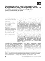
correlated with carbonyl values (r = 0.43; P = 0.007; Fig.
1) but not with AOPP levels (r = 0.007; P = 0.9).
Table 1
Characteristics of systemic sclerosis patients (n = 40)
Characteristic Value
Cutaneous form of the disease: limited / diffuse 23 (57) / 17 (43)
Digital pitting scars 6 (15)
Pulmonary arterial pressure (PAP)
mmHg [mean ± SD (range)] 33 ± 6 (21–48)
>40 mmHg 6 (15)
Pulmonary fibrosis (computed tomography scan) 16 (40)
Forced vital capacity below 75% of normal values 9 (23)
Decreased carbon monoxide diffusing capacity (DLCO/Hb <75%) 16 (40)
Left ventricular ejection fraction (%) (mean ± SD%) 62 ± 8
Antinuclear antibodies ≥1/160 38 (92)
Anti-topoisomerase I antibodies/anti-centromere antibodies 12/7
Creatininaemia (µmol) (mean ± SD) 79 ± 13
Erythrocyte sedimentation rate (mm/1st hour) (mean ± SD) 21 ± 18
C-reactive protein (mg/l) (mean ± SD) 11 ± 16
Ongoing low-dose prednisone (no. patients) (mean mg/day ± SD) 14 (7.4 ± 2)
Ongoing angiotensin-converting enzyme inhibitors (no. patients) 7
Ongoing low-dose aspirin therapy (no. patients) 25
Values are number (%) of patients unless otherwise indicated. DLCO, carbon monoxide diffusion in the lung; Hb, hemoglobin.
Arthritis Research & Therapy Vol 6 No 4 Allanore et al.
R312
Concentrations of VEGF were inversely correlated with dis-
ease duration (r = -0.4; P = 0.01); patients who had had
SSc for less than 5 years had higher median values than
those with longer disease duration (633 [105–1915] vs
424 [26–961]; P = 0.03). VEGF levels were not signifi-
cantly associated with the cutaneous subtypes; patients
with diffuse disease had median VEGF concentrations sim-
ilar to those of patients with limited cutaneous disease
(603.5 [130–1915] vs 570 [26–1594]; P = 0.37). Con-
centrations of sVCAM-1 were not associated with SSc
patient characteristics. We could not discern an influence
on sVCAM-1, VEGF, or sVEGFR-1 concentrations of
inflammation or treatment taken by SSc patients.
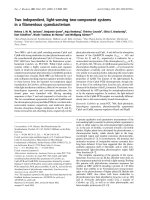
Nifedipine treatment significantly improved the vascular
marker sVCAM-1 and markers of oxidative damage (carbo-
nyls, AOPP) in patients with systemic sclerosis (SSc) but
did not significantly influence VEGF or sVEGFR-1 concen-
trations (Table 3; Figs 2,3). There were significant correla-
tions between individual levels at baseline and after
nifedipine treatment for sVCAM-1 (r = 0.49; P = 0.03), car-
bonyl residues (r = 0.68; P = 0.003), and AOPP (r = 0.67;
P = 0.005). The mean [VEGF]/[sVEGFR-1] ratio was not
statistically different before and after nifedipine treatment
(1100 ± 1190 vs 1450 ± 1280; P = 0.4).
Discussion
We found that 14 days of treatment with nifedipine
decreased serum vascular and oxidative damage markers
in patients with SSc but did not significantly modify the
concentration of VEGF or sVEGFR-1. Our results suggest
that nifedipine can ameliorate endothelium injury in SSc
and that VEGF signalling may by impaired in this disease
without implication of the sVEGFR-1.
Endothelium injury is critical in SSc and is suspected to
occur early in the disease process. Circulating sVCAM-1 is
a recognised marker of disease activity and a possible
marker of disease severity [6,7,21]; the expression of adhe-
Table 2
Serum concentrations of vascular markers in patients with systemic sclerosis (SSc) at baseline and in controls
Serum constituent Controls (n = 20) SSc patients at baseline (n = 40) P (Mann–Whitney test)
sVCAM-1 (pg/ml) 465.5 (378–619) 712 (362–1034) P < 0.0001
Carbonyl residues (nmol/mg protein) 0.34 (0.15–0.64) 0.83 (0.37–1.48) P < 0.0001
AOPP (µmol/l of chloramine-T equivalents) 75.5 (21–91) 109.1 (50–281) P < 0.0001
VEGF (pg/ml) 221 (19–499) 573.5 (26–1915) P = 0.0001
sVEGFR-1 (pg/ml) 19 (8–73) 45 (10–1140) P = 0.0025
Values are median (range) of serum concentrations in patients or controls. AOPP, advanced oxidation protein products; sVCAM-1, soluble
vascular cell adhesion molecule 1; sVEGFR-1, soluble VEGF receptor 1; VEGF, vascular endothelial growth factor.
Figure 1
Correlation between carbonyl residue and vascular endothelial growth factor (VEGF) concentrations in systemic sclerosis patients at baselineCorrelation between carbonyl residue and vascular endothelial growth
factor (VEGF) concentrations in systemic sclerosis patients at baseline
(n = 40; r = 0.43; P = 0.007).
Figure 2
Individual and median values of serum carbonyl concentrations in con-trol subjects (n = 20) and patients with systemic sclerosis (SSc) at baseline and after treatment with 60 mg nifedipine per day (n = 20)Individual and median values of serum carbonyl concentrations in con-
trol subjects (n = 20) and patients with systemic sclerosis (SSc) at
baseline and after treatment with 60 mg nifedipine per day (n = 20).
Available online />R313
sion molecules seems crucial in this disease, which is char-
acterised by early interactions between endothelial cells
and mononuclear cells [22].
We previously demonstrated a sustained major decrease in
oxidative stress in response to treatment with dihydropyrid-
ine-type calcium-channel blockers [17]. The data reported
herein confirm these results with a different design of the
study (midterm effects) and extend the known beneficial
effects of these drugs to a vascular marker (sVCAM). The
mechanism of the beneficial effects of dihydropyridines has
not yet been determined and could result from intrinsic anti-
oxidant properties [23] or from a secondary effect due to
the improvement of the vasospastic disease. Whatever the
mechanism involved, the concomitant decrease of sVCAM-
1 concentration and oxidative stress markers suggests that
these drugs improve endothelium injury. A decrease of
sVCAM-1 by nifedipine was previously reported in SSc
[24]. The improvement of all these markers and the correla-
tion between the values before and after treatment suggest
a powerful general action on endothelial injury. Calcium-
channel blockers of the dihydropyridine type have clearly
been shown to have an effect on Raynaud's syndrome in
SSc and several studies have suggested that they act on
coronary microvascular involvement [25]. Biological data
showing their effects on SSc are scarce, but it has been
suggested that nifedipine has an antiplatelet action [26].
Moreover, experimentally nifedipine may prevent apoptosis
of endothelial cells [27].
Angiogenesis seems to be impaired in SSc and this could
result from excessive angiostatic factors or disrupted
VEGF signalling. We confirm herein the high concentration
of VEGF in serum and its association with the early phase
of the disease [9]. Moreover, we report high concentrations
of the sVEGFR-1 in patients with SSc. sVEGFR-1 has a
strong antagonistic activity and neutralises the effects of
VEGF; it plays a pivotal role in the generation of vascular
diseases such as pre-eclampsia and intrauterine growth
retardation [28]. We hypothesised that the high VEGF con-
centration with impaired angiogenesis could result from
sVEGFR-1 abnormalities; our results do not support this
hypothesis, as the ratio between [VEGF] and [sVEGFR-1]
did not differ between patients with SSc and controls. Sev-
eral factors contribute to VEGF production, including
hypoxic conditions and stimulation by transforming growth
factor, CD40 ligand, interleukin 1, or interleukin 6 [10]. Oxi-
dative stress also promotes angiogenesis [13]. The link
between oxidative stress and angiogenesis is emphasised
in SSc by the baseline correlation between carbonyl resi-
dues and VEGF concentrations. However, although oxida-
tive damage markers were improved by nifedipine
treatment, we did not detect significant changes in VEGF
or sVEGFR-1 concentrations. Whereas it cannot be
excluded that nifedipine does not target the VEGF path-
way, this apparent lack of change supports the hypothesis
Figure 3
Individual and median values of serum sVCAM-1 concentrations in con-trol subjects (n = 20) and patients with systemic sclerosis (SSc) at baseline and after treatment with 60 mg nifedipine per day (n = 20)Individual and median values of serum sVCAM-1 concentrations in con-
trol subjects (n = 20) and patients with systemic sclerosis (SSc) at
baseline and after treatment with 60 mg nifedipine per day (n = 20).
Table 3
Serum concentrations of vascular markers in patients with systemic sclerosis (SSc) at baseline and after nifedipine treatment
Serum constituent Baseline (n = 20) After nifedipine treatment (n = 20) P (Wilcoxon test)
sVCAM-1 (pg/ml) 728.7 (450–1034) 635 (312–890) P = 0.0001
Carbonyl residues (nmol/mg protein) 0.69 (0.37–1.08) 0.57 (0.33–0.86) P = 0.0006
AOPP (µmol/l of chloramine-T equivalents) 111.35 (50.5–257.6) 89.25 (28–186.4) P = 0.004
VEGF (pg/ml) 463 (26–1251) 421.5 (47–2032) P = 0.97
sVEGFR-1 (pg/ml) 46 (10–587) 32 (9–452) P = 0.43
Values are median (range) of serum concentrations in patients. AOPP, advanced oxidation protein products; sVCAM-1, soluble vascular cell
adhesion molecule 1; sVEGFR-1, soluble VEGF receptor 1; VEGF, vascular endothelial growth factor.
Arthritis Research & Therapy Vol 6 No 4 Allanore et al.
R314
that VEGF signalling is impaired in SSc, but more experi-
mental data are needed in order to determine whether the
VEGF pathway is intrinsically defective.
Conclusion
Nifedipine treatment ameliorated endothelium injury in
patients with SSc, as shown by the concentrations of adhe-
sion molecules and oxidative damage markers. The fact that
VEGF and sVEGFR-1 concentrations were not changed
whereas oxidative stress was ameliorated by nifedipine is
consistent with the hypothesis that VEGF signalling is
impaired in SSc. Our results also do not support the impli-
cation of the sVEGFR-1 in the VEGF dysregulation, but
more experimental evidence is needed to determine
whether the VEGF pathway is intrinsically defective in SSc.
Competing interests
None declared.
References
1. LeRoy EC: Systemic sclerosis: a vascular perspective. Rheum
Dis Clin North Am 1996, 22:675-694.
2. Fleiszchmajer R, Perlish JS: Capillary alterations in scleroderma.
J Am Acad Dermatol 1980, 2:161-170.
3. Sgonc R, Gruschwitz MS, Boeck G, Sepp N, Gruber J, Wick G:
Endothelial cell apoptosis in systemic sclerosis is induced by
antibody-dependent cell-mediated cytotoxicity via CD95.
Arthritis Rheum 2000, 43:2550-2562.
4. Cerinic MM, Valentini G, Sorano GG, D'Angelo S, Cuomo G, Fenu
L, Generini S, Cinotti S, Morfini M, Pignone A, Guiducci S, Del
Rosso A, Kalfin R, Das D, Marongiu F: Blood coagulation, fibri-
nolysis, and markers of endothelial dysfunction in systemic
sclerosis. Semin Arthritis Rheum 2003, 32:285-295.
5. Altman RD, Medsger TA Jr, Bloch DA, Michel BA: Predictors of
survival in systemic sclerosis (scleroderma). Arthritis Rheum
1991, 34:403-413.
6. Kahaleh B, Meyer O, Scorza R: Assessment of vascular
involvement. Clin Exp Rheumatol 2003, Suppl 29:S9-S14.
7. Denton CP, Bickerstaff MC, Shiwen X, Carulli MT, Haskard DO,
Dubois RM, Black CM: Serial circulating adhesion molecule
levels reflect disease severity in systemic sclerosis. Br J
Rheumatol 1995, 34:1048-1054.
8. Kikuchi K, Kubo M, Kadono T, Yazawa N, Ihn H, Tamaki K: Serum
concentrations of vascular endothelial growth factor in colla-
gen diseases. Br J Dermatol 1998, 139:1049-1051.
9. Distler O, Del Rosso A, Giacomelli R, Cipriani P, Conforti ML, Gui-
ducci S, Gay RE, Michel BA, Bruhlmann P, Muller-Ladner U, Gay
S, Matucci-Cerinic M: Angiogenic and angiostatic factors in
systemic sclerosis: increased levels of vascular endothelial
growth factor are a feature of the earliest disease stages and
are associated with the absence of fingertip ulcers. Arthritis
Res 2002, 4:R11.
10. Choi JJ, Min DJ, Cho ML, Min SY, Kim SJ, Lee SS, Park KS, Seo
YI, Kim WU, Park SH, Cho CS: Elevated vascular endothelial
growth factor in systemic sclerosis. J Rheumatol 2003,
30:1529-1533.
11. Hebbar M, Peyrat JP, Hornez L, Hatron PY, Hachulla E, Devulder
B: Increased concentrations of the circulating angiogenesis
inhibitor endostatin in patients with systemic sclerosis. Arthri-
tis Rheum 2000, 43:889-893.
12. Shibuya M: Structure and dual function of vascular endothelial
growth factor receptor-1 (Flt-1). Int J Biochem Cell Biol 2001,
33:409-420.
13. Maulik N, Das DK: Redox signaling in vascular angiogenesis.
Free Radic Biol Med 2002, 33:1047-1060.
14. Simonini G, Pignone A, Generini S, Falcini F, Cerinic MM, Gabriele
S, Alberto P, Sergio G, Fernanda F, Marco MC: Emerging poten-
tials for an antioxidant therapy as a new approach to the treat-
ment of systemic sclerosis. Toxicology 2000, 155:1-15.
15. Thompson AE, Shea B, Welch V, Fenlon D, Pope JE: Calcium-
channel blockers for Raynaud's phenomenon in systemic
sclerosis. Arthritis Rheum 2001, 44:1841-1847.
16. Duboc D, Kahan A, Maziere B, Loc'h C, Crouzel C, Menkes CJ,
Amor B, Strauch G, Guerin F, Syrota A: The effect of nifedipine
on myocardial perfusion and metabolism in systemic sclero-
sis. A positron emission tomographic study. Arthritis Rheum
1991, 34:198-203.
17. Allanore Y, Borderie D, Lemaréchal H, Ekindjian OG, Kahan A:
Acute and sustained effects of dihydropyridine-type calcium
channel antagonists on oxidative stress in systemic sclerosis.
Am J Med 2004, 116:595-600.
18. LeRoy EC, Black C, Fleischmajer R, Jablonska S, Krieg T, Medsger
TA Jr, Rowell N, Wollheim F: Scleroderma (systemic sclerosis):
classification, subsets and pathogenesis. J Rheumatol 1988,
1:202-205.
19. Reznick AZ, Packer L: Oxidative damage to proteins: spectro-
photometric method for carbonyl assay. Methods Enzymol
1994, 233:357-363.
20. Witko-Sarsat V, Friedlander M, Capelliere-Blandin C, Nguyen-
Khoa T, Nguyen AT, Zingraff J, Jungers P, Descamps-Latscha B:
Advanced oxidation protein products as a novel marker of oxi-
dative stress in uremia. Kidney Int 1996, 49:1304-1313.
21. Shahin AA, Anwar S, Elawar AH, Sharaf AE, Hamid MA, Eleinin AA,
Eltablawy M: Circulating soluble adhesion molecules in
patients with systemic sclerosis: correlation between circulat-
ing soluble vascular cell adhesion molecule-1 (sVCAM-1) and
impaired left ventricular diastolic function. Rheumatol Int 2000,
20:21-4.
22. Prescott RJ, Freemont AJ, Jones CJ, Hoyland J, Fielding P:
Sequential dermal microvascular and perivascular changes in
the development of scleroderma. J Pathol 1992, 166:255-263.
23. Fukuo K, Yang J, Yasuda O, Mogi M, Suhara T, Sato N, Suzuki T,
Morimoto S, Ogihara T: Nifedipine indirectly upregulates super-
oxide dismutase expression in endothelial cells via vascular
smooth muscle cell-dependent pathways. Circulation 2002,
106:356-361.
24. Dziadzio M, Denton CP, Smith R, Howell K, Blann A, Bowers E,
Black CM: Losartan therapy for Raynaud's phenomenon and
scleroderma: clinical and biochemical findings in a fifteen-
week, randomized, parallel-group, controlled trial. Arthritis
Rheum 1999, 42:2646-2655.
25. Kahan A, Devaux JY, Amor B, Menkes CJ, Weber S, Nitenberg A,
Venot A, Guerin F, Degeorges M, Roucayrol JC: Nifedipine and
thallium-201 myocardial perfusion in progressive systemic
sclerosis. N Engl J Med 1986, 314:1397-1402.
26. Rademaker M, Meyrick Thomas RH, Kirby JD, Kovacs IB: The anti-
platelet effect of nifedipine in patients with systemic sclerosis.
Clin Exp Rheumatol 1992, 10:57-62.
27. Sugano M, Tsuchida K, Makino N: Nifedipine prevents apoptosis
of endothelial cells induced by oxidized low-density
lipoproteins. J Cardiovasc Pharmacol 2002, 40:146-152.
28. Carsten Hornig, Bernhard Barleon, Shakil Ahmad, Piia Vuorela,
Asif Ahmed, Herbert A Weich: Release and complex formation
of soluble VEGFR-1 from endothelial cells and biological
fluids. Lab Invest 2000, 80:443-454.