Báo cáo y học: "Perspectives and limitations of gene expression profiling in rheumatology: new molecular strategies" doc
Bạn đang xem bản rút gọn của tài liệu. Xem và tải ngay bản đầy đủ của tài liệu tại đây (260.4 KB, 7 trang )
140
GCP = granulocyte chemotactic protein; IFN = interferon; IL = interleukin; MCP = monocyte chemotactic protein; OA = osteoarthritis; PBMC =
peripheral blood mononuclear cell; PCR = polymerase chain reaction; RA = rheumatoid arthritis; SLE = systemic lupus erythematosus; TNF =
tumour necrosis factor.
Arthritis Research & Therapy Vol 6 No 4 Häupl et al.
Introduction
Inflammatory rheumatic diseases are among the greatest
diagnostic challenges in modern medicine. Especially in
early cases there are usually no pathognomonic markers
such as distinct clinical features, specific morphological
changes by imaging or typical serological markers.
Similarly to malignant situations, however, early diagnosis
is essential to avoid destructive processes that will lead to
a severely reduced quality of life, early invalidity and
premature death.
In view of the limitations in clinical rheumatology,
expectations of genomics are high. Gene expression
profiling has opened new avenues. Instead of single or a
handful of candidates, tens of thousands of different
genes can be investigated at a given time. This technology
is currently the most advanced and comprehensive
approach to screening gene activity as well as molecular
networks and has already been used in several clinical
studies in rheumatic diseases. Although moving at a
slower pace, proteome analyses are also rapidly improving
and might provide further insight beyond the capabilities
of transcriptome information. Furthermore, genome muta-
tions predisposing for rheumatic diseases might help in
both diagnosis and prognosis of the disease [1].
Clinical questions and expectations focus on molecular
markers or profiles for initial diagnosis [2]. Early diagnosis,
as mentioned, is critical; gene expression profiles at this
initial phase of the disease might provide valuable
Commentary
Perspectives and limitations of gene expression profiling in
rheumatology: new molecular strategies
Thomas Häupl
1
, Veit Krenn
2
, Bruno Stuhlmüller
1
, Andreas Radbruch
3
and Gerd R Burmester
1
1
Department of Rheumatology, Charité, Berlin, Germany
2
Institute for Pathology, Charité, Berlin, Germany
3
German Arthritis Research Centre (DRFZ), Berlin, Germany
Corresponding author: Thomas Häupl,
Received: 13 Feb 2004 Revisions requested: 29 Mar 2004 Revisions received: 27 Apr 2004 Accepted: 12 May 2004 Published: 4 Jun 2004
Arthritis Res Ther 2004, 6:140-146 (DOI 10.1186/ar1194)
© 2004 BioMed Central Ltd
Abstract
The deciphering of the sequence of the human genome has raised the expectation of unravelling the
specific role of each gene in physiology and pathology. High-throughput technologies for gene
expression profiling provide the first practical basis for applying this information. In rheumatology, with
its many diseases of unknown pathogenesis and puzzling inflammatory aspects, these advances
appear to promise a significant advance towards the identification of leading mechanisms of
pathology. Expression patterns reflect the complexity of the molecular processes and are expected to
provide the molecular basis for specific diagnosis, therapeutic stratification, long-term monitoring and
prognostic evaluation. Identification of the molecular networks will help in the discovery of appropriate
drug targets, and permit focusing on the most effective and least toxic compounds. Current
limitations in screening technologies, experimental strategies and bioinformatic interpretation will
shortly be overcome by the rapid development in this field. However, gene expression profiling, by its
nature, will not provide biochemical information on functional activities of proteins and might only in
part reflect underlying genetic dysfunction. Genomic and proteomic technologies will therefore be
complementary in their scientific and clinical application.
Keywords: expression profiling, genomics, molecular strategies, pathway models, signatures
141
Available online />information on triggering mechanisms. Assessment of
disease activity including organ involvement or destruction
is currently limited to general markers of inflammation or
organ function and needs profound improvement. On the
basis of gene expression profiles from an initial molecular
assessment of a patient, we expect to identify subclasses
or different stages of the diseases with relevance to the
therapeutic decision. As in only few other diseases, our
therapeutic anti-rheumatic armamentarium has been
greatly enlarged by modern approaches of combination
therapies, which include the usage of biologics (namely,
cytokine antagonists). Nevertheless, these modern
strategies are effective only in a proportion of patients,
potentially make the patients more prone to infections and
represent an enormous economic burden to the health
care system. Careful diagnostic stratification will therefore
be crucial. Once therapy has been initiated, monitoring of
effectiveness and responsiveness is essential and is
currently dominated by scores derived from physical
examination [3]. Molecular measures are needed that
define the quantity and quality of responsiveness to adjust
the dosage or change the drug. Profiles might also give a
clue to identifying toxic side effects and adverse events
such as infectious complications. Prognostic molecular
markers might arise from long-term studies by correlating
initial expression profiles with the individual outcome.
From a pharmaceutical point of view, unravelling the
molecular puzzle of rheumatic diseases might lead to the
discovery of the dominant pathways in this network and
provide novel targets for drug development. Current
therapies in rheumatic diseases focus predominantly on
the suppression of inflammation. However, destructive
processes and loss of function, as in lupus nephritis or
arthritic cartilage invasion and bone resorption, also
demand the identification of targets to directly inhibit
destruction and/or to induce regeneration and repair. A
deeper knowledge of pathophysiological networks and
gene expression profiling during drug development will
facilitate the selection of the most effective and the least
toxic compounds, thereby reducing costs and bringing
new drugs to clinical application at an earlier stage.
To fulfil all these expectations, systematic analyses,
collating of information and development of molecular
network models will be essential and will provide the basis
for functional interpretation.
Current status of gene expression profiling in
rheumatic diseases
An initial work by Heller and colleagues [4] introduced a
customised array of 96 genes, demonstrating the useful-
ness of arrays in the analysis of inflammatory diseases such
as rheumatoid arthritis (RA). Basing their work on a specific
selection of genes, they identified in synovial tissue
samples from RA the expression of the matrix metallo-
proteinases stromelysin 1, collagenase 1, gelatinase A and
human matrix metallo-elastase, TIMP (tissue inhibitor of
metalloproteinases) 1 and 3, interleukin (IL)-6, vascular cell
adhesion molecule and discernible levels of monocyte
chemotactic protein (MCP)-1, migration inhibitory factor
and RANTES.
More advanced platform technologies with many
thousands of genes up to genome-wide arrays have been
applied in recent studies, aiming for new candidates,
functional mechanisms and diagnostic patterns. Comparing
autoimmune diseases with the response to influenza
vaccination in healthy donors, Maas and colleagues
investigated peripheral blood mononuclear cells (PMBCs)
from patients with RA, systemic lupus erythematosus
(SLE), type I diabetes and multiple sclerosis [5]. Genes
differentially expressed after vaccination were compared
with the profiles of the four autoimmune groups. A panel of
genes was extracted that discriminated between normal
immune and autoimmune responses. However, the
investigators could not identify genes that distinguished
between different autoimmune diseases. Their candidates
were predominantly genes involved in apoptosis, cell cycle
progression, cell differentiation and cell migration, but not
necessarily in the immune response. They further
developed an algorithm to identify patients with these
autoimmune diseases. Because this algorithm also sorted
relatives of patients with autoimmune diseases to the
disease group, the authors speculated that their gene
selection might reflect a genetic trait rather than the
disease process.
Gene expression profiling in lupus was reviewed recently
in detail by Crow and Wohlgemuth [6]. Four different
groups [6–9] have independently identified an interferon
signature by analysing PBMCs. One group [7] confirmed
these findings by comparing the patients’ profiles with in
vitro-induced interferon (IFN)-α, IFN-β or IFN-γ signatures
in PBMCs from healthy donors. This attributed 23 of 161
genes to induction by IFN. In addition to the IFN signature,
Bennett and colleagues [8] found the differential
expression of granulopoietic genes. As Ficoll separation
usually excludes granulocytes, they became aware of a
subpopulation of granular cells, which was co-separated
only in SLE. These were identified as cells of the myeloid
lineage, ranging from promyelocytes to segmented
neutrophils.
Gu and colleagues [10] investigated PBMCs from
spondyloarthropathies, RA and psoriatic arthritis on a 588-
gene commercial platform. Their dominant candidates
included MNDA, a myeloid nuclear differentiation antigen,
two members of the S100 family of proteins, calgranulin A
and B (involved in cellular processes such as cycle
progression and differentiation), JAK3 and mitogen-
activated protein kinase p38, tumour necrosis factor (TNF)
142
Arthritis Research & Therapy Vol 6 No 4 Häupl et al.
receptors, the chemokine receptors CCR1 and CXCR4
and also IL-1β and IL-8. Because stromal cell-derived
factor-1 (SDF-1), the ligand of CXCR4, was found
increased in the synovial fluids of arthritides, the authors
suggested an important role of this chemotactic axis in
spondyloarthropathies and RA. In our studies on highly
purified separated cells, these genes revealed the highest
expression level in neutrophil granulocytes in comparison
with cells positive for CD14, CD4 and CD8. In view of the
findings by Bennett and colleagues [8] that granulocytes
might be co-separated with PBMCs in inflammatory
diseases such as SLE, these data need further
confirmation.
Van der Pouw Kraan and colleagues investigated synovial
tissue samples from RA and osteoarthritis (OA) [11,12].
Basing their decision on molecular profiles, they divided
their RA samples into three subgroups: first, immune-
related processes; second, complement-related activities
with fibroblast dedifferentiation; and third, processes of
tissue remodelling. Their analyses also reflect the
established histological classification of RA into different
subgroups, which is in part based on cellular composition
[13]. Furthermore, the STAT1 pathway was identified as
being associated with immune-related processes. Our
own data on synovial tissues, which were established on a
different technology platform, confirm many of these
findings [14]. We also identified that some of the
processes, especially those associated with tissue
remodelling, are also active in OA compared with normal
tissues [15].
A similar tissue-based approach showed various
inflammatory genes to be upregulated in chronic
inflammation of periprosthetic membranes of RA and OA
patients in the process of prosthetic loosening [16].
To overcome the problem of unspecific dilution and to
allow the histological association of complete profiles,
Judex and colleagues [17] have presented an initial study
on gene expression analysis of laser-microdissected areas
from synovial tissues. They have been able to extract
sufficient RNA from as few as 600 cells to perform
subsequent array analysis.
In contrast, in vitro studies on isolated synovial fibroblasts
from RA patients are well established. Pierer and
colleagues [18] have investigated profiles of synoviocytes
on a functional basis by stimulation through Toll-like
receptor 2 with Staphylococcus aureus peptidoglycan.
Their focus on chemokines revealed a preferential
activation of granulocyte chemotactic protein (GCP)-2,
RANTES, MCP-2, IL-8 and GRO2. Functional
dependence on NF-κB for the induction of MCP-2,
RANTES and GCP-2 was confirmed by inhibition
experiments. Chemotactic importance for monocyte
migration was demonstrated for RANTES and MCP-2,
and for T-cell migration only for RANTES. The expression
of GCP-2 and MCP-2, which have not yet been
investigated in RA, was identified in both synovial tissue
and synovial fluid.
Besides the application in human studies, gene
expression profiling was also performed in arthritis
models. Wester and colleagues [19] investigated the
effect of pristan-induced arthritis in DA rats in comparison
with resistant E3 rats. The authors compared two different
array platforms for a selected number of genes and also
used pooled samples. They demonstrated variable cellular
composition of the lymph nodes by fluorescence-activated
cell sorting and identified only a relatively small number of
genes that were differentially expressed, including mRNA
for major histocompatibility complex class II antigen, immuno-
globulins, CD28, mast cell protease 1, gelatinase B,
carboxylesterase precursor, K-cadherin, cyclin G1, DNA
polymerase and the tumour-associated glycoprotein E4.
By expression profiling in experimental SLE of NZB/W
mice, Alexander and colleagues [20] identified endo-
genous retroviral transcripts in kidney tissue as the highest
differentially expressed genes. Results were confirmed by
in situ hybridisation, demonstrating retroviral transcripts in
renal tubules and also in brain and lung tissue.
Azuma and colleagues used microarrays for the detection
of new candidates in salivary gland tissue from the
MLR/MpJ-lpr/lpr (MRL/lpr) mouse as a model of human
secondary Sjögren’s syndrome [21]. From nine genes,
which were confirmed by reverse transcriptase
polymerase chain reaction (PCR), five had been already
identified in patients with Sjögren’s syndrome.
Firneisz and colleagues [22] used gene expression
profiling in two genetically different arthritis mouse models
[23,24] to identify genes involved in both models.
Subsequently, they computed the spatial autocorrelation
function, a statistical technique used in astrophysics, and
identified critical clustering of selected genes in the two
different genetic backgrounds of these mice.
Aidinis and colleagues [25] investigated immortalised
synovial fibroblasts from human (h)TNF transgenic mice by
microarray and differential display technology. Microarrays
revealed 372 differentially regulated genes, whereas
differential display provided many unknown sequences
and a total of 49 different genes and sequences. Only
20% (n = 11) of these were represented on the mouse
array. The significance of regulation was only partly
confirmed, and one gene (SPARC) was identified as
being regulated in both but in opposite directions.
Functional clustering of all differentially regulated genes in
either of the two methods revealed genes involved in
143
stress response, energy production, transcription, RNA
processing, protein synthesis and degradation, growth
control, adhesion, cytoskeletal organisation, Ca
2+
binding
and antigen presentation.
Limitations to current approaches
As summarised in this short overview, gene expression
profiling with microarrays has been applied in recent work
to the identification of either diagnostic algorithms or new
candidates and pathomechanisms, to functional studies in
mouse and in vitro models, and to the calculation of
potential genomic clusters associated with the disease. In
a few studies different technologies or platforms were
compared. In all studies, confirmation analysis was
possible only for a limited number of genes. Concordance
or divergence of results can therefore be estimated only
from the selection of genes published in more detail.
Up to now, gene expression profiling has given only a first
suggestion of candidates. It is still impossible to interpret
comprehensively this overwhelming flood of data and the
puzzling complexity of as yet insufficiently characterised
molecular networks. Different platform technologies further
complicate comparability. Nevertheless, the publication of
results achieved with the current state of methodology is
essential in the exchange and development of different
approaches to gene expression profiling and in comparing
selected candidates. This will improve our concepts to
overcome the problems and limitations arising from this
technology.
Array technologies and statistical algorithms, as they are
established today, provide measures for signal intensities
and differences on the basis of the abundance of mRNA in
a given sample. In RA, the current results of array analyses
[12] would not necessarily direct drug development
towards the most favourable therapeutic targets such as
TNF and IL-1. In SLE, an interferon signature was
identified; however, indirect signs were detectable but not
the cytokine itself [7]. In contrast, genes of highly
abundant proteins such as immunoglobulins, collagens
and matrix metalloproteinases were readily identified by
array analysis. Furthermore, the mRNA species of many
cell surface receptors were also identified. These
observations suggest that RNA abundance and detection
by array techniques might be related to the functional
category to which a gene belongs. This would be of
special relevance to diagnostic and pathophysiological
interpretation and therefore important to current limitations
and perspectives.
Concerning the lack of detection of TNF, IL-1 or interferon
as candidates for important regulators of patho-
mechanisms, the following possibilities might explain such
limitations: first, the array hybridisation techniques might
not be sensitive enough; second, signals derived from a
defined cell population might be diluted below the
threshold of significance in the complex tissues; or third,
the stimulation might have occurred at a different location
or time, leaving only its signature as an indirect sign of the
activated pathway.
The application of purification or microdissection
techniques might therefore increase sensitivity and
improve our insight into the regulatory networks of
important immune regulators. However, purification
techniques might introduce artefacts. As an alternative
approach, similar to Baechler’s confirmation of the
interferon signature, comparison with cytokine-induced
gene expression signatures might provide an indirect
measure for the activation of the TNF or IL-1 pathway.
Besides the cytokines, genes of the intracellular signalling
cascade are also important for the understanding of
pathophysiology and might be relevant to drug targeting.
Dependent on cell type and function, such proteins might
be expressed from very low to relatively high basal levels.
Upregulation of these genes might not exceed a certain
limit of expression because protein concentration will
quickly increase in the small intracellular compartment,
where they act. Furthermore, the function of these factors
is mostly regulated at the protein level. Therefore, in this
category of molecules, detection of the quantitatively
limited differences is also very difficult. Signals might be
diluted and become undetectable if activation occurred in
a localised manner. Differential expression between
infiltrating and tissue cells might also confuse inter-
pretation and falsely indicate regulation, especially when
cellular composition is variable. This might also be crucial
for separation procedures, when variable quantities of
cells with different profiles remain as contaminants.
On the basis of these findings and general
considerations, it is currently almost impossible for many
signalling processes to become readily obvious as being
truly regulated. A different cellular composition resulting
from infiltration is inherent in the inflammatory processes
analysed in rheumatology. Parameters that reflect this
cellular composition and functional components might
need to be introduced into the analysis to improve
interpretation. The fact that molecular profiles enabled
the identification of an unexpected subpopulation in
PBMCs by Bennett and colleagues encourages one to
believe in the possibility of identifying parameters for a
molecular differential blood count or tissue composition.
Thus, many of the currently published data will merit re-
evaluation when improved technologies of interpretation
become available.
Recent developments in array technology
An extensive review of microarrays by Grant and
colleagues [26] describes the general features of spotting
Available online />144
and photolithography array technology as well as the
general tools for bioinformatic analysis of these arrays.
Rapid advances in this field have brought new
technologies to the market. These include PCR arrays
[27], bead arrays [28] and bioelectronic sensors [29–31].
Concerning the different slide or wafer-based array
technologies, reproducibility and quality have undergone
constant improvement for all platforms. Although
photolithographic technology is currently highly efficient
for genome-wide array analysis, new surfaces provided in
the context of spotting technology might improve
sensitivity [32]. Gene expression profiles of only up to a
few hundred genes might be determined more rapidly,
with increased sensitivity and less expense, with the use
of real-time PCR technology prefabricated on a card
system with up to 384 different reactions.
In addition, with a relatively low investment, with less
working time and with applicability to DNA [33] as well as
protein or antibody screening, bead array systems can
currently detect many hundreds of different products even
from very small sample volumes. For example, Cook and
colleagues [34] have applied this system to the detection
of six different cytokines at the protein level in tears from
allergic patients.
The new evolving detection methods based on bio-
electronic sensors are forming an electronic circuit
mediated only by nucleic acid hybridisation. This very
intriguing approach, which is currently applicable to DNA
detection and mutation analysis, might soon become
applicable to the quantification of cDNA. This system is
currently established for only a few DNA species. With
low investment and convenient application, this system
inherits the potential to be developed for a cost-effective
bedside test.
Bioinformatics
Molecular profiles of previously published experiments are
extremely complex. Bioinformatics has long been focusing
on the technical challenges and the enormous amount of
data from image analysis (millions of pixels per image) and
comparisons of genes (several hundreds of thousands).
Many efforts to distinguish signals from background and
to identify and eliminate artefacts have now created high-
quality platforms. Many algorithms to identify differential
gene expression and to group similarities together have
been established, using different types of distance
measures, statistics and cluster methods [35]. Supervised
clustering, neuronal networks and classification algorithms
might provide astonishing results [36–38].
However, these technologies are also regarded as black
boxes by many clinical investigators, as leading away from
understanding the principles of gene selection and
disregarding established clinical experience or previous
molecular knowledge. It is now becoming more than obvious
that bioinformatics depends essentially on a basic
knowledge of biology. ‘Systems biology’, ‘molecular
networks’, ‘biochemical systems theory’ [39] and other
meaningful terms have been used to express this basic need
for a functional understanding of molecular mechanisms in
biology. Our molecular knowledge – especially of a
rheumatological background – has to be systematically
collected and organised to make this information retrievable.
Gene ontologies (GO) and functional networks in the KEGG
or GenMAPP databases are still in their infancy.
Interpretation in rheumatology is restricted to the personal
knowledge and investigative capacity of the scientists and is
susceptible to misinterpretation.
In the face of our limited knowledge of the role and
function of most of the genes that we discover in our
experiments, strategies for systematic investigation are
essential. Gene expression profiling will essentially
depend on valid statistical methods for estimating the
reliability of gene selection. A combined analysis of
molecular and clinical data will be necessary. Functional
data need to be integrated into our interpretation to
identify the key molecules that connect the network and
define the boundaries of different phenotypes of the
system [40]. These will allow models to develop and will
reduce our screening and analysis efforts to the principal
components and actors.
Strategies
Suggestions by Firestein and Pisetsky [41] underline the
importance of an understandable and reproducible
bioinformatics approach. Most software packages have
now reached a level that provides enough statistical
power for basic comparative analyses. Platform technolo-
gies are becoming increasingly available from professional
suppliers and are achieving high reproducibility. Currently
evolving high-throughput technologies that confirm gene
expression profiling on a functional basis, such as protein,
tissue [42] or cell arrays, are still limited to a few
representative candidates. Analysis of defined cell
populations will provide cornerstones to our view of
systems biology but will not provide sufficient insight into
the networks of functional units consisting of different
interacting cells and organ systems.
Intelligent strategies will therefore be necessary, making
use of the currently most advanced capabilities in gene
expression profiling. Besides the principal limitations of
mRNA quantification in comparison with proteomics and
of functional interpretation, there are currently two general
hurdles: a mixture of profiles from different cell types, and
a mixture of profiles derived from different stimuli or
functional processes. As in routine laboratory analysis,
standards and ranges need to be defined to distinguish
Arthritis Research & Therapy Vol 6 No 4 Häupl et al.
145
between different molecular phenotypes on an individual
basis.
Defining signatures as specific patterns derived from
singular functional or cellular entities, signatures of highly
purified leucocyte cell types [43] and precisely defined
cellular stimulation (for example, stimulation by Toll-like
receptor 2 in synovial fibroblasts [18]) contribute to the
establishment of such a systematic data collection for
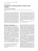
referencing. On the basis of such referenced information,
algorithms have to be established that are able to identify
the contribution of each cellular and functional component
to the complex profile of an individual sample (Fig. 1).
To achieve such a general approach, it is indispensable to
adhere to the standardisation of techniques, to intensify
collaborations between different expert groups, to share
array data as raw data and to define guidelines of good
scientific practice to respect and honour individual
contributions to such databases, which must be made
publicly accessible [44].
Conclusions
Gene expression profiling provides a completely new
approach to rheumatology research. As an interdisciplinary
technology it has stimulated fruitful collaboration between
experts in array technology, bioinformatics, immunology
and rheumatology. The molecular overview by genome-
wide profiles has revealed that many questions arise that
demand careful standardisation and validation. The
complexity of clinical samples has also initiated new
experimental strategies to dissect cellular and functional
signatures and to enable the interpretation of profiles from
each patient individually. To accomplish this approach,
data sharing and collectively developed knowledge bases
in rheumatology for data mining will markedly accelerate
this time-consuming process and will also open new
avenues for many established models and many as yet
unanalysed disease entities in rheumatology.
Competing interests
None declared.
Acknowledgements
We thank Christian Kaps PhD, Oligene GmbH, Berlin, for fruitful dis-
cussion. Perspectives and strategies result from our current work,
which is supported by the BMBF grants 01GS0110 and 01GS0160.
References
1. Suzuki A, Yamada R, Chang X, Tokuhiro S, Sawada T, Suzuki M,
Nagasaki M, Nakayama-Hamada M, Kawaida R, Ono M, et al.:
Functional haplotypes of PADI4, encoding citrullinating
enzyme peptidylarginine deiminase 4, are associated with
rheumatoid arthritis. Nat Genet 2003, 34:395-402.
2. Visser H, le Cessie S, Vos K, Breedveld FC, Hazes JM: How to
diagnose rheumatoid arthritis early: a prediction model for
persistent (erosive) arthritis. Arthritis Rheum 2002, 46:357-
365.
3. Furst DE, Breedveld FC, Kalden JR, Smolen JS, Burmester GR,
Dougados M, Emery P, Gibofsky A, Kavanaugh AF, Keystone EC,
et al.: Updated consensus statement on biological agents for
the treatment of rheumatoid arthritis and other immune medi-
ated inflammatory diseases (May 2003). Ann Rheum Dis 2003,
62 (Suppl 2):ii2-ii9.
4. Heller RA, Schena M, Chai A, Shalon D, Bedilion T, Gilmore J,
Woolley DE, Davis RW: Discovery and analysis of inflammatory
disease-related genes using cDNA microarrays. Proc Natl
Acad Sci USA 1997, 94:2150-2155.
5. Maas K, Chan S, Parker J, Slater A, Moore J, Olsen N, Aune TM:
Cutting edge: molecular portrait of human autoimmune
disease. J Immunol 2002, 169:5-9.
6. Crow MK, Wohlgemuth J: Microarray analysis of gene expres-
sion in lupus. Arthritis Res Ther 2003, 5:279-287.
7. Baechler EC, Batliwalla FM, Karypis G, Gaffney PM, Ortmann
WA, Espe KJ, Shark KB, Grande WJ, Hughes KM, Kapur V, et al.:
Interferon-inducible gene expression signature in peripheral
blood cells of patients with severe lupus. Proc Natl Acad Sci
USA 2003, 100:2610-2615.
8. Bennett L, Palucka AK, Arce E, Cantrell V, Borvak J, Banchereau
J, Pascual V: Interferon and granulopoiesis signatures in sys-
temic lupus erythematosus blood. J Exp Med 2003, 197:711-
723.
9. Han GM, Chen SL, Shen N, Ye S, Bao CD, Gu YY: Analysis of
gene expression profiles in human systemic lupus erythe-
matosus using oligonucleotide microarray. Genes Immun
2003, 4:177-186.
10. Gu J, Marker-Hermann E, Baeten D, Tsai WC, Gladman D, Xiong
M, Deister H, Kuipers JG, Huang F, Song YW, et al.: A 588-gene
microarray analysis of the peripheral blood mononuclear cells
of spondyloarthropathy patients. Rheumatology (Oxford) 2002,
41:759-766.
11. van der Pouw Kraan TC, van Gaalen FA, Kasperkovitz PV, Verbeet
NL, Smeets TJ, Kraan MC, Fero M, Tak PP, Huizinga TW, Pieter-
man E, et al.: Rheumatoid arthritis is a heterogeneous
disease: evidence for differences in the activation of the
STAT-1 pathway between rheumatoid tissues. Arthritis Rheum
2003, 48:2132-2145.
12. van der Pouw Kraan TC, van Gaalen FA, Huizinga TW, Pieterman
E, Breedveld FC, Verweij CL: Discovery of distinctive gene
expression profiles in rheumatoid synovium using cDNA
microarray technology: evidence for the existence of multiple
pathways of tissue destruction and repair. Genes Immun
2003, 4:187-196.
13. Krenn V, Morawietz L, Häupl T, Neidel J, König A: Grading of
chronic synovitis – a histopathological grading system for
molecular and diagnostic pathology. Path Res Pract 2002,
198:317-325.
Available online />Figure 1
Illustration of a strategy to characterise cellular composition, extract
regulated genes and identify the dominant triggers in complex profiles
of blood and tissue samples.
146
Arthritis Research & Therapy Vol 6 No 4 Häupl et al.
14. Häupl T, Stuhlmuller B, Janitz M, Kiesslich O, Rohrlach T, Kaps C,
Tandon N, Morawietz L, Girnus A, Wachtel A, et al.: Gene expres-
sion profiling in rheumatoid arthritis. Z Rheumatol 2003,
62(Suppl 1):PDo-50.
15. Häupl T, Kaps C, Kiesslich O, Rohrlach T, Morawietz L, Neidel J,
Pruss A, Krenn V, Gursche A, Zacher J, et al.: Synovial gene
expression profiles in chronic joint diseases. Osteoarthritis
Cartilage 2003, 11(Suppl 1):S101.
16. Morawietz L, Gehrke T, Frommelt L, Gratze P, Bosio A, Moller J,
Gerstmayer B, Krenn V: Differential gene expression in the
periprosthetic membrane: lubricin as a new possible patho-
genetic factor in prosthesis loosening. Virchows Arch 2003,
443:57-66.
17. Judex M, Neumann E, Lechner S, Dietmaier W, Ballhorn W, Grifka
J, Gay S, Scholmerich J, Kullmann F, Muller-Ladner U: Laser-
mediated microdissection facilitates analysis of area-specific
gene expression in rheumatoid synovium. Arthritis Rheum
2003, 48:97-102.
18. Pierer M, Rethage J, Seibl R, Lauener R, Brentano F, Wagner U,
Hantzschel H, Michel BA, Gay RE, Gay S, et al.: Chemokine
secretion of rheumatoid arthritis synovial fibroblasts stimu-
lated by Toll-like receptor 2 ligands. J Immunol 2004,
172:1256-1265.
19. Wester L, Koczan D, Holmberg J, Olofsson P, Thiesen HJ, Holm-
dahl R, Ibrahim S: Differential gene expression in pristane-
induced arthritis susceptible DA versus resistant E3 rats.
Arthritis Res Ther 2003, 5:R361-R372.
20. Alexander JJ, Saxena AK, Bao L, Jacob A, Haas M, Quigg RJ:
Prominent renal expression of a murine leukemia retrovirus in
experimental systemic lupus erythematosus. J Am Soc
Nephrol 2002, 13:2869-2877.
21. Azuma T, Takei M, Yoshikawa T, Nagasugi Y, Kato M, Otsuka M,
Shiraiwa H, Sugano S, Mitamura K, Sawada S, et al.: Identifica-
tion of candidate genes for Sjogren’s syndrome using MRL/
lpr mouse model of Sjogren’s syndrome and cDNA micro-
array analysis. Immunol Lett 2002, 81:171-176.
22. Firneisz G, Zehavi I, Vermes C, Hanyecz A, Frieman JA, Glant TT:
Identification and quantification of disease-related gene clus-
ters. Bioinformatics 2003, 19:1781-1786.
23. Courtenay JS, Dallman MJ, Dayan AD, Martin A, Mosedale B:
Immunisation against heterologous type II collagen induces
arthritis in mice. Nature 1980, 283:666-668.
24. Glant TT, Mikecz K, Arzoumanian A, Poole AR: Proteoglycan-
induced arthritis in BALB/c mice. Clinical features and
histopathology. Arthritis Rheum 1987, 30:201-212.
25. Aidinis V, Plows D, Haralambous S, Armaka M, Papadopoulos P,
Kanaki MZ, Koczan D, Thiesen HJ, Kollias G: Functional analysis
of an arthritogenic synovial fibroblast. Arthritis Res Ther 2003,
5:R140-R157.
26. Grant EP, Pickard MD, Briskin MJ, Gutierrez-Ramos JC: Gene
expression profiles: creating new perspectives in arthritis
research. Arthritis Rheum 2002, 46:874-884.
27. Haque KA, Pfeiffer RM, Beerman MB, Struewing JP, Chanock SJ,
Bergen AW: Performance of high-throughput DNA quantifica-
tion methods. BMC Biotechnol 2003, 3:20.
28. Dunbar SA, Vander Zee CA, Oliver KG, Karem KL, Jacobson JW:
Quantitative, multiplexed detection of bacterial pathogens:
DNA and protein applications of the Luminex LabMAP system.
J Microbiol Methods 2003, 53:245-252.
29. Bernacki SH, Farkas DH, Shi W, Chan V, Liu Y, Beck JC, Bailey
KS, Pratt VM, Monaghan KG, Matteson KJ, et al.: Bioelectronic
sensor technology for detection of cystic fibrosis and heredi-
tary hemochromatosis mutations. Arch Pathol Lab Med 2003,
127:1565-1572.
30. Lenigk R, Liu RH, Athavale M, Chen Z, Ganser D, Yang J, Rauch
C, Liu Y, Chan B, Yu H, et al.: Plastic biochannel hybridization
devices: a new concept for microfluidic DNA arrays. Anal
Biochem 2002, 311:40-49.
31. Vernon SD, Farkas DH, Unger ER, Chan V, Miller DL, Chen YP,
Blackburn GF, Reeves WC: Bioelectronic DNA detection of
human papillomaviruses using eSensor trade mark : a model
system for detection of multiple pathogens. BMC Infect Dis
2003, 3:12.
32. Ramakrishnan R, Dorris D, Lublinsky A, Nguyen A, Domanus M,
Prokhorova A, Gieser L, Touma E, Lockner R, Tata M, et al.: An
assessment of Motorola CodeLink microarray performance
for gene expression profiling applications. Nucleic Acids Res
2002, 30:e30.
33. Oliphant A, Barker DL, Stuelpnagel JR, Chee MS: BeadArray
technology: enabling an accurate, cost-effective approach to
high-throughput genotyping. Biotechniques 2002, Suppl 56–
58:60-61.
34. Cook EB, Stahl JL, Lowe L, Chen R, Morgan E, Wilson J, Varro R,
Chan A, Graziano FM, Barney NP: Simultaneous measurement
of six cytokines in a single sample of human tears using
microparticle-based flow cytometry: allergics vs. non-
allergics. J Immunol Methods 2001, 254:109-118.
35. Herzel H, Beule D, Kielbasa S, Korbel J, Sers C, Malik A, Eickhoff
H, Lehrach H, Schuchhardt J: Extracting information from cDNA
arrays. Chaos 2001, 11:98-107.
36. van ‘t Veer LJ, Dai H, van de Vijver MJ, He YD, Hart AA, Mao M,
Peterse HL, van der Kooy K, Marton MJ, Witteveen AT, et al.:
Gene expression profiling predicts clinical outcome of breast
cancer. Nature 2002, 415:530-536.
37. Staunton JE, Slonim DK, Coller HA, Tamayo P, Angelo MJ, Park J,
Scherf U, Lee JK, Reinhold WO, Weinstein JN, et al.: Chemosen-
sitivity prediction by transcriptional profiling. Proc Natl Acad
Sci USA 2001, 98:10787-10792.
38. Ringner M, Peterson C: Microarray-based cancer diagnosis
with artificial neural networks. Biotechniques 2003, Suppl:30-
35.
39. Voit EO, Radivoyevitch T: Biochemical systems analysis of
genome-wide expression data. Bioinformatics 2000, 16:1023-
1037.
40. Somogyi R, Greller LD: The dynamics of molecular networks:
applications to therapeutic discovery. Drug Discov Today
2001, 6:1267-1277.
41. Firestein GS, Pisetsky DS: DNA microarrays: boundless tech-
nology or bound by technology? Guidelines for studies using
microarray technology. Arthritis Rheum 2002, 46:859-861.
42. Krenn V, Petersen I, Morawietz L, Häupl T, Köpenick A, Dietel M,
Konthur Z, Skriner K: Tissue arrays in proteomics. Path Res
Pract 2004, 200:95-103.
43. Grützkau A, Grün J, Rudwaleit M, Goldschmidt L, Janitz M,
Burmester GR, Radbruch A, Häupl T: Gene expression profiling
of monocytes in rheumatoid arthritis. Z Rheumatol 2003, 62
(Suppl 1):FE1-FE4.
44. Geschwind DH: Sharing gene expression data: an array of
options. Nat Rev Neurosci 2001, 2:435-438.