Báo cáo y học: "Autoantibody profile in systemic lupus erythematosus with psychiatric manifestations: a role for anti-endothelial-cell antibodies" docx
Bạn đang xem bản rút gọn của tài liệu. Xem và tải ngay bản đầy đủ của tài liệu tại đây (206.22 KB, 7 trang )
Open Access
Available online />R366
Vol 6 No 4
Research article
Autoantibody profile in systemic lupus erythematosus with
psychiatric manifestations: a role for anti-endothelial-cell
antibodies
Fabrizio Conti
1
, Cristiano Alessandri
1
, Daniela Bompane
1
, Michele Bombardieri
1
,
Francesca Romana Spinelli
1
, Anna Carlotta Rusconi
2
and Guido Valesini
1
1
Cattedra di Reumatologia, Dipartimento di Clinica e Terapia Medica Applicata – Università degli Studi di Roma 'La Sapienza', Rome, Italy
2
Dipartimento di Scienze Psichiatriche e Medicina Psicologica, Università degli Studi di Roma 'La Sapienza', Rome, Italy
Corresponding author: Guido Valesini,
Received: 29 Mar 2004 Revisions requested: 16 Apr 2004 Revisions received: 7 May 2004 Accepted: 18 May 2004 Published: 17 Jun 2004
Arthritis Res Ther 2004, 6:R366-R372 (DOI 10.1186/ar1198)
http://arthr itis-research.com/conte nt/6/4/R366
© 2004 Conti et al.; licensee BioMed Central Ltd. This is an Open Access article: verbatim copying and redistribution of this article are permitted in
all media for any purpose, provided this notice is preserved along with the article's original URL.
Abstract
This study was performed to determine the correlation between
psychiatric manifestations and several autoantibodies that might
participate in the pathogenesis of psychiatric disorders in the
course of systemic lupus erythematosus (SLE). Fifty-one
unselected outpatients with SLE were enrolled. Psychiatric
evaluation was performed according to the Diagnostic and
Statistical Manual of Mental Disorders, 4th edition. The
prevalence of antibodies against endothelial cells (AECA),
cardiolipin, β2 glycoprotein I, Ro, Ro52, La, glial fibrillary acidic
protein, ribosomal P protein, dsDNA, and nucleosomes was
assessed by experimental and commercial enzyme-linked
immunosorbent assays. According to the cutoff value, AECA
were present in 11 of 17 (64.7%) SLE patients with psychosis
and mood disorders and in 10 of 34 (29.4%) patients without
psychiatric manifestations other than anxiety (P = 0.03).
Moreover, the AECA binding index was significantly higher in the
first group (P = 0.03). Conversely, no significant correlation was
found between the presence of the other autoantibodies studied
and psychiatric involvement. The results of this study suggest a
relationship between AECA and psychosis and mood disorders
in SLE, supporting the hypothesis of a biological origin of these
disturbances.
Keywords: anti-endothelial-cell antibodies, autoantibodies, mood disorders, psychiatric disorders, systemic lupus erythematosus
Introduction
Systemic lupus erythematosus (SLE) is a chronic autoim-
mune disease characterized by multisystemic involvement
with a broad spectrum of clinical manifestations.
Neuropsychiatric SLE (NPSLE) includes neurological syn-
dromes of the central, peripheral, and autonomic nervous
system as well as the psychiatric syndromes observed in
patients with SLE. These manifestations can precede the
onset of SLE or occur at any time during the course of the
disease. In 1999, the American College of Rheumatology
(ACR) proposed a standard nomenclature for this condi-
tion, with case definitions for 19 neuropsychiatric syn-
dromes associated with SLE [1].
In the course of SLE, a variety of psychiatric disturbances
are reported, including mood disorders (depressive symp-
toms), psychosis, and anxiety [2]. The reported prevalence
of psychiatric disorders in SLE varies widely, ranging from
17% to 75% [3,4]. The diagnosis of psychiatric syndromes
in SLE is difficult and depends on the exclusion of compli-
cations due to an iatrogenic effect of drugs, to metabolic
abnormalities, or to infections [5-8]. Moreover, the diagno-
sis requires a careful psychiatric evaluation to exclude
merely reactive psychological disturbances; in particular,
anxiety may reflect a reactive process rather than a feature
of NPSLE [1,2,9-11].
It has been suggested that several autoantibody specifici-
ties play a role in the pathogenesis of NPSLE. Potential
ACR = American College of Rheumatology; AECA = anti-endothelial-cell antibodies; β2-GPI = β2 glycoprotein I; BI = binding index; CL = cardiolipin;
CNS = central nervous system; DSM-IV = Diagnostic and Statistical Manual of Mental Disorders, 4th edition; ELISA = enzyme-linked immunosorbent
assay; FCS = fetal calf serum; GFAP = glial fibrillary acidic protein; HBSS = Hank's balanced salt solution; NPSLE = neuropsychiatric systemic lupus
erythematosus; OD = optical density; P = ribosomal P protein; PBS = phosphate-buffered saline; SD = standard deviation; SLE = systemic lupus
erythematosus; SLEDAI = SLE Disease Activity Index; SSc = systemic sclerosis.
Arthritis Research & Therapy Vol 6 No 4 Conti et al.
R367
pathogenic relevance has been attributed to, among oth-
ers, antineuronal, antiphospholipid, antiganglioside, and
anti-ribosomal P protein (anti-P) antibodies [reviewed [12]].
However, particularly regarding psychiatric syndromes,
conflicting results have been reported on the association
between serum autoantibodies and symptoms. For exam-
ple, the association between serum antibodies to ribosomal
P proteins and lupus psychosis has not always been con-
firmed and is still debated [13-18]. This high variability
among different studies is probably related to differences in
the populations of patients studied and the laboratory tests
used to detect serum autoantibodies.
The aim of our study was to determine the correlation of
psychiatric manifestations and several autoantibodies
(those against endothelial cells, cardiolipin (CL), β2 glyco-
proteinI (β2-GPI), Ro, La, glial fibrillary acidic protein
(GFAP), ribosomal P protein, dsDNA, and nucleosomes)
that might participate in the pathogenesis of psychiatric
disorders in the course of SLE.
Materials and methods
Patients
This study included 51 unselected outpatients with SLE
(44 women, 7 men; mean age 36.8 years, range 22–54
years; mean disease duration 9.4 years, range 0.5–26
years) attending the Rheumatology Division of the Univer-
sity of Rome 'La Sapienza'. All patients fulfilled the ACR
revised criteria for the classification of SLE [19]. Informed
consent was obtained from each patient and the local eth-
ics committee approved the study protocol. A blood sam-
ple was taken from each patient and was stored at -20°C
until assay.
Psychiatric diagnosis was assigned in accordance with the
Diagnostic and Statistical Manual of Mental Disorders, 4th
edition (DSM-IV) [20]. The Structured Clinical Interview for
DSM-IV axis I Disorders [21] was administered to all
patients by the same psychiatrist. Patients were catego-
rized in group A or B on the basis of clinical psychiatric
examination. Those with more severe psychopathology
such as psychosis and mood disorders (recurrent major
depressive disorder, dysthymic disorder, or depressive dis-
order not otherwise specified) were included in group A.
Group B included patients without psychiatric manifesta-
tions other than anxiety. We did not include in group A
patients with anxiety disturbance alone because in most
SLE patients anxiety is considered a secondary stress
reaction and not a direct manifestation of NPSLE [1,2,9-
11].
Current SLE disease activity was measured using the SLE
Disease Activity Index (SLEDAI) [22]. The rheumatologist
responsible for assessment of SLEDAI was blind to the
psychiatric evaluation.
ELISA for anti-endothelial-cell antibodies
Human umbilical-vein endothelial cells were isolated by col-
lagenase perfusion from normal-term umbilical cord veins
as previously described [23] and were cultured in M199
medium (Sigma Chemical Co, St. Louis, MO, USA) supple-
mented with 20% FCS. These cells (third to fourth pas-
sage) were used to detect anti-endothelial-cell antibodies
(AECA) of IgG isotype using a cell-surface ELISA on living
cells allowed to grow to confluence in microtiter plates.
After three washes with Hank's balanced salt solution
(HBSS), nonspecific binding sites were blocked for 2
hours at room temperature with 3% bovine serum albumin/
HBSS. After two washes with HBSS, the wells were incu-
bated in duplicate with 100 µl of the sera diluted 1:50 in
HBSS for 2 hours at room temperature. After three washes
with HBSS, the bound antibodies were detected with alka-
line-phosphatase-conjugated goat antibodies antihuman
IgG (Sigma), using 1 mg/ml p-nitrophenylphosphate. Opti-
cal density (OD) was measured at 405 nm wavelength and
AECA were expressed as a binding index (BI), equal to 100
× (S-A)/(B-A), where S is the OD of the sample tested, A
is the OD of a negative control, and B that of a positive ref-
erence serum. AECA were considered positive when the BI
was higher than the cutoff value (mean + 2 SD of 66
healthy controls) corresponding to 50% of a positive refer-
ence serum from an SLE patient [24]. Finally, we evaluated
AECA IgG reactivity in a disease control group of 34 con-
secutive patients with systemic sclerosis (SSc) (32
women, 2 men; mean age 51.1 years, range 21–77; mean
disease duration 9.5 years, range 1.2–35; 8 with diffuse
SSc and 26 with limited SSc) attending the Rheumatology
Division of the University of Rome 'La Sapienza'. The diag-
nosis of SSc was made in accordance with the criteria of
the American Rheumatism Association [25].
ELISA for anti-glial fibrillar acidic protein
Human glial fibrillary acidic protein (GFAP) purified from
human brain was purchased from Biogenesis (Poole, UK;
BH 17 7 DA). GFAP at 5 µg/ml in carbonate-bicarbonate
buffer (pH 9.6) was used to coat 96-well microtiter plates
overnight at 4°C. After three washes with PBS, plates were
blocked for 2 hours at room temperature with 3% FCS–
0.05% Tween 20 in PBS. After four washes in 1% FCS–
0.1% Tween 20 in PBS, plates were incubated for 1 hour
at room temperature with sera of patients diluted 1/100 in
0.1% Tween 20 in PBS. Mouse monoclonal IgG
1
anti-
GFAP (Sigma) diluted 1/1000 in 0.1% Tween 20 in PBS
was used as positive control. Subsequently, after four
washes, plates were incubated for 1 hour at room temper-
ature with alkaline phosphatase conjugated antihuman and
antimouse IgG from goat and rabbit respectively (Sigma)
diluted 1/1000 in 0.1% Tween 20 in PBS. After four
washes, p-nitrophenylphosphate tablets in ethanolamine
were used for the enzyme reaction. Plates were read at 405
nm wavelength and results were expressed as OD. All
Available online />R368
assays were performed in duplicate. Anti-GFAP assays
were considered positive when OD was higher than the
cutoff value (mean ± 3 SD of 44 healthy controls).
ELISA for the other autoantibodies studied
Anti-Ro, anti-Ro52, anti-La, anti-CL, anti-P (P0, P1, P2),
and anti-β
2
-GPI ELISA kits were obtained from Diamedix
(Miami, FL, USA). Anti-dsDNA and anti-nucleosome anti-
bodies were obtained from Orgentec Diagnostika (Mainz,
Germany). ELISA was performed in accordance with the
manufacturer's instructions. All assays were performed in
duplicate. A positive control and several normal human sera
were run in the same assay to confirm the specificity of the
results.
Statistical analysis
Qualitative differences between subgroups were analysed
by the chi-squared and Fisher's exact tests. The Wilcoxon
unpaired test was used to compare quantitative variables in
different groups. Spearman's rank correlation coefficient
was applied for calculation of the correlation between par-
allel variables in single patients. A P value less than 0.05
was considered statistically significant.
Results
The prevalence of psychiatric disturbances in our SLE
patients was 3.9% (2 of 51) for psychosis, 29.4% (15 of
51) for mood disorders, and 31.4% (16 of 51) for anxiety.
In Table 1, we report descriptive statistics and clinical data
of SLE patients grouped as specified in the Patients sec-
tion. Group A was composed of patients with more severe
psychopathology, such as psychosis (two patients had a
severe delusional paranoid disorder, 1 of them with the per-
secutory type and the other with the erotomanic type) or
mood disorders. We identified 15 patients with mood dis-
orders: 11 patients with a major depressive syndrome (5
with a moderate single episode and 6 with a moderate
recurrent disturbance), 1 patient with dysthymic disorder of
early onset, and 3 patients with depressive disorder not
otherwise specified. Group B included patients without
psychiatric manifestations other than anxiety. The two
groups did not differ significantly in mean age, gender, dis-
ease duration, steroid dosage, disease activity, or neuro-
logical manifestations.
The prevalence of the autoantibodies tested in our patients
with SLE is summarized in Table 2. AECA IgG was present
Table 1
Demographic and clinical findings in patients with systemic lupus erythematosus grouped according to their psychiatric symptoms
Characteristic Group A
a
(n = 17) Group B
a
(n = 34)
Age (years) (mean, range) 37, 23–52 36.7, 14–70
Sex (males/females) 3/14 4/30
Disease duration (years) (mean, range) 9.6, 0.5–21 10, 0.5–24
SLEDAI (mean ± SD) 5.0 ± 5.5 5.1 ± 7.5
Neurological manifestations [no. (%)] 5 (29.4) 13 (38.2)
Cutaneous manifestations [no. (%)] 13 (76.5) 21 (61.8)
Arthritis [no. (%)] 14 (82.3) 23 (67.6)
Renal manifestations [no. (%)] 2 (11.8) 12 (35.3)
Cytopenia [no. (%)] 12 (70.6) 20 (58.8)
Serositis [no. (%)] 4 (23.5) 10 (29.4)
Prednisone [no. (%); mean mg/day] 12 (70.6); 10.0 28 (82.3); 12.3
Hydroxychloroquine [no. (%)] 5 (29.4) 16 (47)
Azathioprine [no. (%)] 4 (23.5) 8 (23.5)
Methotrexate [no. (%)] 2 (11.8) 2 (5.9)
Cyclosporin A [no. (%)] 3 (17.6) 4 (11.8)
Cyclophosphamide [no. (%)] none 3 (8.8)
a
Group A, patients with mood disorders and psychosis; group B, patients without psychiatric manifestations other than anxiety. Differences
between the groups as measured by the chi-squared or Wilcoxon unpaired tests were not statistically significant. SLEDAI, systemic lupus
erythematosus Disease Activity Index.
Arthritis Research & Therapy Vol 6 No 4 Conti et al.
R369
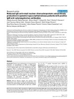
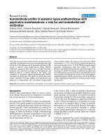
in 11 of 17 (64.7%) in group A and 10 of 34 (29.4%) in
group B (P = 0.03). Moreover, AECA BI was significantly
higher in group A (P = 0.03) (Fig. 1), even though we
observed in group B four SLE patients with high AECA BI
(>80%), without any correlation with the SLEDAI score.
Finally, a significant difference between the AECA BI of
both groups versus normal donors was observed (P <
0.0001). According to the cutoff value, AECA prevalence
in SSc population was 14.7% (5 of 34) and the AECA BI
was significantly higher in SSc patients than in healthy con-
trols (P = 0.002) (Fig. 1). A significant difference between
the AECA BI of group A and group B of SLE patients
versus SSc patients was observed (P = 0.0002 and P =
0.01 respectively) (Fig. 1).
No significant correlation was found between anti-CL, anti-
β2-GPI, anti-dsDNA, anti-Ro, anti-Ro52, anti-La, anti-P,
anti-nucleosome, or anti-GFAP antibodies and the pres-
ence of psychiatric disorders. However, a higher preva-
lence, even though not significant, of anti-Ro52 in group A
than in group B (35% vs 17%) was observed (see Table 2
for details). In our SLE patients, a significant correlation
between AECA and anti-dsDNA antibodies was observed
(P = 0.009). Interestingly, considering the two groups of
patients separately, this association was detected only in
group A (P = 0.03).
No correlation between the autoantibodies tested and the
other clinical features of SLE and SLEDAI was observed.
Discussion
This study provides the first evidence of an association
between serum AECA and psychiatric disorders in patients
with SLE. In 1999, the ACR nomenclature for NPSLE pro-
vides case definitions for 19 neuropsychiatric syndromes
seen in SLE, with reporting standards and recommenda-
tions for laboratory and imaging tests [1]. Regarding psy-
chiatric disorders, the committee adopted the terminology
of DSM-IV [20]. Accordingly, in this study psychiatric diag-
nosis was assigned following the DSM-IV. In our series,
mood disorders and psychosis were observed in 29.4%
and 3.9% of patients, respectively. Mood disorders have
been diagnosed frequently in SLE patients, with a preva-
lence ranging from 16% to 51%, whereas the reported
prevalence of psychosis has varied from 2% to 7% [26-
28]. Therefore, our estimation of the rates of psychiatric
disorders corroborates the previous findings.
Despite an overall impressive amount of investigations, the
pathogenesis of NPSLE is poorly understood. Although a
direct pathogenic role of autoantibodies in NPSLE through
induction of neural dysfunction, vasculopathy, and coagu-
lopathy has been suggested [12], the clinical association
between autoantibodies, detected both in serum and cere-
brospinal fluid, with NPSLE remains unclear.
AECA antibodies are a group of heterogeneous antibodies
that react with different endothelial-cell antigens. These
autoantibodies have been detected in several autoimmune
Table 2
Autoantibody profile of patients with systemic lupus erythematosus grouped according to their psychiatric symptoms
Autoantibodies Group A
a
(n = 17) No. (%) Group B
a
(n = 34) No. (%)
AECA IgG
b
11 (64.7) 10 (29.4)
Anti-GFAP IgG 2 (11.7) 6 (17.6)
Anti-Ro IgG 8 (47) 15 (44.1)
Anti-Ro52 IgG 6 (35.3) 6 (17.6)
Anti-La IgG 5 (31.2)
c
9 (26.4)
Anti-dsDNA IgG 9 (52.9) 19 (55.9)
Anti-nucleosome IgG 12 (70.6) 27 (79.4)
Anti-CL IgG 3 (17.6) 4 (11.7)
Anti-CL IgM 2 (11.8) 8 (23.5)
Anti-β2-GPI IgG 3 (17.6) 5 (14.7)
Anti-β2-GPI IgM 3 (23) 7 (28)
Anti-P IgG 2 (11.8) 2 (5.9)
a
Group A, patients with mood disorders and psychosis; group B, patients without psychiatric manifestations other than anxiety.
b
P = 0.03 on the
χ
2
test; differences between groups A and B in other comparisons were not statistically significant.
c
Measured in 16 of 17 patients. AECA, anti-
endothelial-cell antibodies; β2-GPI, β2 glycoprotein I; CL, cardiolipin; GFAP, glial fibrillar acidic protein; P, ribosomal P protein.
Available online />R370
diseases and have been associated with nephritis and vas-
culitis in SLE patients [29]. The proportion of AECA-posi-
tive sera ranges from 15% to 80% of cases in SLE, and
recent data suggest their involvement in endothelial dys-
function and a pathogenic role of AECA in SLE [30-34].
Interestingly, some in vitro and in vivo reports have demon-
strated an increased expression of adhesion molecules on
human umbilical-vein endothelial cells, mediated by AECA
IgG purified from sera of SLE patients [35-37]. Despite
these findings, the role of AECA in NPSLE has not been
fully investigated. Song and co-workers [38] showed the
clinical association of AECA with disease activity in a
cohort of 41 SLE patients. Interestingly, they described the
association of AECA with NPSLE. Recently, Meroni and
co-workers found AECA positivity in 5 of 14 SLE patients
with involvement of the central nervous system (CNS) [39].
The results of our study, demonstrating a significant asso-
ciation between AECA and psychiatric involvement in SLE
patients, a feature not previously reported, strengthen the
view of a possible implication of AECA in the development
of NPSLE.
On the other hand, we did not find any correlation between
psychopathology and the other antibody specificities
investigated.
The relation between antiphospholipid antibodies and CNS
involvement in SLE, first reported in 1984, has now been
confirmed; thrombosis associated with these antibodies is
considered the main pathogenic mechanism [40-43].
Moreover, antiphospholipid antibodies may contribute to
neurological damage by reacting with brain cells by means
of β2-GPI interactions. In this regard, we previously demon-
strated the expression of β2-GPI mRNA by astrocytes, neu-
ronal cells, and endothelial cells, suggesting that these
cells can represent a target of autoantibodies in the
antiphospholipid antibody syndrome [44,45]. Since it has
been postulated that AECA reactivity may be due in part to
the binding to a complex of β2-GPI with phospholipids on
endothelial cells [32,33], we evaluated anti-β2-GPI and
anti-CL antibodies by means of ELISA. No significant asso-
ciations were found between these autoantibodies and
AECA reactivity.
Conversely, in line with previous reports [46-48], we found
a significant correlation between AECA reactivity and anti-
dsDNA antibodies in SLE patients. Interestingly, when the
two groups of patients were considered separately, this
correlation was detected only in those with mood disorders
or psychosis.
The prevalence of anti-P antibodies in SLE patients ranges
from 6% to 36% [49], presenting ethnic differences. More-
over, a close association between lupus psychosis and the
presence of anti-P antibodies has been described [13,15-
17], and serum levels of these antibodies decrease after
successful treatment [16]. In contrast, other researchers
did not find any significant correlation between anti-P anti-
bodies and NPSLE including psychosis [18,50]. Yoshio
and colleagues suggested that in SLE patients there is a
strong association of IgG and IgM antiribosomal P0 protein
antibodies with CNS disease, excluding lupus psychosis
[51]. Interestingly, the ribosomal P protein P0 has been
identified by molecular cloning strategy as an endothelial
autoantigen in SLE patients [52]. In the present study, we
estimated a prevalence of 7.8% for anti-P antibodies in SLE
patients without any association with either psychiatric dis-
turbance or AECA reactivity. Remarkably, in our series the
two patients with lupus psychosis were both seronegative
for this autoantibody specificity. This variability among dif-
ferent studies was probably related to the differences in
selection of NPSLE patients as well as to the laboratory
methods used to detect anti-P autoantibodies.
Figure 1
Anti-endothelial-cell antibody serum levels in systemic lupus erythema-tosus (SLE) patients, systemic sclerosis (SSc) patients, and normal healthy donorsAnti-endothelial-cell antibody serum levels in systemic lupus erythema-
tosus (SLE) patients, systemic sclerosis (SSc) patients, and normal
healthy donors. Box–whisker plot of anti-endothelial-cell antibodies
(AECA) IgG binding index (BI) in SLE patients with psychosis and
mood disorders (group A, n = 17), in SLE patients without psychiatric
involvement other than anxiety (group B, n = 34), in SSc patients (n =
34), and in 66 normal healthy donors. AECA were expressed as a BI,
equal to 100 × (S-A)/(B-A), where S is the optical density (OD) of the
sample tested, A is the OD of a negative control, and B that of a posi-
tive reference serum. AECA were considered positive when the BI was
higher than the cutoff value (mean + 2 SD for 66 healthy controls), cor-
responding to 50% of a positive reference serum from an SLE patient.
Median, quartiles, range, and possibly extreme values are indicated. The
broken line represents the cutoff. †, group A vs Group B, P = 0.03; ‡,
group A vs SSc, P = 0.0002; *, group A vs normal healthy donors,
P < 0.0001; ¶ group B vs SSc, P = 0.01; Φ, group B vs normal healthy
donors, P < 0.0001; §, SSc vs normal healthy donors, P = 0.002.
Arthritis Research & Therapy Vol 6 No 4 Conti et al.
R371
A high positive predictive value of anti-GFAP antibodies for
NPSLE has been described [53]. The intermediate filament
protein of astrocytes, GFAP, is up-regulated in gliotic
hypertrophy and perivascular inflammation of Alzheimer's
disease and multiple sclerosis where anti-GFAP antibodies
were also described [54-56]. Recently, Trysberg and co-
workers [57] showed increased levels of GFAP in the
cerebrospinal fluid of patients with NPSLE. Moreover, suc-
cessful therapy with cyclophosphamide in six NPSLE
patients resulted in significantly decreased CSF levels of
GFAP [57]. In our cohort of SLE patients, we found an anti-
GFAP prevalence of 15.7% without any correlation
between anti-GFAP antibodies and psychiatric morbidity.
Nevertheless, the exact role of anti-GFAP in the pathogen-
esis of NPSLE should be clarified by prospective studies
with both intrathecal and serum determination.
Finally, anxiety and depression are frequently detected in
SLE patients and it has been suggested that these disor-
ders are predominantly psychoreactive [10,11]. However,
in a large retrospective study, SLE patients with depression
presented CNS involvement more often than patients with-
out, supporting the view that mood disorders are not merely
a response to stress [58]. Our data demonstrating the
presence of serum AECA in 60% of patients with depres-
sion and only in 25% of patients with anxiety alone support
the hypothesis that depression in SLE could have a biolog-
ical origin.
Conclusion
This study provides evidence of an association between
serum AECA and psychiatric disorders (i.e. psychosis and
mood disturbances) in patients with SLE. Conversely, we
could not demonstrate any correlation between psychopa-
thology and the other antibody specificities investigated.
The high prevalence of serum AECA in patients with psy-
chosis and mood disorders supports the hypothesis of a
biological origin of these disturbances.
Competing interests
None declared.
References
1. ACR ad hoc committee on Neuropsychiatric Lupus Nomenclature:
The American College of Rheumatology nomenclature and
case definitions for neuropsychiatric lupus syndromes. Arthri-
tis Rheum 1999, 42:599-608.
2. Ainiala H, Hietaharju A, Loukkola J, Peltola J, Korpela M, Metsanoja
R, Auvinen A: Validity of the new American College of Rheuma-
tology criteria for neuropsychiatric lupus syndromes: a popu-
lation-based evaluation. Arthritis Rheum 2001, 45:419-423.
3. Buchbinder R, Hall S, Littlejohn GO, Ryan PF: Neuropsychiatric
manifestations of systemic lupus erythematosus. Aust N Z J
Med 1988, 18:679-684.
4. Johnson RT, Richardson EP: The neurological manifestations of
systemic lupus erythematosus. Medicine 1968, 47:337-369.
5. Wekking EM: Psychiatric symptoms in systemic lupus ery-
thematosus: an update. Psychosom Med 1993, 55:219-228.
6. Iverson GL, McCracken LM: Attributing psychopathology to sys-
temic lupus erythematosus: some methodological
considerations. Ann Rheum Dis 1992, 51:134-135.
7. Lewis DA, Smith RE: Steroid induced psychiatric syndromes: a
report of 14 cases and review of literature. J Affect Disord
1983, 5:319-332.
8. Hay EM, Black D, Huddy A, Creed F, Tomenson B, Bernstein RM,
Holt PJ: Psychiatric disorder and cognitive impairment in sys-
temic lupus erythematosus. Arthritis Rheum 1992, 35:411-416.
9. Lindal E, Thorlacius S, Steinsson K, Stefansson JG: Psychiatric
disorders among subjects with systemic lupus erythematosus
in an unselected population. Scand J Rheumatol 1995,
24:346-351.
10. Segui J, Ramos-Casals M, Garcia-Carrasco M, de Flores T, Cer-
vera R, Valdes M, Font J, Ingelmo M: Psychiatric and psychoso-
cial disorders in patients with systemic lupus erythematosus:
a longitudinal study of active and inactive stages of the
disease. Lupus 2000, 9:584-588.
11. Shortall E, Isenberg D, Newman SP: Factors associated with
mood and mood disorders in SLE. Lupus 1995, 4:272-279.
12. Greenwood DL, Gitlits VM, Alderuccio F, Sentry JW, Toh BH:
Autoantibodies in neuropsychiatric lupus. Autoimmunity 2002,
35:79-86.
13. Bonfa E, Elkon KB: Clinical and serologic associations of the
antiribosomal P protein antibody. Arthritis Rheum 1986,
29:981-985.
14. Teh LS, Isenberg DA: Antiribosomal P protein antibodies in sys-
temic lupus erythematosus. A reappraisal. Arthritis Rheum
1994, 37:307-315.
15. Bonfa E, Golombek SJ, Kaufman LD, Skelly S, Weissbach H, Brot
N, Elkon KB: Association between lupus psychosis and anti-
ribosomal P protein antibodies. N Engl J Med 1987,
317:265-271.
16. Isshi K, Hirohata S: Association of anti-ribosomal P protein anti-
bodies with neuropsychiatric systemic lupus erythematosus.
Arthritis Rheum 1996, 39:1483-1490.
17. Schneebaum AB, Singleton JD, West SG, Blodgett JK, Allen LG,
Cheronis JC, Kotzin BL: Association of psychiatric manifesta-
tions with antibodies to ribosomal P proteins in systemic
lupus erythematosus. Am J Med 1991, 90:54-62.
18. Teh LS, Bedwell AE, Isenberg DA, Gordon C, Emery P, Charles PJ,
Harper M, Amos N, Williams BD: Antibodies to protein P in sys-
temic lupus erythematosus. Ann Rheum Dis 1992, 51:489-494.
19. Hochberg MC: Updating the American College of Rheumatol-
ogy revised criteria for the classification of systemic lupus
erythematosus. Arthritis Rheum 1997, 40:1725.
20. American Psychiatric Association: Diagnostic and Statistical Man-
ual of Mental Disorders 4th edition. Washington, DC: American
Psychiatric Association; 1994.
21. First MB, Spitzer RL, Gibbon M, Williams JB: Structured Clinical
Interview for DSM-IV Axis I Disorders (SCID-I/P), Computer Pro-
gram Handbook. Version 2.0 New York: New York State Psychiat-
ric Institute, Biometrics Research Department; 1996.
22. Bombardier C, Gladman DD, Urowitz MB, Caron D, Chang CH:
Derivation of the SLEDAI. A disease activity index for lupus
patients. The Committee on Prognosis Studies in SLE. Arthritis
Rheum 1992, 35:630-640.
23. Vismara A, Meroni PL, Tincani A, Harris EN, Barcellini W, Brucato
A, Khamashta M, Hughes GR, Zanussi C, Balestrieri G: Relation-
ship between anti-cardiolipin and anti-endothelial cell anti-
bodies in systemic lupus erythematosus. Clin Exp Immunol
1988, 74:247-253.
24. van der Zee JM, Siegert CE, de Vreede TA, Daha MR, Breedveld
FC: Characterization of anti-endothelial cell antibodies in sys-
temic lupus erythematosus (SLE). Clin Exp Immunol 1991,
84:238-244.
25. Subcommittee for Scleroderma Criteria of the American Rheuma-
tism Association Diagnostic and Therapeutic Criteria Committee:
Preliminary criteria for the classification of systemic sclerosis
(scleroderma). Arthritis Rheum 1980, 23:581-590.
26. Brey RL, Holliday SL, Saklad AR, Navarrete MG, Hermosillo-Romo
D, Stallworth CL, Valdez CR, Escalante A, del Rincon I, Gronseth
G, Rhine CB, Padilla P, McGlasson D: Neuropsychiatric syn-
dromes in lupus: prevalence using standardized definition.
Neurology 2002, 58:1214-1220.
27. Mok CC, Lau CS, Wong RWS: Neuropsychiatric manifestations
and their clinical associations in Southern Chinese patients
Available online />R372
with systemic lupus erythematosus. J Rheumatol 2001,
28:766-771.
28. Ainiala H, Loukkola J, Peltola J, Korpela M, Hietaharju A: The prev-
alence of neuropsychiatric syndromes in systemic lupus
erythematosus. Neurology 2001, 57:496-500.
29. D'Cruz DP, Houssiau FA, Ramirez G, Baguley E, McCutcheon J,
Vianna J, Haga HJ, Swana GT, Khamashta MA, Taylor JC: Anti-
bodies to endothelial cells in systemic lupus erythematosus: a
potential marker for nephritis and vasculitis. Clin Exp Immunol
1991, 85:254-261.
30. Carvalho D, Savage CO, Black CM, Pearson JD: IgG antien-
dothelial cell autoantibodies from scleroderma patients
induce leukocyte adhesion to human vascular endothelial
cells in vitro. Induction of adhesion molecule expression and
involvement of endothelium-derived cytokines. J Clin Invest
1996, 97:111-119.
31. Bordron A, Dueymes M, Levy Y, Jamin C, Leroy JP, Piette JC,
Shoenfeld Y, Youinou PY: The binding of some human antien-
dothelial cell antibodies induces endothelial cell apoptosis. J
Clin Invest 1998, 101:2029-2035.
32. Del Papa N, Guidali L, Spatola L, Bonara P, Borghi MO, Tincani A,
Balestrieri G, Meroni PL: Relationship between anti-phospholi-
pid and anti-endothelial cell antibodies III: beta 2 glycoprotein
I mediates the antibody binding to endothelial membranes
and induces the expression of adhesion molecules. Clin Exp
Rheumatol 1995, 13:179-185.
33. Del Papa N, Guidali L, Sala A, Buccellati C, Khamashta MA,
Ichikawa K, Koike T, Balestrieri G, Tincani A, Hughes GR, Meroni
PL: Endothelial cells as target for antiphospholipid antibodies.
Human polyclonal and monoclonal anti-beta 2-glycoprotein I
antibodies react in vitro with endothelial cells through adher-
ent beta 2-glycoprotein I and induce endothelial activation.
Arthritis Rheum 1997, 40:551-561.
34. Papa ND, Raschi E, Moroni G, Panzeri P, Borghi MO, Ponticelli C,
Tincani A, Balestrieri G, Meroni PL: Anti-endothelial cell IgG frac-
tions from systemic lupus erythematosus patients bind to
human endothelial cells and induce a pro-adhesive and a pro-
inflammatory phenotype in vitro. Lupus 1999, 8:423-429.
35. Wellicome SM, Kapahi P, Mason JC, Lebranchu Y, Yarwood H,
Haskard DO: Detection of a circulating form of vascular cell
adhesion molecule-1: raised levels in rheumatoid arthritis and
systemic lupus erythematosus. Clin Exp Immunol 1993,
92:412-418.
36. Belmont HM, Buyon J, Giorno R, Abramson S: Up-regulation of
endothelial cell adhesion molecules characterizes disease
activity in systemic lupus erythematosus. The Shwartzman
phenomenon revisited. Arthritis Rheum 1994, 37:376-383.
37. Carson CW, Beall LD, Hunder GG, Johnson CM, Newman W:
Serum ELAM-1 is increased in vasculitis, scleroderma, and
systemic lupus erythematosus. J Rheumatol 1993, 20:809-814.
38. Song J, Park YB, Lee WK, Lee KH, Lee SK: Clinical associations
of anti-endothelial cell antibodies in patients with systemic
lupus erythematosus. Rheumatol Int 2000, 20:1-7.
39. Meroni PL, Tincani A, Sepp N, Raschi E, Testoni C, Corsini E,
Cavazzana I, Pellegrini S, Salmaggi A: Endothelium and the brain
in CNS lupus. Lupus 2003, 12:919-928.
40. Harris EN, Gharavi AE, Asherson RA, Boey ML, Hughes GR: Cer-
ebral infarction in systemic lupus: association with anticardiol-
ipin antibodies. Clin Exp Rheumatol 1984, 2:47-51.
41. Asherson RA, Khamashta MA, Gil A, Vazquez JJ, Chan O, Baguley
E, Hughes GR: Cerebrovascular disease and antiphospholipid
antibodies in systemic lupus erythematosus, lupus-like dis-
ease, and the primary antiphospholipid syndrome. Am J Med
1989, 86:391-399.
42. Herranz MT, Rivier G, Khamashta MA, Blaser KU, Hughes GR:
Association between antiphospholipid antibodies and epi-
lepsy in patients with systemic lupus erythematosus. Arthritis
Rheum 1994, 37:568-571.
43. Sanna G, Bertolaccini ML, Cuadrado MJ, Laing H, Khamashta MA,
Mathieu A, Hughes GR: Neuropsychiatric manifestations in sys-
temic lupus erythematosus: prevalence and association with
antiphospholipid antibodies. J Rheumatol 2003, 30:985-992.
44. Caronti B, Calderaro C, Alessandri C, Conti F, Tinghino R, Pini C,
Palladini G, Valesini G: Serum anti-beta2-glycoprotein I anti-
bodies from patients with antiphospholipid antibody syn-
drome bind central nervous system cells. J Autoimmun 1998,
11:425-429.
45. Caronti B, Calderaro C, Alessandri C, Conti F, Tinghino R, Pal-
ladini G, Valesini G: Beta2-glycoprotein I (beta2-GPI) mRNA is
expressed by several cell types involved in anti-phospholipid
syndrome-related tissue damage. Clin Exp Immunol 1999,
115:214-219.
46. Chan TM, Yu PM, Tsang KL, Cheng IK: Endothelial cell binding
by human polyclonal anti-DNA antibodies: relationship to dis-
ease activity and endothelial functional alterations. Clin Exp
Immunol 1995, 100:506-513.
47. Chan TM, Cheng IK: Identification of endothelial cell membrane
proteins that bind anti-DNA antibodies from patients with sys-
temic lupus erythematosus by direct or indirect mechanisms.
J Autoimmun 1997, 10:433-439.
48. Moscato S, Pratesi F, Bongiorni F, Scavuzzo MC, Chimenti D,
Bombardieri S, Migliorini P: Endothelial cell binding by systemic
lupus antibodies: functional properties and relationship with
anti-DNA activity. J Autoimmun 2002, 18:231-238.
49. Arnett FC, Reveille JD, Moutsopoulos HM, Georgescu L, Elkon KB:
Ribosomal P autoantibodies in SLE. Arthritis Rheum 1996,
39:1833-1839.
50. Gerli R, Caponi L, Tincani A, Scorza R, Sabbadini MG, Danieli MG,
De Angelis V, Cesarotti M, Piccirilli M, Quartesan R, Moretti P,
Cantoni C, Franceschini F, Cavazzana I, Origgi L, Vanoli M, Boz-
zolo E, Ferrario L, Padovani A, Gambini O, Vanzulli L, Croce D,
Bombardieri S: Clinical and serological associations of ribos-
omal P autoantibodies in systemic lupus erythematosus: pro-
spective evaluation in a large cohort of Italian patients.
Rheumatology 2002, 41:1357-1366.
51. Yoshio T, Masuyama J, Ikeda M, Tamai K, Hachiya T, Emori T,
Mimori A, Takeda A, Minota S, Kano S: Quantification of antiri-
bosomal P0 protein antibodies by ELISA with recombinant P0
fusion protein and their association with central nervous sys-
tem disease in systemic lupus erythematosus. J Rheumatol
1995, 22:1681-1687.
52. Frampton G, Moriya S, Pearson JD, Isenberg DA, Ward FJ, Smith
TA, Panayiotou A, Staines NA, Murphy JJ: Identification of candi-
date endothelial cell autoantigens in systemic lupus ery-
thematosus using a molecular cloning strategy: a role for
ribosomal P protein P0 as an endothelial cell autoantigen.
Rheumatology 2000, 39:1114-1120.
53. Sanna G, Piga M, Terryberry JW, Peltz MT, Giagheddu S, Satta L,
Ahmed A, Cauli A, Montaldo C, Passiu G, Peter JB, Shoenfeld Y,
Mathieu A: Central nervous system involvement in systemic
lupus erythematosus: cerebral imaging and serological profile
in patients with and without overt neuropsychiatric
manifestations. Lupus 2000, 9:573-583.
54. Kato S, Gondo T, Hoshii Y, Takahashi M, Yamada M, Ishihara T:
Confocal observation of senile plaques in Alzheimer's dis-
ease: senile plaque morphology and relationship between
senile plaques and astrocytes. Pathol Int 1998, 48:332-340.
55. Tanaka J, Nakamura K, Takeda M, Tada K, Suzuki H, Morita H,
Okado T, Hariguchi S, Nishimura T: Enzyme-linked immuno-
sorbent assay for human autoantibody to glial fibrillary acidic
protein: higher titer of the antibody is detected in serum of
patients with Alzheimer's disease. Acta Neurol Scand 1989,
80:554-560.
56. Newcombe J, Gahan S, Cuzner ML: Serum antibodies against
central nervous system proteins in human demyelinating
disease. Clin Exp Immunol 1985, 59:383-390.
57. Trysberg E, Nylen K, Rosengren LE, Tarkowski A: Neuronal and
astrocytic damage in systemic lupus erythematosus patients
with central nervous system involvement. Arthritis Rheum
2003, 48:2881-2887.
58. Utset TO, Golden M, Siberry G, Kiri N, Crum RM, Petri M: Depres-
sive symptoms in patients with systemic lupus erythemato-
sus: association with central nervous system lupus and
Sjogren's syndrome. J Rheumatol 1994, 21:2039-2045.