Báo cáo Y học: Domain organization, folding and stability of bacteriophage T4 fibritin, a segmented coiled-coil protein docx
Bạn đang xem bản rút gọn của tài liệu. Xem và tải ngay bản đầy đủ của tài liệu tại đây (416.5 KB, 9 trang )
Domain organization, folding and stability of bacteriophage T4
fibritin, a segmented coiled-coil protein
Sergei P. Boudko
1,2
, Yuri Y. Londer
1
, Andrei V. Letarov
1
, Natalia V. Sernova
1
, Juergen Engel
2
and Vadim V. Mesyanzhinov
1
1
Shemyakin-Ovchinnikov Institute of Bioorganic Chemistry, Moscow, Russia;
2
Biozentrum der Universitaet Basel, Switzerland
Fibritin is a segmented coiled-coil homotrimer of the
486-residue product of phage T4 gene wac.Thisprotein
attaches to a phage particle by the N-terminal region and
forms fibrous whiskers of 530 A
˚
, which perform a chaperone
function during virus assembly. The short C-terminal region
has a b-ann ulus-like structure. We engineered a set of fibritin
deletion mutants sequentially truncated from the N-termini,
and the mutants were s tudied by differential scanning
calorimetry (DSC) and CD measurements. The analysis
of DSC curves indicates that full-length fibritin exhibits
three thermal-heat-absorption peaks centred at 321 K
(DH ¼ 1390 kJÆmol trimer
)1
), at 336 K (DH ¼ 7600 kJÆmol
trimer
)1
), and at 345 K (DH ¼ 515 kJÆmo l trimer
)1
). These
transitions were assigned to the N-terminal, segmented
coiled-coil, and C-terminal functional domains, respectively.
The coiled-coil region, containing 13 segments, melts
co-operatively as a single domain with a mean enthalpy
DH
res
¼ 21 kJÆmol residue
)1
.TheratioofDH
VH
/DH
cal
for
the coiled-coil part of the 120-, 182-, 258- and 281-residue per
monomer mutants, truncated from the N-termini, and for
full-length fibritin are 0.91, 0.8 8, 0.42, 0.39, and 0.13,
respectively. This gives an indication of the d ecrease of the
Ôall-or-noneÕ character of the transition with increasing
protein s ize. The deletion o f the 12-residue-long loop in the
120-residue fibritin increases the thermal stability of the
coiled-coil region. According to CD data, full-length fibritin
and all the m ut ants t runcated f rom the N- termini ref old
properly after heat denaturation. In contrast, fibritin XN,
which is deleted for the C-terminal domain, forms aggregates
inside the cell. The XN protein can be partially refolded by
dilution from urea and does not refold after heat denatur-
ation. These results confirm that the C-terminal domain is
essential for correct fibritin assembly both in vivo and in vitro
and a cts as a foldon.
Keywords: bacteriophage; foldon; microcalorimetry; protein
engineering; segmente d coiled coil.
Fibritin, a structural protein of bacteriophage T4 encoded
by gene wac (named for whisker’s antigen con trol), belongs
to a specific class of accessory proteins that act in the virus
assembly process. Six fibritin molecules form the collar/
whisker complex that consists of a ring embracing the phage
neck with thin filaments (whiskers) protruding from the
collar [1]. T his complex is a sensing device that controls th e
retraction of the long tail fibers in adverse environments and
thus prevents undesirable infection [2]. The whiskers act also
as a chaperone and help the proximal and distal parts of the
long tail fibers to join correctly by increasing the effective
target sizes and thereby increasing the rates of otherwise
slow diffusion–limited bimolecular interactions [3].
The structure of fibritin was predicted from sequence and
biochemical analyses to be mainly a parallel segmented
triple-helical coiled-coil [4,5]. Fibritin is a homotrimer of 486
residues per monomer and consists of three functional parts.
Its predominant central region has 1 3 consecutive a helical
coiled-coil segments linked by loops. The protein is attached
to a phage particle by the N-terminal part that does not
have heptad periodicity [6], and the short C-termini is
essential for in vivo protein folding and trimerization [5].
Functional activities of fibritin can be related to the
exposure of hydrophobic patches in the c oiled-coil [7].
The full-length fibritin of 530 A
˚
could not be crystallized,
probably because of its inherent flexibility. However, a set of
smaller fibritin mutants was engineered and expressed in the
soluble trimeric forms in an Escherichia coli system [5,8,9].
The structures of the E and M fibritins, which are truncated
for the last 120 and 75, respectively, C-terminal residues per
monomer were solved to atomic resolution by X-ray
crystallography [8,9]. Three identical subunits form a
trimeric p arallel coiled-coil domain and a small a structural
C-terminal domain. The coiled-coil part of fibritin E is
divided into three segments separated by short sequences
called insertion loops. The C-terminal domain, which
consists of 30 residues from e ach monomer, contains a
b-annulus-like structure with a hydrophobic interior.
Residues within the C-terminal domain make extensive
hydrophobic and some polar inter–subunit interactions [8].
This is consistent with the C-terminal domain being
important for the correct assembly of fibritin, as shown by
mutational studies ([5] and S. P. Boudko, unpublished
results). Tight interactions between C-terminal residues of
adjacent subunits counteract the latent instability that is
Correspondence to V. V. Mesyanzhinov, Howard Hughes Medical
Institute, Shemyakin-Ovchinnikov Institute of Bioorganic Chemistry,
Miklukho-Maklaya Street 16/10, 117997 Moscow, Russia.
Fax: + 7 095 336 6022, Tel.: + 7 095 335 5 588,
E-mail:
Abbreviations: DSC, differential scanning calorimetry;
IPTG, isopropyl thio-b-
D
-galactoside.
(Received 20 July 2001, revised 6 December 2 001, accepted
11 December 2001)
Eur. J. Biochem. 269, 833–841 (2002) Ó FEBS 2002
suggested by the structural properties o f the coiled-coil
segments [8]. Trimerization is likely to begin with the
formation of the C-terminal domain that acts as a folding
nucleus domain (foldon) and subsequently initiates the
assembly of the coiled c oil [8,10]. The interplay between the
stabilizing effect of the C-terminal domain and the labile
coiled-coil domain may be essential for the fibritin function
and for the correct functioning of many other a helix fibrous
proteins as well.
In the present work, we o btained a set of fibritin mutants
sequentially truncated from the N-termini. We engineered
also mutant S1 that have deleted for one loop of 12 residues
in fibritin E. To characterize the thermodynamic properties,
stability, and domain organizations, we analysed these
fibritin mutants by d ifferential scanning calorimetry (DSC)
and CD measurements. The analysis of DSC curves
indicates that full-length fibritin has three thermal heat-
absorption transitions that were reasonably assigned to the
N-terminal, segmented coiled-coil, and C-terminal func-
tional domains, respectively.
Full-length fibritin and all the mutants truncated from the
N-termini refold p roperly a fter heat denaturation. We
designed also the XN mutant, a full-length fibritin that has
no C-terminal domain (Fig. 1) that forms aggregates inside
the cell. The XN protein can be partially refolded by fast
dilution from urea and does not refold after heat denatur-
ation. The XN protein can be refolded by fast dilution from
urea and does not refold after heat d enaturation.
MATERIALS AND METHODS
E. coli
strains and plasmids
The Top10 E. coli strain (Invitrogen, USA) was used for the
selection of recombinant clones and plasmid DNA purifi-
cation. Protein expression was performed in the BL21
(DE3) strain (Promega, USA) containing the T7 RNA
polymerase gene under lac UV5 control in the E. coli
chromosome. DNA fragments encoding truncated fibritin
mutants were cloned in the pET19b (+) and pET23d (+)
expression vectors containing the ribosome-binding site for
effective translation (Novagen, U SA), that allow transcrip-
tion from the T7 RNA polymerase promoter.
Design of fibritin mutants
We used previously designed expression vectors for a full-
length fibritin [8], fibritin XN [10,11], E, M [8,9], F (V. V.
Mesyanzhinov, unpublished results), and the S1 fibritin
[12,13]. To create the B1, SM1, SM4 mutants, we
amplified the DNA fragments of interest by PCR and
introduce the NcoI and BamHI restriction sites for
subsequent cloning into plasmid vectors. Cloning was
performed using the common t echniques described in [14].
The S1 mutant that lacks 12 residues of the L11 loop
(residues Asn-Gly-Thr-Asn-Pro-Asn-Gly-Ser-Thr-Val-Glu-
Glu, Asn404-Glu415) was c onstructed on the basis of
fibritin E. We have used an overlap ping PCR method to
delete the DNA piece en coding this loop [13]. Sequencing
was carried out by the d ideoxy chain termination method
using a DNA sequencing kit/BigDye terminator cycle
sequencing ready reaction (Applied Biosystems) a nd an
automated DNA sequencer.
Expression and purification of fibritin mutants
The cell culture of the E. coli BL21 (DE3) strain carrying
the respective vector was grown at 37 °C in 500 mL of
2 · tryptone/yeast medium [14] until the density reached a
D
600
value of 0 .6. Protein expression was induced by 1 m
M
IPTG with subsequent incubation for 3 h at 37 °Cwith
vigorous aeration. We used a modification of the previously
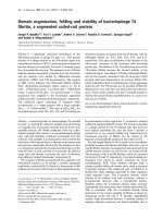
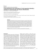
Fig. 1. Schematic presentations and amino-acid sequence of fibritin.
(a) Schematic presen tation of th e fibritin mutants used in this work:
full-length fibritin ( wac), XN, B1, SM1, SM4, E, S1, M, and F. For
each mutant, the range of amino-acid sequence that it comprises of the
full-length fibritin sequence is given. The N-terminal domain is a broad
box; coiled-coil regions are narrow boxes; the loops, separating coiled-
coil segments, are hexamers; the C-terminal domain (foldon) is a
sphere. (b) Amino-acid sequence of full-length fibritin and heptad
scheme of the fibritin coiled coil part. The hydrophobic residues in the
a and d positions are shown in bold. The coiled-coil segments are
indicated by roman (I–XIII), and the loops are marked [L1–L11]. The
bacteriophage T4 gene wac nucleotide sequence is deposited in the
EMBL Ge ne Data Ba nk: accession number X12888. Atomic c oordi-
nates of fibritin E and fibritin M, deposited in PDB, are 1AA0 and
1AVY, respectively.
834 S. P. Boudko et al. (Eur. J. Biochem. 269) Ó FEBS 2002
described method for purification of fibritin mutants [5].
The pellet from 500 mL of the E. coli culture was
resuspended in 10 mL of Tris/EDTA buffer (50 m
M
Tris/HCl, pH 8.0, 1 m
M
EDTA) and sonicated with
cooling. The cell debris was removed by centrifugation at
25 000 g for 20 min. T o precipitate nucleic acids, 1 mL of
30% (w/v) streptomycin sulfate (Sigma, USA) solution in
Tris/EDTA buffer was added; the concentrated protein
solution was kept on ice for 15 min. After centrifugation,
ammonium sulfate was added to the supernatant to a final
concentration of 20–50% saturation, depending on the
particular mutant, and the mixture was incubated overnight
at 4 °C. Protein precipitate was collected by low-speed
centrifugation, and resuspended in 3–10 mL o f Tris/EDTA
buffer. Nucleic acid and protein precipitation procedures
were skipped f or protein S1. After ammonium sulfate
precipitation, the protein solution was applied to a 10-mL
hydroxyapatite column (Bio-Rad; DNA grade) equilibrated
with 10 m
M
Na phosphate (pH 8.0) and was hed with
10 m
M
Na phosphate. The flow-through fractions, contain-
ing recombinant proteins, were dialysed against Tris/EDTA
buffer and stored at 4 °C. The E, S1 and F proteins were
additionally applied to a 15-mL DEAE–Sephacryl column
and eluted w ith a linear gradient o f NaCl. Fractions
containing proteins were dialysed against Tris/EDTA buffer
andstoredat4°C.
The protein purity was judged by denaturing SDS/PAGE
using two systems: for proteins with M
r
larger than 12 kDa
we used the Lae mmli system [15]; f or smaller ones we
applied the S chaegger and Jagow system [ 16]. Protein
concentration was determined by measuring the absorbency
at 280 nm in 6
M
GdnHCl, and the extinction coefficient
was c alculated a s described in [17]. For the DSC procedure
the proteins were dialysed against NaCl/P
i
[10 m
M
Na
phosphate (pH 8 .0), 150 m
M
NaCl or 10 m
M
Na phosphate
(pH 8.0)], centrifuged at 10 000 g for 30 min, and degassed
for 5 min.
Purification and refolding of the XN fibritin
The pellet from 500 mL of the E. coli cells expressing fibritin
XN was suspended in 10 mL of Tris/EDTA buffer ( 50 m
M
Tris/HCl (pH 8.0), 1 m
M
EDTA) and sonicated under
cooling. The cell extract was centrifuged at 3500 g for
30 min and supernatant was r emoved. The pellet was
resuspended in 0.5 mL of 8
M
urea for 10 min and the
suspension was centrifuged at 10 000 g for 30 min to
remove insoluble particles. The supernatant was mixed with
50 mL of the refolding buffer (50 m
M
Tris/HCl, pH 8 .0,
2m
M
EDTA, 2 m
M
phenylmethanesulfonyl fluoride),
incubated at 4 °C for 3–4 days and then concentrated to
2 m L. The protein solution was further purified on the
hydroxyapatite column as described a bove. The yield of the
soluble protein was % 15% o f initial concentration i ndicat-
ing weak refolding.
DSC
Calorimetric measurements were performed using a
VP-DSC Microcalorimeter (Microcal Inc.) equipped with
a cell (covered with Tantaloy 61
TM
) of 0.5 mL volume at
a heating rate of 1 KÆmin
)1
. Baseline subtraction, calcu-
lation of DH
cal
for different peaks and determination of
absolute heat capacity were performed using the MicroCal
ORIGIN
5.0 program. To determine absolute heat capacity
of proteins, we used the following parameters in the
equation:
DC
p
¼ g
0
qðtÞV
0
ð1 þ 0:00002tÞ C
Abs
p
ðtÞÀvð1 þ atÞC
W
p
ðtÞ
hi
where DC
p
is the sample-buffer baseline minus the buffer-
buffer baseline, g
0
is the c oncentration o f protein (g ÆmL
)1
),
q(t) is the relative density of water (stored in the
ORIGIN
program [18]), V
0
is the nominal volume (0.5194 mL) of the
sample cell, t is temperature in °C, C
Abs
p
(t)istheabsolute
heat capacity (calÆdeg
)1
Æg
)1
) o f the protein i n solution, v is
the partial specific volume of the protein (0.717 mLÆmg
)1
),
a is the coefficient of thermal expansion of the protein
(0.0007 1/ a °C), and C
W
p
(t) is the unit-volume heat capacity
of water (calÆdeg
)1
ÆmL
)1
) (stored in Origin). The thermal
coefficient of cubic expansion of tantalum is 0.00002.
The values of the van’t Hoff enthalpy of the process for
the peaks representing the melting of coiled coil region were
calculated as for a first ord er reaction [19]:
D
1
0
H
vh
¼
4RT
2
max
ðhDC
p
i
max
À D
1
0
C
p
=2Þ
D
1
0
H
cal
where D
1
0
H
vh
is the van’t Hoff enthalpy for transition from
state 0 to state 1, D
1
0
H
cal
is the calorimetric enthalpy, T
max
is
the temperature of the m aximum heat c apacity, ÆDC
p
æ
max
is
theexcessheatcapacityofproteinsinthemaximumofthe
peak, and D
1
0
C
p
is the difference between he at capacities for
state 1 and 0 (after and before t he transition).
CD measurements
CD spectra of mutant proteins were recorded with an Aviv
62DS circular dichroism spectrometer (Aviv Inc., USA),
equipped with a thermostatic quartz cell having a 1-mm
path length. CD data were analysed using the
CONTIN
program [20].
RESULTS
Engineering and properties of fibritin deletion mutants
To investigate the stability and thermodynamic properties
of T4 fibritin, a set of recombinant truncated mutants was
designed and analysed. All these molecules contained an
intact C-terminal part and had different numbers of coiled-
coil segments and separating segments loops (Fig. 1 and
Table 1). Fibritin S1, based on fibritin E with 120 resides per
chain, had a deleted loop L11 of 12 residues, and fibritin XN
hadnoC-terminalregionof30residues.
To enhance the prote in stability, five mutations were
introduced into the 74 residues of fibritin M that forms
the last coiled-coil segment (5,5 heptad repeats) and the
complete C-terminal domain [9]. Particularly, the Ser421
residue was substituted for Lys to test the possible
formation of interchain salt bridge with Glu426. The
substitutions Asn428 to Asp and Thr433 to Arg were
designed to create a similar interchain salt bridge b etween
these two residues. Residue 425, an Asp in a d position,
was replaced by an Ile, which is generally a favourable
residue in this position for a trimeric coile d coil [21].
Ó FEBS 2002 Thermodynamics of segmented coiled coil protein (Eur. J. Biochem. 269) 835
The crystal structure of two fibritin truncated mutants,
E and M, that have 120 and 74 residues per monomer,
respectively, have been determined to a tomic resolution [ 8].
X-ray crystallography confirmed that both m utants are
trimeric, parallel, co iled coils with a small C-terminal
domain that has a b-annulus structure. In addition, we
were able to obtain crystals of fibritin B1, that has 281
residues per monomer. Crystals belong to space group P2
1
,
and existence o f threefold noncrystallographic symmetry
pattern in observed X-ray diffraction data indicates that the
B1 protein is a trimer too (N. V. Sernova, unpublished
results). These data and the repetitive segmented structure
of fibritin suggest that other fibritin mutants studied that
have b-annulus C-terminal domain mentioned above also
should have a parallel t rimeric coiled-coil structure.
Indeed, all these recombinant m utants, except fibritin
XN, expressed from the plasmids in E. coli cells were
soluble and proteins were purified by ammonium sulfate
precipitation followed by chromatography on hydroxy-
apatite. Fibritin XN was refolded from inclusion bodies as
described in Materials and methods. It is known that full-
length fibritin, as well as some N-terminally truncated
mutants, are resistant to 1% SDS [5,10,13]. These proteins
do not dissociate to the monomer chains in the presence o f
SDS at room temperature, and they migrate on SDS/PAGE
as trimers. All the mutants used in this r esearch have such a
resistance to SDS again e xcept fibritin XN ( data n ot
shown) .
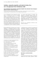
Figure 2 shows the CD spectra of the purified fibritin
mutants. These spectra indicate that all mutants, except the
shortest fibritin F, exhibited properties characteristic of a
high content of a helicity. The a helical contents slightly
decreased with decre asing size of the mutants. The mean
residue elliptic ity at 220 nm was )32 800 degÆcm
2
Ædmol
)1
for full-length fibritin and )25 800 and )21 900 degÆcm
2
Æ
dmol
)1
for fibritin B1 and fibritin SM4, respectively.
Interestingly, fibritin M e xhibited more a helicity than
fibritin E, probably due to the absence of insertion loops.
The CD spectrum of fibritin F represented mostly the
secondary structure of t he C-terminal domain, which is in a
good agreement with published data [22].
Assignment of the fibritin thermal transitions
to functional domains
The full-length fibritin, and the N-te rminally truncated B1,
SM1, SM4, E, M, and F mutants were a nalyzed by DSC.
The DSC data were also collected for fibritin XN that had
no C-terminal domain. Our goal was to answer a question
about how many thermodynamically independent domains
fibritin has, and to assign the thermal transitions to
individual functional regions. M easurements were per-
formed in 10 m
M
Na pho sphate buffer, pH 8.0 with
0.15
M
NaCl. In these conditions, th e endotherm for a
full-length fibritin exh ibited three well-resolved heat-
absorption peaks centred at 321 K (DH ¼ 1390 kJÆmol
trimer
)1
), 336 K (DH ¼ 7600 kJÆmol trimer
)1
), and 345 K
(DH ¼ 515 kJÆmol trimer
)1
), respectively (Fig. 3A).
The transition at 321 K can be assigned to the N-terminal
region (residues 1–50), which has no heptad periodicity, and
most probably to the first adjacent downstream putative
coiled-coil segment (residues 51–83) and the large loop L1
(residues 8 4–96) (Fig. 1B). A ll the fibritin mutants, of
different length, truncated from the N-termini had n o
corresponding peak. Additionally, fibritin XN, that con-
tained the N-terminus, had a heat absorption peak at 321 K
of the same enthalpy as wild-type fibritin (see b elow).
The transition at 345 K was clearly related to the
C-terminal domain. The DSC endotherm showed that all
truncated fibritin molecules, containing the C-terminal
domain, had the heat absorption peak (Fig. 3A,B). Its
enthalpy was approximately equal for all studied fibritin
mutants (Fig. 3A, internal) as well as for the isolated
C-termini [22]. The highest transition temperature of
the different oligomeric protein domains was usually
Table 1. Thermodynamic properties of fibritin truncated mutants.
No of amino-acid
residues
DH
cal
of all transitions
(total) (JÆmol
)1
)
DH
cal
coiled-coil
transition (JÆmol
)1
)
DH
cal
folding
nucleus (JÆmol
)1
)
DH
vh
/DH
cal
coiled-coil
transition
Wac 486 )9280 )7600 – 0.13
B1 281 )3610 )3080 )530 0.39
SM1 258 )3170 )2640 )530 0.42
SM4 182 )1660 )1170 )490 0.88
E 120 – )687 – 0.91
S1 108 )1216 )656 )560 0.81
M75 )630 – – –
F58 )515 – )515 –
Fig. 2. Far C D spectra of wac, B1, SM1, E , M and F fibritins.
836 S. P. Boudko et al. (Eur. J. Biochem. 269) Ó FEBS 2002
concentration dependent [22]. Indeed, t he 345 K transition
of fibritin was concentration dependent (data not shown) as
was found for the isolated C-termini [ 22].
In addition, the CD spectrum of fibritin SM4 indicated
that the secondary structure of the C-terminal domain melts
between 335 and 358 K (Fig. 4A). The DSC endotherms for
B1, SM1, and SM4 mutants (all containing the C-terminal
domain) revealed that the 330 K h eat adsorption t ransition
was almost accomplished at 335 K, while the 345 K
transition was just beginning. According to the CD data,
the SM4 protein was completely unfolded at 358 K. The
CD spectrum of fibritin’s C-terminal domain was calculated
as the difference of spectra at 335 K and 358 K. It had a
characteristic positive peak centered at 229 nm with molar
ellipticity h
molar
¼ 12 000 degÆcm
2
Ædmol
)1
(Fig. 4 B) that
was in agreement with the CD spectrum of the purified
C-terminal domain [22].
The major heat absorption peak at 336 K, observed for a
full-length fibritin, had an enthalpy that was four times
larger than the other two transitions at 321 K and 345 K,
and it definitely can be assigned to the coiled-coil part. The
occurrence of only a single transition strongly supports
co-operative heat-induced unfolding of all coiled coil
segments. Unfolding of the coiled coil of fibritin XN gave
two heat a bsorption peaks centred at 330 K and at 336 K
(see below). The appearance of the 330 K transition can be
explained b y the structure destabilization at the C-terminus
due to the elimination of 30 l ast residues.
Besides the 345 K peak, fibritin B1, which consisted
about half of a full-length molecule (Fig. 1), as well as
shorter SM1 and SM4 mutants all had another heat
absorption peak with a midpoint at 330 K. (Fig. 3A).
However, for fibritin E this peak was centred at 320 K, and
the smallest fibritin M and F showed no separation of
melting between the C-terminal domain and the coiled-coil
region (Fig. 3A). Significant stabilization of fibritin M, in
comparison with a wild-type fibritin, can be explained
mainly by two residues substitutions. As confirmed by
X-ray crystallography [9]
,
the mutation Ser421 to Lys
created a new salt bridge between residues Lys421 and
Glu426. These residues occupy the g and e heptad’s
positions in different chains within fibritin M trimer. It is
known that interchain salt bridges have a stabilizing effect
on the coiled coil [23]. Anoth er mutation, Asn425 to Ile,
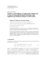
Fig. 4. The calorimetric enthalpy plots for the full-length fibritin (wac), B 1, SM1, SM4 , and F proteins in 0.01
M
Na phosphate buffer (pH 8.0) and
0.15
M
NaCl. T he enthalpy a ssigned to t he coiled-coil part represent a lin ear dependence with the slope o f 21 kJÆmol res
)1
.
Fig. 3. Temperature dependence o f the partial heat capacity of fi britin mu tants i n 0 .01
M
Na phosphate buffer (pH 8.0) and 0.15
M
NaCl. Protein
concentration was 16 m
M
chain
)1
for the full-length fibritin, and 50 m
M
chain
)1
for the others. (a) Thermal transition profiles of the wac, B1, SM1,
SM4, M, and F m utants. (b) Thermal transition curves for the E, S1, a nd F fi britins.
Ó FEBS 2002 Thermodynamics of segmented coiled coil protein (Eur. J. Biochem. 269) 837
eliminates an unusual interaction between the Asp in a d
position that is mediated in fibritin E by a chloride ion
located on the threefold axis [8]. This interaction, also found
in other coiled-coil proteins, i s c onsidered to be important
for the correct alignment of polypeptide chains upon a
coiled-coil formation [23,24]. However, in fibritin, its
C-terminal domain governs such an assembly alignment.
Furthermore, Ile425 is well accommodated at its d position
in the trimeric coiled-coil struc ture [9], and this mutation
also seems to increase the stability o f fibritin M.
The DH
cal
values of the 336 K peak of full-length fibritin,
andofthe330KpeaksoftheB1,SM1,SM4,andE
truncated molecules were proportional t o their size (Fig. 5).
The m ean enthalpy, calculated from the slope of the graph,
was DH
res
¼ 21 kJÆmol residue
)1
. The singularity a nd pro-
portionality of that transition are consistent with the
thermal unfolding of a uniform do main. By varying the
ionic strength of the sample buffer, no discrete melting of
subdomains was found for the short coiled-coil segments
(data not shown).
The melting temperature of the coiled-coil region of the
B1,SM1,SM4(T
m
¼ 33 0 K), and E (T
m
¼ 320 K)
mutants was lower than that for the respective part of a
wild-type fibritin (T
m
¼ 336 K). This was an indication that
the deletion of the N-terminal sequence of fibritin had a
destabilizing influence. The ratio of DH
VH and
DH
cal
for the
E, SM4, SM1, B1 mutants, and for a full-length fibritin were
0.91, 0.88, 0.42, 0.39 and 0.13, respectively (Table 1),
indicating a decrease of the all-or-none transition character
with increasing domain size. A plot of total DH
cal
against
the number of residues for all mutants, truncated from the
N-termini, yielded a homogeneous curve with an i nitial
slope of 6.5 ± 0.5 and a final slope of 27.5 ± 2 kJÆ(mol
residue)
)1
(Fig. 5).
Preliminary results indicate that at low ionic strength
(10 m
M
sodium phosphate buffer, pH 8.0) full-length fibr-
itin exhibited two heat absorption peaks (T
1m
¼ 326 K,
and T
2m
¼ 334 K) that are probably related to the
transition of the coiled-coil region. The position of the
326 K peak approximately matched the position of a single
transition peak of the B1, SM1, and SM4 mutants
(T
m
¼ 327–328 K) (data not shown). At the present, by
varying pH and ionic strength conditions, we are trying to
detect subdomain transitions of the coiled-coil region.
Stability of the S1 fibritin
Three coiled coil segments of fibritin E are separated by
two loops: r esidues Gly386–Gly391 form the first one
(L10) and the second one (L11) contains the residues
Asn404–Gly417 [9] (Fig. 1). To clarify the role of the loop
regions in protein stability, we designed fibritin S1 lacking
the Asn-Gly-Thr-Asn-Pro-Asn-Gly-Se r-Thr-Val-Glu-Glu
sequence of loop L11 [13]. The two last L11 loop residues,
ArgandGly,werepreservedinS1tomadethecoiledcoil
continuous (Fig. 1B).
The calorimetric transitions for the coiled-coil regions of
the E and S 1 mutants differed by 10 K ( Fig. 3B). The
coiled-coil part, which lacked the loop sequence, melted at
330 K while fibritin E had a transition at 320 K. The
enthalpy of this transition was DH
cal
¼ 656 kJÆ(mol
trimer)
)1
for fibritin S1 and 687 kJÆ(mol trimer)
)1
for
fibritin E. Most probably, the stability of S1 increased due
to the f ormation of uniform coiled coil containing two
segments, XI and XII. Also, e limination of loop 11 might
have helped to form of additional salt bridge between
residues Glu435 and Lys440, at the g and e positions,
respectively. That bridge was initially proposed [5], but it
was not found in fibritin E crystal structure [8]. Crystallo-
graphic investigations of fibritin S1 structure are in progress.
Refolding of the XN fibritin
Due to aberrant folding, fibritin XN, lacking the C-terminal
domain, was not soluble during in vivo expression and it
formed aggregates [10]. We were able to purify and dissolve
these aggregates in 8
M
urea. Then the protein was partially
refolded by the fast 100-fold dilution from 8
M
to 0.08
M
urea in 50 m
M
Tris/HCl buffer, pH 8.0 a nd purified on a
hydroxyapatite column. The CD spectrum of an in vitro
refolded fibritin XN was similar to the spectrum of a full-
length fibritin (data not shown). However, the DSC
endotherm of the refolded XN fibritin did not reveal a
heat-adsorption 345 K-peak characteristic for the C-termi-
nal domain, and the protein had three thermal transition
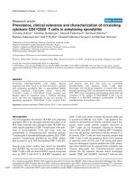
peaks centred at 321 K, 329 K, and 336 K (Fig. 6A).
The main d ifference between fibritin XN and other
truncated fibritin molecules, which contained the C-terminal
domain, was lack of ability of the XN molecule to refold
after temperature-induced denaturation. After one round of
heating to 340 K and subsequent slow cooling t o 293 K for
60 min, the protein revealed a complete lack of refolding
(Fig. 6 A). In contrast, all fibritin mutants containing the
C-terminal domain exhibited reversible refolding under t he
Fig. 5. Far CD spectra for the SM4, and F proteins and folding nucleus
alone in a solution of 0.01
M
Na phosphate buffer (pH 8.0) and 0.15
M
NaCl. (a) Spectra of the SM4 fibritin (182 residues per monomer)
were registered at 298, 335, and 358 K. The protein has the native
conformation at 298 K, and is completely unfolded at 358 K. The
335 K spectrum is the spectrum of the partially unfolded state in
which the coiled-coil part is disordered and the folding nucleus
domain still has its sec ondary structure. T his may b e seen at 229 nm:
the 335 K spectrum has a more positive h-value than the 358 K
spectrum. The difference of the signals for these two spectra assigned
only for the folding n ucleus (30 residues) is presented in (b) in
comparison with the isolated the C-terminal part spectra [22]. The
C-termini peak, centred at 229 nm, can easily be detected also for
fragment F th at h as o nly 5 8 residues p er mo no mer ( a).
838 S. P. Boudko et al. (Eur. J. Biochem. 269) Ó FEBS 2002
same conditions. As an example, Fig. 6B shows the results
of heat denaturation of fibritin B1. After heating to 336 K,
the transition curves for second and third rounds differed
from the first one by only a few percent. The d ifferences
were even smaller f or shorter fibritin fragments. Significant
flattening of th e peaks corresponding to the coiled-coil
region was observed only a fter heating to 369 K (see
Fig. 6B, f or fibritin B1). Prolonged heating led to a further
decrease of the extent o f refolding. I ndependent of temper-
ature and time of heat exposure, refolding of the C-terminal
domain was completely reversible as indic ated by identical
DH°-values, sharpness and h eight of the 345 K peak.
DISCUSSION
Previous work has demonstrated that a full-length fibritin
has a complex pattern of heat-induced transitions [5] that
were difficult to assign to individual domains. Also it was
not possible to determine calorimetric parameters for the
individual steps in transition curve and to investigate the
interactions between individual segments in the three-
stranded coiled-coil domain. A more detailed analysis was
performed now with the help of truncated fibritin molecules.
The C -terminal domain has the highest me lting temper-
ature a nd it melts independently from all the other regions.
Due to its trimeric nature, the midpoint temperature of the
C-terminal domain transition is slightly concentration
dependent, an observation which is in agreement with the
results for purified domain [22]. It acts as a cross-linker
between the three chains and, as it was proposed earlier
[5,8,10], i t helps to align t hree chains and serves as a fo ldon
by increasing local chain concentration at the C-terminus.
In addition , t he C-terminal domain of fibritin, like other
oligomerization domains [25,26], stabilizes adjacent
upstream coiled-coil segments.
For the coiled-coil region of fibritin B1, which contains
about half of a fibritin sequence, only a single transition was
observed. The assignment of the 330 K transition is evident
from the loss of a helicity at this temperature and changes in
the magnitude of the accompanying enthalpy. The ratio o f
the van’t Hoff enthalpy to calorimetric enthalpy of 0.39
indicates that the nine putative segments of the coiled-coil
domain of fibritin B1 do not unfold in an all-or-none
manner. ÔNon all-or-none transitionÕ means that we do have
intermediates, but in the case of fibritin and other fibrous
proteins these intermediates do not have fixed structures
because these proteins have a zipper-like mechanism of
folding-unfolding [27]. Nevertheless, the sharpness of the
transition and t he failure to detect a splitting of the
transition profile i nto individu al subpeaks suggests that
loop regions, connecting B1 coiled-coil segments, serve as
co-operative linkers between the segments. According to
equilibrium criteria, the unfolding and reversible refolding
of the nine segments therefore occurs in a s ingle step.
The s ingularity of the coiled-coil transition, midpoint
temperature and peak sharpness are maintained also for the
SM1 and S M4 fibritins in which the nu mber of coiled-coil
segments is reduced to eight and five, respectively. The all-
or-none approximation is better f ulfilled for these p roteins,
which is expe cted for th eir smaller size and more limited
contacts. Interestingly, the enthalpy of the transition f or the
E, SM4, SM1 and B1 fibritins increases linearly with an
increasing number of amino-acid r esidues in the coiled-coil
region. In contrast to the independent melting of the coiled-
coil segments of different stability, this is additional evidence
for the co-operative transition of the e ntire coiled-coil
region. The ratio of the van’t Hoff enthalpy to calorimetric
enthalpy for fibritin E is 0.91, is very close to 1 for the all-
or-none approximation. This finding, which is in accordance
with the crystallographic observation [8] that two coiled-coil
segments of fibritin E is a repetitive structured domain with
loop regions as a part of the structure. The e nthalpy change
per residue in the c oiled-coil domain of all the fibritin
mutants (DH
res
¼ )21 JÆmol
)1
) has the same magnitude as
for a three-stranded coiled-coil domain of laminin [28], and
for a two-stranded coiled coil of leucine zippers [29,30].
According to CD data, we were able to refold fibritin
XN, which was solved in urea, by rapid dilution. During the
Fig. 6. Consequent DSC scans performed for the XN and B1 fibritin mutants in 0.01
M
Na phosphate buffer (pH 8.0) with 0.15
M
NaClwithascanrate
of 1 KÆmin
)1
. The a bsolute h eat capacity vs. th e temperatu re is shown. (a) The XN fibritin scans: t he first is of the folded fragment, the second i s after
treating the fragment at 340 K for 5 min and cooling down to room t emperature for more than 1 h. (b) Consequent scans of the B1 fragment
(without refilling the cells): the first two s cans were performed u ntil 336 K followed by cooling down to 298 K for 1 h; the others scans were
performed until 369 K.
Ó FEBS 2002 Thermodynamics of segmented coiled coil protein (Eur. J. Biochem. 269) 839
first round of DSC, the refolded XN protein exhibits several
heat absorption peaks, one of which was assigned to the
N-terminal domain. Following the first round of heat
denaturation, it was impossible to r efold of the m olecule by
slow cooling to low temperature. In contrast, full revers-
ibility has been observed for all fragments containing the
C-terminal domain. These results strongly confirm our
previous con clusion [8,10] that the C -termini is essential for
fibritin assembly in vivo and in vitro and act as a f oldon.
Foldon is a protein unit that forms on the initial steps of
folding [31,32] which frequently perform a specific, distinct
function that remains intact even after isolated or trans-
ferred into other proteins [22,33–35]. The stabilizing and
assembly of the trimeric T4 fibritin foldon has been
demonstrated recently by protein engineering for several
chimera proteins [22,36,37].
ACKNOWLEDGEMENTS
We thank Dr Kyle Tanner for critical reading of the manuscript,
and Dr Sergei Yu. Venyaminov for providing the CONTIN
program. This work was supported in part by HHMI (grants
75195–52080, and 55000324), Russian Foundation for Basic
Research (grant 99-04-4843 0), and by the ÔUniversities of Ru ssiaÕ
grant to V. V. M, and by Swiss National Science Foundation (grant
31-49281.96) to J. E.
REFERENCES
1. Coombs, D.H. & Eiserling, F.A. ( 1977) Studies on the structure,
protein composition and assembly of the neck of bacteriophage
T4. J. Mol. Biol. 116, 375–405.
2. Conley, M.P. & Wood, W.B. (1975) Bacteriophage T4 whiskers: a
rudimentary environment-sensing device. Proc.NatlAcad.Sci.
USA 72, 3701–3705.
3. Terzaghi, B.E., Terzaghi, E. & Coombs, D. (1979) The role of the
collar/whisker complex in bacteriophage T4D tail fiber attach-
ment. J. M ol. Biol. 127, 1–14.
4. Sobolev, B.N. & Mesyanzhinov, V.V. (1991) The wac gene
product of bacteriop hage T4 c ontains coiled-coil structural
patterns. J. Biomol. Struct. Dyn. 8, 953–965.
5. Efimov, V.P., Nepluev, I.V., Sobolev, B.N., Zurabishvili, T.G.,
Schulthess, T., Lustig, A., Engel, J., Haener, M ., Aebi, U.,
Venyaminov, S., Yu, Potekhin, S.A. & Mesyanzhinov, V.V. (1994)
Fibritin encoded by bacteriophage T4 gene wac has a parallel
triple-stranded alpha-helical coiled-coil structure. J. Mol. Biol.
242, 470–486.
6. Cohen, C. & P arry, D .A. (1994) Alpha-helical coiled co ils: m ore
facts and better predictions. Science 263, 488–489.
7. Siegert, R., Leroux, M.R., Scheufler, C., Hartl, F.U. & Moarefi , I.
(2000) Structure of the molecular chaperone prefoldin. Unique
interaction of multiple coiled coil tentacles with unfolded proteins.
Cell 103, 621–632.
8. Tao, Y., Strelkov, S.V., Mesyanzhinov, V.V. & Rossmann, M.G.
(1997) Structure of b acteriophage T4 fibritin: a segmented coiled
coil and the role of t he C-terminal domain. Structure 5, 789–798.
9. Strelkov, S.V., Tao, Y., Shneider, M.M., M esyanzhin ov, V.V. &
Rossmann, M.G. (1998) Structure of bacteriophage T4 fibritin M:
atroublesomepackingarrangement.Acta. Crystallogr. D54,
805–816.
10. Letarov, A.V., Lo nder, Y.Y., B oudko, S.P. & Mesyanzhinov,
V.V. (1999) The carboxy-terminal domain initiates trimerization
of bacteriophage T4 fibritin. Biochemistry (Moscow). 64, 817–823.
11. Letarov, A.V. (1999) Examination of the Folding Pathways of the
Bacteriophage T4 Fibritin. PhD Thesis. Ivanovsky Institute of
Virology, Academy of Me dic al Sciences, Moscow, Russia.
12. Londer, Y.Y. (1999) Folding, Assembly, and Stability o f Bacte-
riophage T4 Fibritin. PhD Thesis. Bakh Institute of Biochemistry,
Russian Academy of Sciences, Moscow, Russia.
13. Londer, Y. & Mesyanzhinov, V.V. (1999) Thermostability of
bacteriophage T4 fibritin and i ts deletion m utants. Bioorg. Khim.
25, 257–263.
14. Ausubel, F .M., Brent, R., Kingst on, R.E., Moore, D.D.,
Seidman, J.G., Smith, J.A. & Struhl, K. (1992) Short Protocols in
Molecular Biology. John Wiley and Sons, Toronto, C anada.
15. Laemmli, U.K. (1970) Cleavage of structural proteins during
the assembly of the head o f bacteriophage T4. Nature 227,
680–685.
16. Schagger, H. & Jagow, G. (1987) Tricine-sodium dodecyl
sulfate-polyacrylamide gel e lectrophoresis for t he separation
of pro teins in the range from 1 to 100 kDa. Ana l. Biochem. 166,
368–379.
17. Edelhoch, H. (1967) Spectroscopic determination of tryptophan
andtyrosineinproteins.Biochemistry 6, 1948–1954.
18. MicroCal Inc. (1998) DSC Data Analysis in OriginÒ.Tutorial
guide. MicroCal Inc., Northampton, MA, USA.
19. Privalov, P.L. & Potekhin, S.A. (1986) Scanning microcalorimetry
in studying temperature -induced changes in proteins. Methods
Enzymol. 131, 4–51.
20. Provencher, S.W. & Glockner, J. (1981) Estimation of globular
protein secondary structure from circular dichroism. Biochemistry
20, 33–37.
21. Harbury, P.B., K im, P.S. & Alber, T. (1994) Crystal structure of
an isoleucine-zipper t rimer. Nature 371, 80–83.
22. Frank,S.,Kammerer,R.A.,Mechling,D.,Schulthess,T.,Land-
wehr,R.,Bann,J.,Guo,Y.,Lustig,A.,Baechinger,H.&Engel,J.
(2001) Stabilisation o f short c ollagen-like triple helices by protein
engineering. J. Mol. Biol. 30 8, 1081–1089.
23. Lumb, K.J. & Kim, P.S. (1995) Measurement of interhelical
electrostatic interactions in the GCN4 leucine zipper. Science 268,
436–439.
24. Lumb, K.J. & Kim, P.S. (1995) A buried polar interaction imparts
structural uniqueness in a designed heterodimeric coiled coil.
Biochemistry 34, 8642–8648.
25. Engel, J. & Kammerer, R.A. (2000) What are oligomerization
domains good for? Matrix Biol. 19, 283– 288.
26. Cohen, C. & Parry, D.A. (1998) A conserved C-terminal assembly
region in paramyosin and myosin rods. J. Struct. Biol. 122,
180–187.
27. Engel, J. & Schwarz, G. (1970) Co-operative conformational
transitions of linear biopolymers. Angew. Chem. Int. 9, 389–400.
28. Kammerer, R.A., Antonsson, P., Schulthess, T., Fauser, C. &
Engel, J. (1995) Selective chain recognition in the C-terminal
alpha-helical coiled-coil region of laminin. J. Mol. Biol. 250,
64–73.
29. Bosshard, H.R., Durr, E., Hitz, T. & Jelesarov, I. (2001)
Energetics of coiled coil folding: the nature of the transition states.
Biochemistry 40, 3544–3552.
30. Yu, M H. & King, J. (1984) Single amino acid substitutions
influencing th e f olding p athway of the phage P22 t ailspike
endorhamnosidase. Proc. Natl Acad. Sci. USA 81, 6584–6588.
31. Panchenko, A.R., Luthey-Schulten, Z. & Wolynes, P.G. (1996)
Foldons, protein structural mo dules and exons. Proc.NatlAcad.
Sci. USA 93, 2008–2013.
32. Inaba, K., Kobayashi, N. & Fersht, A.R. (2000) Conversion of
two-state to m ulti-state folding o n fusion o f two protein foldons.
J. Mol. Biol. 302, 219–233.
33. Yanagawa, H., Yoshida, K., Torigoe, C., Prak, J.S., Sato, K.,
Shirai, T. & Go, M. (1993) Protein anatomy; functional r oles o f
barnase module. J. Biol. Chem. 268, 5861–5865.
34. Wakasugi, K., Isimori, K ., Im ai, K ., W ada, Y. & Morishima, I.
(1994) ÔModuleÕ substitution in hemoglo bin subunits. J. Biol.
Chem. 269, 18750–18756.
840 S. P. Boudko et al. (Eur. J. Biochem. 269) Ó FEBS 2002
35. Miroshnikov, K.A., Sernova, N.V., Shneider, M.M. &
Mesyanzhinov, V.V. (2000) Transformation of a fragment of
beta-structural bacteriophage T4 adhesin to stable alpha-helical
trimer. Biochemistry (Moscow) 65, 1346–1351.
36. Krasnykh, V., Belousova, N., Korokhov, N., Mikheeva, G. &
Curiel, D.T. (2001) Genetic targeting o f an a denovirus vector v ia
replacement of th e fiber protei n with the phage T4 fi britin. J. Virol.
75, 4176–4183.
Ó FEBS 2002 Thermodynamics of segmented coiled coil protein (Eur. J. Biochem. 269) 841