Báo cáo y học: "Diosgenin, a plant steroid, induces apoptosis in human rheumatoid arthritis synoviocytes with cyclooxygenase-2 overexpression" pdf
Bạn đang xem bản rút gọn của tài liệu. Xem và tải ngay bản đầy đủ của tài liệu tại đây (546.96 KB, 11 trang )
Open Access
Available online />R373
Vol 6 No 4
Research article
Diosgenin, a plant steroid, induces apoptosis in human
rheumatoid arthritis synoviocytes with cyclooxygenase-2
overexpression
Bertrand Liagre
1
, Pascale Vergne-Salle
2
, Cecile Corbiere
1
, Jean L Charissoux
3
and
Jean L Beneytout
1
1
Laboratoire de Biochimie, UPRES EA 1085, Faculté de Pharmacie, 2 rue du Docteur Marcland, 87025 Limoges Cedex, France
2
Service de Rhumatologie, CHRU Dupuytren, 2 avenue Martin Luther King, 87042 Limoges Cedex, France
3
Service d'Orthopédie-Traumatologie, CHRU Dupuytren, 2 avenue Martin Luther King, 87042 Limoges Cedex, France
Corresponding author: Bertrand Liagre,
Received: 19 Mar 2004 Revisions requested: 16 Apr 2004 Revisions received: 4 May 2004 Accepted: 18 May 2004 Published: 17 Jun 2004
Arthritis Res Ther 2004, 6:R373-R383 (DOI 10.1186/ar1199)
http://arthr itis-research.com/conte nt/6/4/R373
© 2004 Liagre et al.; licensee BioMed Central Ltd. This is an Open Access article: verbatim copying and redistribution of this article are permitted in
all media for any purpose, provided this notice is preserved along with the article's original URL.
Abstract
In the present study, we have shown for the first time that a plant
steroid, diosgenin, causes an inhibition of the growth of
fibroblast-like synoviocytes from human rheumatoid arthritis,
with apoptosis induction associated with cyclooxygenase-2
(COX-2) up-regulation. Celecoxib, a selective COX-2 inhibitor,
provoked a large decrease in diosgenin-induced apoptosis even
in the presence of exogenous prostaglandin E
2
, whereas
interleukin-1β, a COX-2 inducer, strongly increased diosgenin-
induced apoptosis of these synoviocytes. These findings
suggest that the proapoptotic effect of diosgenin is associated
with overexpression of COX-2 correlated with overproduction of
endogenous prostaglandin E
2
. We also observed a loss of
mitochondrial membrane potential, caspase-3 activation, and
DNA fragmentation after diosgenin treatment.
Keywords: apoptosis, cyclooxygenase-2, diosgenin, human synoviocyte, rheumatoid arthritis
Introduction
Rheumatoid arthritis (RA) is an inflammatory joint disease in
which perpetuation of chronic synovitis leads to bone and
cartilage degradation. Inflammatory cytokines or soluble
factors are essential in the pathogenesis of RA. IL-1 and
tumor necrosis factor-α are the principal mediators of tis-
sue destruction in many immunoinflammatory diseases
such as RA [1-3]. The two cytokines induce, in synergy, the
production of high levels of matrix metalloproteinases by
synovial cells and chondrocytes [4]. IL-6 and IL-8 also par-
ticipate in the pathogenesis of RA; for example, IL-6 sup-
ports the proliferation of synovial cells [5], while IL-8
promotes the formation of new blood vessels in synovial
membrane [6].
RA is characterized by the proliferation of synoviocytes,
which also produce prostanoids. Eicosanoids and prosta-
noids are important lipid mediators that are produced at
elevated levels in inflamed tissues including rheumatoid
synovium and in cultured human RA fibroblast-like synovio-
cytes (FLS) [7-11]. Cyclooxygenase (COX), which con-
verts arachidonic acid into prostaglandin endoperoxides, is
the rate-limiting enzyme in prostanoid synthesis [12]. At
least two forms of COX have been identified and their
genes have been cloned [13,14]. COX-1 is constitutively
expressed in most cells and tissues. In contrast, COX-2 is
highly inducible by serum, growth factors, lipopolysaccha-
rides, and cytokines, especially interleukin-1 (IL-1), in cer-
tain cell types involved in inflammatory processes, e.g.
fibroblasts and macrophages [15,16]. Crofford and co-
workers [17] showed that IL-1β enhanced de novo synthe-
sis of COX-2 – but not of COX-1 – mRNA and protein
either in rheumatoid synovial explants or in cultured rheu-
matoid synoviocytes. These observations suggest that
Ac-DEVD-AMC = N-acetyl-Asp-Glu-Val-Asp–7-amino-4-methylcoumarin; Ac-DEVD-CHO = N-acetyl-Asp-Glu-Val-Asp-aldehyde; COX = cyclooxyge-
nase; ∆ψm = mitochondrial membrane potential; DAPI = 4',6-diamidino-2-phenylindole; DMEM = Dulbecco's modified Eagle's medium; ELISA =
enzyme-linked immunosorbent assay; FCS = fetal calf serum; FLS = fibroblast-like synoviocytes; IL = interleukin; JC-1 = 5,5',6,6'-tetrachloro-1,1',3,3'-
tetraethylbenzimidazole carbocyanide iodide; MTT = 3-(4,5-dimethylthiazol-2-yl)-2,5-diphenyltetrazolium bromide; PBS = phosphate-buffered saline;
PGE
2
= prostaglandin E
2
; RA = rheumatoid arthritis.
Arthritis Research & Therapy Vol 6 No 4 Liagre et al.
R374
COX-2 may play an important part in the overproduction of
prostaglandin E
2
(PGE
2
) by rheumatoid synovia.
Recent reports have outlined the role of COX-2 and pros-
taglandins in cell apoptosis, particularly in cancer cells [18-
20]. Overexpression of the COX-2 gene protects cancer
cells from apoptosis, and drugs that inhibit COX-2 have
been shown to induce programmed death in these cells
[21,22]. In addition, the use of nonsteroidal anti-inflamma-
tory drugs (specific or nonspecific COX-2 inhibitors) has
been shown to reduce the size and number of neoplastic
polyps in patients with familial polyposis [23,24]. Altera-
tions in the apoptosis of synovial cells have been described
in resident synoviocytes as well as in inflammatory cells and
are associated with the pathogenesis of RA [25]. These
changes constitute hallmarks of synovial cell activation and
contribute to both chronic inflammation and hyperplasia.
RA FLS are affected most prominently, and their resistance
to apoptosis has been linked closely to the progressive
destruction of articular cartilage. The role of COX-2 and
prostaglandins in synoviocyte death is still under
investigation.
We have investigated for the first time the effect of dios-
genin, a plant steroid, on the proliferation rate and apopto-
sis in the human RA FLS. Particular attention was paid to
the modulation of COX-2 expression and activity in RA syn-
oviocyte viability.
Materials and methods
Materials
Dulbecco's modified Eagle's medium (DMEM), fetal calf
serum (FCS), and penicillin–streptomycin were supplied by
Gibco-BRL (Cergy Pontoise, France). Collagenase was
obtained from Worthington Biochemical Corporation
(Freehold, NJ, USA). Dispase, hyaluronidase, DNase I,
diosgenin ([25R]-5α-spirosten-3β-ol), 4',6-diamidino-2-
phenylindole (DAPI), 3-(4,5-dimethylthiazol-2-yl)-2,5-diphe-
nyltetrazolium bromide (MTT), and monoclonal antibody β-
actin were purchased from Sigma (Saint Quentin Fallavier,
France). 5B5 and JC/70A monoclonal antibodies and sec-
ondary polyclonal antibody conjugated with peroxidase
were purchased from Dako (Trappes, France). RMO52
monoclonal antibody and fluorescein (DTAF)-conjugated
goat anti-mouse antibody were purchased from Immu-
notech (Marseilles, France). COX-2 monoclonal antibody
was supplied by Santa Cruz Biotechnology (TEBU; Le Per-
ray en Yvelines, France). JC-1 (5,5',6,6'-tetrachloro-
1,1',3,3'-tetraethylbenzimidazole carbocyanide iodide) was
supplied by Molecular Probes (Leiden, The Netherlands).
CaspACE™ Assay System Fluorometric was supplied by
Promega (Charbonnieres, France). Cell Death Detection
ELISA
plus
was supplied by Roche Diagnostics (Meylan,
France). Celecoxib was obtained from Pharmacia (Skokie,
IL, USA). PGE
2
and ELISA kits for PGE
2
were purchased
from Cayman Chemical (SpiBio, Massy, France). Recom-
binant human IL-1β and Quantikine
®
human IL-6 and IL-8
immunoassay kits were purchased from R&D Systems
(Lille, France).
Preparation of human synovial cells
RA synoviocytes were isolated from fresh synovial biopsies
obtained from six RA patients undergoing hip arthroplasty.
All patients fulfilled the 1987 American Rheumatism Asso-
ciation criteria for RA [26]. The mean age of the patients
was 62.2 ± 4.6 years (range 55–68 years). The mean dis-
ease duration was 9.3 ± 2.2 years. At the time of surgery,
the disease activity score (DAS 28) was greater than 3.2.
These activities were approved by local institutional review
boards, and all subjects gave written informed consent.
Synovia were minced and digested with 1.5 mg/ml colla-
genase-dispase, 1 mg/ml hyaluronidase, and 0.15 mg/ml
DNase I for 3–4 hours at 37°C as previously described [9].
After centrifugation, cells were resuspended in DMEM sup-
plemented with 10% FCS, 4.5 g/l D-glucose, 25 mM
Hepes, 100 U/ml penicillin, and 100 µg/ml streptomycin
(Gibco BRL) in a humidified atmosphere containing 5% (v/
v) CO
2
at 37°C. After 48 hours, nonadherent cells were
removed. Adherent cells (macrophage-like and FLS) were
cultured in complete medium, and, at confluence, cells
were trypsinized and only the FLS were passed. These
cells were used between passages 4 and 8, when they
morphologically resembled FLS after indirect immunofluo-
rescence study (see Culture of human RA FLS). RA FLS
were cultured 45–60 days before experimentation. This
delay allowed the elimination of all possible interactions
resulting from any preoperative treatment (with nonsteroi-
dal anti-inflammatory drugs, analgesics, disease-modifying
antirheumatic drugs, or steroids).
Culture of human RA FLS
Between passages 4 and 8, RA FLS were trypsinized. Cell
count and viability were determined and cells were plated
in culture plates or flasks (Falcon, Oxnard, CA, USA). Via-
bility, measured by trypan blue dye exclusion [27] at the
start and the end of culture, was always greater than 95%.
FLS (10
5
) from RA patients were used for indirect immun-
ofluorescence study [28]. The following monoclonal anti-
bodies were used: 5B5 (anti-prolyl hydroxylase) for
fibroblasts at 1/50 dilution (Dako, Burlingame, CA, USA),
JC/70A (anti-CD31) for endothelial cells at 1/50 (Dako),
and RMO52 (anti-CD14) for macrophages at 1/50 (Immu-
notech). The negative control was a mouse antibody of the
same isotype (Immunotech). Incubations were performed
at room temperature for 30 min. Binding of monoclonal
antibodies was visualized using fluorescein (DTAF)-conju-
gated goat anti-mouse antibody (Immunotech) at 1/50
dilution.
Available online />R375
For all experiments, RA FLS were allowed to adhere and
grow for 48 hours in culture medium before exposure to
diosgenin. A stock solution of 10
-2
M diosgenin was pre-
pared in ethanol and diluted in culture medium to give a
final concentration of 10–80 µM. The same amount of eth-
anol (<0.4%) was added to control cells. The culture
medium was not changed during the entire study.
Human RA FLS proliferation and light microscopy
Cell proliferation was measured using the MTT assay. Cells
(10
3
cells/well) were plated in 96-well culture plates and
grown for 48 hours before treatment with 10–80 µM dios-
genin for 24–96 hours. MTT was carried out daily as previ-
ously described [29] and experiments were performed in
sextuple assays.
For light microscopy, after 24–72 hours of treatment, RA
FLS cultured cells were fixed in PBS (pH 7.4) containing
4% paraformaldehyde for 20 min at room temperature and
washed in PBS for 15 min. Observations were made with
phase-contrast microscopy.
Mitochondrial membrane potential (∆ψm) and DAPI
staining
∆ψm was estimated using JC-1 (Molecular Probes). This is
a fluorescent compound that exists as a monomer at low
concentrations. At higher concentrations, it forms aggre-
gates. Fluorescence of the JC-1 monomer is green,
whereas that of the aggregate is red. Mitochondria with
intact membrane potential concentrate JC-1 into aggre-
gates, which fluoresce red, whereas de-energized mito-
chondria cannot concentrate it and fluoresce green [30].
Human RA FLS were grown for 48 hours before treatment
with 40µM diosgenin for 24 hours. Control cells were
grown in medium containing the same amount of ethanol as
treated cells. Adherent cells were incubated in 1 ml of
medium containing JC-1 (1 µg/ml) for 30 min at 37°C and
pictures were taken with a Nikon microscope ECLIPSE
E800 (Nikon Corporation, Champigny sur Marne, France).
Moreover, human RA FLS were stained with DAPI (0.5 µg/
ml) for 5 min at room temperature in the dark and the cells
were examined by fluorescence microscopy.
Caspase-3 activity
After 40 µM diosgenin treatment for 24 or 48 hours, human
RA FLS were homogenized in lysis buffer in accordance
with the manufacturer's protocol (CaspACE™ Assay Sys-
tem Fluorometric, Promega). Fluorometric assays were
conducted in white, opaque tissue-culture plates (Falcon,
Becton Dickinson Labware, NJ, USA) and all measure-
ments were carried out in triplicate. First, 100 µl of assay
buffer (10 mM dithiothreitol, dimethyl sulfoxide, caspase
buffer) (Promega) was added to each well. Peptide sub-
strate for caspase-3 (Ac-DEVD–AMC [N-acetyl-Asp-Glu-
Val-Asp–7-amino-4-methylcoumarin]) was added to each
well to a final concentration of 2.5 mM. Caspase inhibitor
(Ac-DEVD-CHO [N-acetyl-Asp-Glu-Val-Asp-aldehyde]) at
2.5 mM was also used just before the addition of the sub-
strate. The supernatant of each cell lysate collected was
added to each well to start the reaction. Background fluo-
rescence was determined in wells containing assay buffer
and substrate without cell lysate. Assay plates were incu-
bated at 37°C for 1 hour for the measurement of caspase-
3 activity. Fluorescence was measured with a microplate
reader (Fluorolite 1000, Dynex Technologies, Chantilly, VA,
USA) using 360 nm excitation and 460 nm emission filters.
Raw data (relative units of fluorescence) corresponded to
the concentrations of 7-amino-4-methylcoumarin released
[31].
Apoptosis quantification: DNA fragmentation
Human RA FLS were cultured in six-well culture plates (2 ×
10
5
cells/well). After diosgenin treatment (40 µM for 24 or
48 hours), apoptosis was quantified on pooled cells (float-
ing and adherent) using the 'cell death' ELISA (Cell Death
Detection ELISA
plus
, Roche Diagnostics). Cytosol extracts
were obtained in accordance with the manufacturer's pro-
tocol and apoptosis was measured as previously described
[32]. Other conditions are represented by cells pretreated
for 4 hours at 37°C with IL-1β (1 ng/ml) or celecoxib (1 µM)
and then 40 µM diosgenin was added in each flask for 24
or 48 hours in DMEM containing 10% (v/v) FCS in an
atmosphere of 5% CO
2
. We verified that under our experi-
mental conditions 1 µM celecoxib was not proapoptotic. To
examine the role of exogenously added PGE
2
, we preincu-
bated cells for 4 hours with COX-2 inhibitor (celecoxib 1
µM) in the absence or presence of PGE
2
(10 nM), followed
by incubation with 40 µM diosgenin for an additional 24
hours.
COX-2 expression analysis
Human RA FLS were cultured in 150-cm
2
tissue-culture
flasks. After treatment with 40 µM diosgenin for 24 or 48
hours or with IL-1β (1 ng/ml) for 24 hours, adherent cells
were trypsinized and pooled with the floating cell fraction.
Western blot analysis was performed as previously
described [32], using the primary monoclonal antibodies β-
actin (mouse anti-human β-actin [1:5000], Sigma), COX-2
(mouse anti-human COX-2 [1:100], Santa Cruz Biotech-
nology), and secondary polyclonal antibody conjugated
with peroxidase (Dako). Blots were visualized using
enhanced chemiluminescence reagents (Amersham Bio-
sciences, Orsay, France) and immediately exposed to x-ray
film.
Assay of PGE
2
production
Human RA FLS were grown in 25-cm
2
tissue-culture flasks
for 48 hours before treatment. After washing with PBS (pH
7.4), cells were pretreated for 4 hours at 37°C with IL-1β
Arthritis Research & Therapy Vol 6 No 4 Liagre et al.
R376
(1 ng/ml) or celecoxib (1 µM) and then 40 µM diosgenin
was added in each flask for 24 or 48 hours in DMEM con-
taining 10% (v/v) FCS in an atmosphere of 5% CO
2
. Other
conditions are represented by cells incubated with 40 µM
diosgenin alone, IL-1β (1 ng/ml) alone, or celecoxib (1 µM)
alone for 24 or 48 hours. The PGE
2
concentration in the
medium was measured using an ELISA kit in accordance
with the instructions of the manufacturer (Cayman Chemi-
cal) and was normalized with respect to the number of via-
ble cells present in the particular culture at the time of
sampling.
IL-6 and IL-8 assay conducted on conditioned medium
Human RA FLS were grown in 25-cm
2
tissue-culture flasks
for 48 hours before treatment. After washing with PBS (pH
7.4), cells were pretreated for 4 hours at 37°C with IL-1β
(1 ng/ml) and then 40 µM diosgenin was added to each
flask for 24 or 48 hours in DMEM containing 10% (v/v)
FCS in an atmosphere of 5% CO
2
. Other conditions are
represented by cells incubated with 40 µM diosgenin alone
or IL-1β (1 ng/ml) alone for 24 or 48 hours. The cytokine
concentration in the medium was measured using a Quan-
tikine
®
human IL-6 or IL-8 immunoassay kit in accordance
with the instructions of the manufacturer (R&D Systems)
and was normalized with respect to the number of viable
cells present in the particular culture at the time of
sampling.
Statistical analysis
The median and standard deviation (SD) were calculated
using Excel (Microsoft Office, Version 98). Statistical anal-
ysis of differences was carried out by analysis of variance
(ANOVA) using StatView Version 5.0 (SAS Institute Inc,
Cary, NC, USA). A P-value of less than 0.05 (Fisher's pro-
tected-least-significant-difference test) was considered to
indicate significance.
Results
Effect of diosgenin on human RA FLS proliferation and
morphological modifications
Cells were cultured in 10% FCS medium with or without
10–80 µM diosgenin for 24–96 hours and cell proliferation
was evaluated by the MTT test. Under our experimental
conditions, a dramatic decrease in proliferation was
observed until 24 hours after diosgenin treatment (40 and
80 µM) (Fig. 1), especially at 24 hours for 40 µM diosgenin,
when the percentage of inhibition was 76% (P < 0.05). As
the percentage of inhibition did not strongly increase at 80
µM (79%; P < 0.05) (Fig. 1), we chose 40 µM for subse-
quent experiments.
Direct observation with phase-contrast microscopy demon-
strated that human RA FLS treated with 40 µM diosgenin
showed numerous morphological differences from control
cells (Fig 2a). Cell shrinkage, cytoplasm condensation, and
formation of cytoplasmic filaments appeared after 40 µM
diosgenin treatment for 24, 48, and 72 hours (Fig.
2b,2c,2d respectively).
Diosgenin-induced disruption of ∆ψm in human RA FLS
To ascertain potential mechanisms by which diosgenin
inhibited the human RA FLS proliferation rate, we studied
the effect of diosgenin on ∆ψm, because alterations in mito-
chondrial structure and function have been shown to play a
crucial role in apoptosis.
∆ψm was analyzed in adherent RA FLS after 24 hours of
treatment with diosgenin using the potential-dependent,
aggregate-forming lipophilic cation JC-1. Fluorescence,
seen in Fig. 3, showed ∆ψm differences. We found that
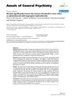
Figure 1
Effect of diosgenin on proliferation of human rheumatoid arthritis (RA) fibroblast-like synoviocytes (FLS)Effect of diosgenin on proliferation of human rheumatoid arthritis (RA)
fibroblast-like synoviocytes (FLS). Cells were cultured in 10% FCS
medium for 48 hours and then incubated (time 0) for 24–96 hours with
diosgenin at 10–80 µM. RA FLS proliferation was evaluated by the
MTT [3-(4,5-dimethylthiazol-2-yl)-2,5-diphenyltetrazolium bromide] test.
Measurements were made on FLS from six different patients. Repre-
sentative results from six independent experiments are shown; values
are the mean ± SD from triplicate cultures. * A P value of less than 0.05
(Fisher's protected-least-significant-difference test) was considered to
indicate significance in comparison with controls. OD, optical density.
0
0.02
0.04
0.06
0.08
0.1
0.12
024487296
Time (hours)
Mean of OD (550 nm)
control
diosgenin 10 µM
diosgenin 20 µM
diosgenin 40 µM
diosgenin 80 µM
∗
∗
∗
∗∗
∗
∗
∗∗
∗
∗
∗
∗
∗∗
∗
∗
∗∗
∗
Available online />R377
diosgenin induced a decrease of ∆ψm in RA FLS, shown
by the incorporation of JC-1 monomers into the mitochon-
dria (fluorescence in green, Fig. 3b), compared with
cytosolic J-aggregate formation at high membrane potential
in control cells (fluorescence in red, Fig. 3a).
Moreover, the morphology of treated human RA FLS was
examined by fluorescence microscopy after DAPI staining.
Diosgenin treatment of human RA FLS altered the extracel-
lular and nuclear membrane permeability, as is shown by
the DAPI nuclear localisation (Fig. 3b) in comparison with
untreated cells (Fig. 3a).
Caspase-3 activity and DNA fragmentation analysis
It is well known that apoptosis is characterized by chroma-
tin condensation and DNA fragmentation and is mediated
by the cysteine protease family called caspases, such as
caspase-3, which is the major executioner of apoptosis.
In our study, caspase-3 activity and DNA fragmentation
were analyzed in human RA FLS treated or not with 40 µM
diosgenin for 24 or 48 hours. Caspase-3 activity was sig-
nificantly increased over time (2-fold at 24 hours and 2.6-
fold at 48 hours in the diosgenin-treated cells versus con-
trols; P < 0.05) (Fig. 4).
Quantitative determination of cytoplasmic histone-associ-
ated DNA fragments (mononucleosomes and oligonucleo-
somes) was performed with ELISA. Results showed that
DNA fragmentation was enhanced 7-fold (P < 0.05) in
treated cells at 24 hours and strongly induced at 48 hours
(19-fold; P < 0.05) in comparison with controls (Table 1).
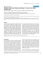
Figure 2
Morphologic changes in human rheumatoid arthritis fibroblast-like synoviocytesMorphologic changes in human rheumatoid arthritis fibroblast-like syn-
oviocytes. Cells were incubated without (a) or with 40 µM diosgenin
for 24 hours (b), 48 hours (c), or 72 hours (d). Original magnification
×400.
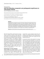
Figure 3
Analysis of mitochondrial membrane potential (∆ψm) after diosgenin treatmentAnalysis of mitochondrial membrane potential (∆ψm) after diosgenin treatment. Human rheumatoid arthritis (RA) fibroblast-like synoviocytes (FLS)
were cultured in 10% FCS medium for 48 hours and then treated (b) or not (a) with 40 µM diosgenin. ∆ψm was analyzed in adherent RA FLS after
24 hours of treatment, using the potential-dependent aggregate-forming lipophilic cation JC-1 (5,5',6,6'-tetrachloro-1,1',3,3'-tetraethylbenzimidazole
carbocyanide iodide). Red fluorescence (a) represents mitochondria with intact membrane potential whereas green fluorescence (b) represents de-
energized mitochondria. Staining with DAPI (4',6-diamidino-2-phenylindole), showed that diosgenin treatment of cells altered the extracellular and
nuclear membrane permeability, as is shown by the nuclear localization of the DAPI (b, white arrows) in comparison with untreated cells (a). Pictures
were taken with a Nikon microscope ECLIPSE E800 (original magnification ×400). One of three representative experiments from three different
patients is shown.
Arthritis Research & Therapy Vol 6 No 4 Liagre et al.
R378
Up-regulation of COX-2 expression and activity in
diosgenin-induced RA FLS death
Numerous studies have shown that COX-2 expression pre-
vents apoptosis in cancer cells, especially in colon cancer,
in contrast to other cell types, for which the effects of COX-
2 and related PGE
2
in the regulation of apoptosis could
very well be cell-type-specific. Here we show that dios-
genin induced overexpression of COX-2 over time (Fig. 5).
This overexpression was correlated with COX-2 activity.
Indeed, PGE
2
production was increased over time after
diosgenin treatment: 2.3- and 4.7-fold (P < 0.05) at 24 or
48 hours, respectively, in comparison with controls (Fig. 6).
To further develop these results, it would be interesting to
know if COX-2 was directly associated with diosgenin-
Figure 4
Effect of diosgenin on caspase-3 activation in human rheumatoid arthri-tis fibroblast-like synoviocytesEffect of diosgenin on caspase-3 activation in human rheumatoid arthri-
tis fibroblast-like synoviocytes. After 48 hours' adherence, cells were
cultured in 10% FCS medium and treated or not with 40 µM diosgenin
for 24 or 48 hours, and then caspase-3 activity was measured with cas-
pase-3 substrate (Ac-DEVD-AMC [N-acetyl-Asp-Glu-Val-Asp–7-amino-
4-methylcoumarin]) in accordance with the manufacturer's protocol
(see Materials and methods). Data are the mean ± SD of three experi-
ments from three different patients and are expressed as relative units
of fluorescence (RUF). *
,#
A P-value of less than 0.05 (Fisher's pro-
tected-least-significant-difference test) was considered to indicate sig-
nificance in comparison with the relevant controls. Ac-DEVD-CHO, N-
acetyl-Asp-Glu-Val-Asp-aldehyde.
Table 1
DNA fragmentation in human rheumatoid arthritis fibroblast-
like synoviocytes after diosgenin treatment
Cells Time (h)
24 48
Control 1 1
Diosgenin-treated 7.0 ± 1.4* 19.0 ± 3.8
#
Apoptosis was quantified on floating and adherent cells using ELISA
(see Materials and methods). The fold induction of DNA
fragmentation is shown relative to the value for the control culture,
which is taken as 1. Data are expressed as mean ± SD of three
experiments from three different patients. *
,#
A P value less than 0.05
(Fisher's protected-least-significant-difference test) was considered
to indicate significance in comparison with controls.
Figure 5
Cyclooxygenase-2 (COX-2) western blot analysis in human rheumatoid arthritis fibroblast-like synoviocytesCyclooxygenase-2 (COX-2) western blot analysis in human rheumatoid
arthritis fibroblast-like synoviocytes. Cells were cultured without agents
(lanes 1 and 5, controls at 24 and 48 hours, respectively) or were incu-
bated with 40 µM diosgenin for 24 hours (lane 2) or 48 hours (lane 3)
or with IL-1β (1 ng/ml) for 24 hours (lane 4). Protein extracts prepared
from the cells were subjected to western blotting and cellular expres-
sions of COX-2 and β-actin were estimated using mouse anti-human
COX-2 and β-actin antibodies, respectively, as described in Materials
and methods. Each band was quantified by densitometry analysis soft-
ware. One of three representative experiments from three different
patients is shown.
Figure 6
Effect of diosgenin on prostaglandin E
2
(PGE
2
) production by human rheumatoid arthritis fibroblast-like synoviocytes (FLS)Effect of diosgenin on prostaglandin E
2
(PGE
2
) production by human
rheumatoid arthritis fibroblast-like synoviocytes (FLS). Cells were cul-
tured in 10% FCS medium for 48 hours and then treated or not with 40
µM diosgenin for 24 or 48 hours. The PGE
2
levels in the culture
medium were measured by enzyme immunoassay. Measurements were
made on FLS from four different patients. Data are expressed as mean
± SD of four experiments. *
,#
A P-value of less than 0.05 (Fisher's pro-
tected-least-significant-difference test) was considered to indicate sig-
nificance in comparison with the relevant controls.
Available online />R379
induced human RA FLS apoptosis. In order to clarify this
point, we used a specific inhibitor of COX-2 activity to verify
DNA fragmentation after diosgenin treatment and exam-
ined the effect of exogenously added PGE
2
on diosgenin-
induced synoviocyte death. We also studied whether
COX-2 induction with IL-1β could have a synergistic effect
with diosgenin on DNA fragmentation.
Endogenously produced PGE
2
associated with
diosgenin-induced human RA FLS apoptosis
Our results showed that pretreatment with celecoxib
before diosgenin treatment inhibited COX-2 activity over
time: PGE
2
production was decreased by 73% and 95%
(P < 0.05) at 24 and 48 hours, respectively, in comparison
with diosgenin alone (Table 2). DNA fragmentation was
also studied using the same conditions, and we found that
pretreatment with celecoxib before diosgenin treatment
reduced the production of mononucleosomes and oligonu-
cleosomes by 47% and 46% at 24 and 48 hours, respec-
tively; P < 0.05) in comparison with diosgenin alone (Fig.
7a). In contrast, exogenous PGE
2
did not have the same
effect as endogenous PGE
2
: we showed that the addition
of 10 µM exogenous PGE
2
alone or in the presence of
celecoxib before diosgenin treatment reduced DNA frag-
mentation in comparison with diosgenin alone (Fig. 7b).
Effect of diosgenin after selective COX-2 overexpression
induced by IL-1β on DNA fragmentation
Stimulation of RA FLS with IL-1β before diosgenin treat-
ment dramatically enhanced COX-2 activity over time: we
showed that PGE
2
synthesis increased 38- and 139-fold (P
< 0.05) over 24 and 48 hours, respectively, in comparison
with diosgenin alone (Table 2). The addition of IL-1β alone
had no effect on human RA FLS apoptosis (Fig. 8). On the
contrary, DNA fragmentation increased significantly after
stimulation of the cells with IL-1β before diosgenin treat-
ment (2.7- and 3.6-fold at 24 or 48 hours respectively; P <
0.05) in comparison with diosgenin alone (Fig. 8).
Table 2
Effect of diosgenin on prostaglandin E2 concentration after
cyclooxygenase-2 inhibition or induction in human rheumatoid
arthritis fibroblast-like synoviocytes
Treatment Time (h)
24 48
Diosgenin 3.1 ± 0.7 5.71 ± 1.79
Celecoxib + diosgenin 0.86 ± 0.16* 0.29 ± 0.08
#
IL-1β + diosgenin 120.40 ± 17.44* 792.24 ± 56.33
#
Human rheumatoid arthritis (RA) fibroblast-like synoviocytes (FLS)
were preincubated with or without celecoxib (1 µM) or IL-1β (1 ng/
ml) for 4 hours, and then 40 µM diosgenin was added for 24 or 48
hours. The concentration of prostaglandin E
2
(PGE
2
) was measured
by enzyme immunoassay and is expressed asng/ml for 10
5
cells. Data
are PGE
2
concentrations expressed as mean ± SD of three
experiments from three different patients. The PGE
2
concentrations
under the other conditions at 24 or 48 hours, respectively, were 1.39
± 0.41 and 1.21 ± 0.26 for culture with medium alone, 0.73 ± 0.19
and 0.66 ± 0.12 for celecoxib alone, and 20.26 ± 3.19 and 17.42 ±
3.03 for IL-1β alone. *
,#
A P value of less than 0.05 (Fisher's
protected-least-significant-difference test) was considered to
indicate significance in comparison with diosgenin alone.
Figure 7
Effect of diosgenin on DNA fragmentation after incubation with celecoxib (an inhibitor of cyclooxygenase-2 [COX-2]) (a) or exogenous prostaglandin E
2
(PGE
2
) (b)Effect of diosgenin on DNA fragmentation after incubation with
celecoxib (an inhibitor of cyclooxygenase-2 [COX-2]) (a) or exogenous
prostaglandin E
2
(PGE
2
) (b). (a) Human rheumatoid arthritis fibroblast-
like synoviocytes (FLS) were preincubated with or without celecoxib (1
µM) for 4 hours and then 40 µM diosgenin was added for 24 or 48
hours. Measurements were made on FLS from four different patients.
Data are expressed as mean ± SD of four experiments. *
,#
A P value of
less than 0.05 (Fisher's protected-least-significant-difference test
[PLSD]) was considered to indicate significance in comparison with
diosgenin alone. (b) Cells were preincubated with or without celecoxib
(1 µM) in the absence or presence of PGE
2
(10 nM) for 4 hours, fol-
lowed by incubation with 40 µM diosgenin for an additional 24 hours.
Measurements were made on FLS from four different patients. Data are
expressed as mean ± SD of four experiments. *A P-value of less than
0.05 (Fisher's PLSD test) was considered to indicate significance in
comparison with diosgenin alone. OD, optical density.
Arthritis Research & Therapy Vol 6 No 4 Liagre et al.
R380
IL-6 and IL-8 secretion in response to diosgenin
treatment
Human RA FLS are major producers of IL-6 and IL-8 in syn-
ovium and these proinflammatory cytokines participate in
the pathogenesis of RA. Moreover, it is known that
proinflammatory cytokine production can be activated dur-
ing apoptosis of other cell types.
Our results showed that only IL-8 production (Fig. 9b) was
significantly increased after 48 hours of diosgenin treat-
ment (3.3-fold; P < 0.05) in comparison with control. IL-6
secretion was not modified over time with 40 µM diosgenin
in comparison with controls (Fig. 9a). Moreover, we
showed that diosgenin had a synergistic effect with IL-1β
stimulation on the production of IL-8 by human RA FLS
(Fig. 9b) but not of IL-6 (Fig. 9a). IL-1β-stimulated IL-8 pro-
duction was increased 1.5- and 2.2-fold (P < 0.05) after
diosgenin treatment for 24 and 48 hours, respectively, in
comparison with IL-1β alone (Fig. 9b).
Discussion
Diosgenin is a steroidal saponin, which is extracted from
the root of wild yam (Dioscorea villosa). It has been
reported to have various effects, such as a hypocholester-
olemic action in the rat [33], or an antioxidant activity in HIV
patients with dementia [34]. This steroid was used for our
work because we recently showed that it alters cell cycle
distribution and induces apoptosis in the human osteosar-
coma 1547 cell line, with up-regulation of COX-2 activity
[35,36].
It has been previously suggested that both COX-1 and
COX-2 are expressed by human RA FLS and that the
expression of COX-2 messenger RNA and protein is
enhanced by proinflammatory cytokines such as IL-1β and
tumor necrosis factor α [17]. Our report is the first work on
the induction of apoptosis in human RA FLS by diosgenin.
Diosgenin caused a dramatic increase in COX-2 expres-
sion over time, correlated with a strong production of
PGE
2
. These results suggest that the induction of apopto-
sis by diosgenin was associated with an up-regulation of
COX-2.
This study also showed that human RA FLS treated with
diosgenin became rounder, shrank, and became separated
from adjacent cells.
Apoptosis is a highly orchestrated and controlled form of
cell death, distinct from the pathologic process of necrosis
that occurs as a result of cellular damage. Apoptosis
involves specific initiating stimuli and intracellular signals
and requires expression of a well-defined set of genes that
accomplish the cellular program. In general, apoptosis
involves sequential activation of a proteolytic cascade of
enzymes called caspases [37]. Caspase-3 activation is
considered a convenient marker of apoptosis and is
regarded as the point of no return in the proapoptotic
signalling cascade [38]. In our study, RA FLS death was
clearly related to the activation of the caspase cascade, as
diosgenin increased the caspase-3 activity over time. This
finding provides other important information, particularly
about the relevance of the mitochondrial pathway [39] in
this phenomenon. Indeed, diosgenin induced also a loss of
∆ψm, suggesting the important role of mitochondria in dios-
genin-mediated apoptosis of human RA FLS. Recently, Itoh
and co-workers [40] showed a crucial involvement of mito-
chondria in Fas-mediated apoptosis of RA synovial fibrob-
lasts associated with the activation of caspase-3. RA FLS
death induced by diosgenin was quantified by the determi-
nation of cytoplasmic histone-associated DNA fragments.
The apoptotic ratio, determined by ELISA, significantly
increased over time for cells treated with diosgenin.
Curiously, we showed that the level of apoptosis in RA FLS
treated with diosgenin seemed to be associated with up-
regulation of COX-2 (Fig. 10), in contrast with most data
from other cell types such as cancer cells, in which COX-2
expression has been shown to prevent apoptosis. Indeed,
overexpression of COX-2 in several pathological condi-
tions, such as colon carcinoma, has pointed to a causative
role of COX-2 in tumor initiation and/or promotion [20,23].
On the other hand, increased PGE
2
has been shown to be
Figure 8
Effect of diosgenin on DNA fragmentation after stimulation with IL-1β (an inducer of cyclooxygenase-2 [COX-2])Effect of diosgenin on DNA fragmentation after stimulation with IL-1β
(an inducer of cyclooxygenase-2 [COX-2]). Human rheumatoid arthritis
fibroblast-like synoviocytes (FLS) were preincubated with or without IL-
1β (1 ng/ml) for 4 hours and then 40 µM diosgenin was added for 24
or 48 hours. Measurements were made on FLS from four different
patients. Data are expressed as mean ± SD of four experiments. *
,#
A P
value of less than 0.05 (Fisher's protected-least-significant-difference
test) was considered to indicate significance in comparison with dios-
genin alone. OD, optical density.
Available online />R381
related to the induction of apoptosis in chondrocytes in the
growth plate [41], and more recently Pelletier and co-work-
ers [42] found that the in situ increase in chondrocyte
death/apoptosis in experimental osteoarthritis was mainly
caspase dependent and was influenced by up-regulation of
the level of COX-2. Concerning the studies on
synoviocytes, it was shown that nitric oxide induced synovi-
ocyte death through COX-2 expression and PGE
2
synthe-
sis, with a significant change in ∆ψm associated with the
activation of caspase-3 [43]. These results concerning the
effects of nitric oxide are in harmony with our study on the
effects of diosgenin on RA FLS. Recently, we reported that
diosgenin-induced apoptosis in human 1547 osteosar-
coma cells was associated with an increase of PGE
2
pro-
duction [35]. All these works show that the effects of COX-
2 and related PGE
2
in the regulation of apoptosis reflect
differences in cellular responses. For this reason, our study
was focused on COX-2, to find out whether its up-regula-
tion was a cause or a consequence of diosgenin-induced
apoptosis in human RA FLS.
The recently developed selective COX-2 inhibitors (coxibs)
are now being used as anti-inflammatory agents to treat
patients with RA. A large-scale clinical trial of celecoxib has
provided evidence that this coxib can reduce the incidence
of severe upper gastrointestinal toxicity in patients with RA
[44,45]. However, at high concentrations (>10 µM),
celecoxib caused apoptosis of RA FLS [46] and also of
cancer cells [22]. By using a selective inhibitory concentra-
tion (1 µM) [47], our study demonstrated that COX-2 inhi-
bition by celecoxib provoked a large decrease in diosgenin-
induced apoptosis of human RA FLS even in the presence
of exogenous PGE
2
. These results are in agreement with
the study of Jovanovic and co-workers [43], in which the
authors reported that, in human osteoarthritic synoviocytes,
selective inhibition of COX-2 by NS-398 significantly inhib-
ited sodium nitroprusside-induced apoptosis, even in the
presence of exogenously added PGE
2
. On the other hand,
after stimulation of cells by IL-1β, which dramatically
enhanced COX-2 expression and activity, our work showed
that consecutive diosgenin treatment induced a large
increase in apoptosis of RA FLS over time, with an increase
in COX-2 activity in comparison with diosgenin alone (Fig.
10). These new investigations provide strong evidence that
modulation of COX-2 is associated with diosgenin-induced
human RA FLS death but, as exogenous PGE
2
alone did
not induce synoviocyte apoptosis, the exact mechanism by
which endogenous PGE
2
sensitizes human RA FLS to cell
death is still not clear. One hypothesis could be that,
because endogenous PGE
2
is synthetized by endogenous
arachidonic acid, this fatty acid may participate in the effect
of diosgenin. This could explain the different effects
observed between endogenous and exogenous PGE
2
.
Conclusion
Our study shows for the first time that diosgenin, a plant
steroid, induces an inhibition of human RA FLS cell growth
with apoptosis induction. We show that diosgenin-induced
apoptosis is associated with an increase of endogenous
COX-2 activity: celecoxib, a selective COX-2 inhibitor,
Figure 9
Effect of diosgenin on the production of IL-6 (a) and IL-8 (b) by human rheumatoid arthritis (RA) fibroblast-like synoviocytes (FLS)Effect of diosgenin on the production of IL-6 (a) and IL-8 (b) by human rheumatoid arthritis (RA) fibroblast-like synoviocytes (FLS). Cells were stimu-
lated or not with IL-1β (1 ng/ml) for 4 hours and then 40 µM diosgenin was added for 24 or 48 hours. The control bars represent unstimulated cells.
The concentrations of IL-6 and IL-8 were determined in culture supernatants by ELISA. Measurements were made on FLS from three different
patients. Data are expressed as mean ± SD of three experiments.
#
A P-value of less than 0.05 (Fisher's protected-least-significant-difference
[PLSD] test) was considered to indicate significance in comparison with controls, and **
, ##
P value of less than 0.05 (Fisher's PLSD test) was con-
sidered to indicate significance in comparison with IL-1β alone.
Arthritis Research & Therapy Vol 6 No 4 Liagre et al.
R382
provoked a large decrease of apoptosis whereas IL-1β, a
COX-2 inducer, significantly increased diosgenin-induced
apoptosis of human RA FLS. Moreover, the effect of dios-
genin is associated with the disruption of ∆ψm, caspase-3
activation, and DNA fragmentation (Fig. 10). Although the
excess endogenous production of PGE
2
appears to be
associated with the induction of RA FLS death, the exact
mechanism by which this compound brings about this phe-
nomenon remains to be elucidated.
Competing interests
None declared.
Acknowledgements
This study was supported by grants from Pharmacia Laboratory and
Ministère de l'Education nationale, de la Recherche et de la Technolo-
gie. The authors acknowledge Dr Raphaël Duval for his excellent techni-
cal assistance.
References
1. Chu CQ, Field M, Feldmann M, Maini RN: Localization of tumor
necrosis factor alpha in synovial tissues and at the cartilage-
pannus junction in patients with rheumatoid arthritis. Arthritis
Rheum 1991, 34:1125-1132.
2. Dinarello CA: Interleukin-1 and tumor necrosis factor: effector
cytokines in autoimmune diseases. Semin Immunol 1992,
4:133-145.
3. Pelletier JP, Faure MP, DiBattista JA, Wilhelm S, Visco D, Martel-
Pelletier J: Coordinate synthesis of stromelysin, interleukin-1,
and oncogene proteins in experimental osteoarthritis. An
immunohistochemical study. Am J Pathol 1993, 142:95-105.
4. Dayer JM, Burger D: Interleukin-1, tumor necrosis factor and
their specific inhibitors. Eur Cytokine Netw 1994, 5:563-571.
5. Mihara M, Moriya Y, Kishimoto T, Ohsugi Y: Interleukin-6 (IL-6)
induces the proliferation of synovial fibroblastic cells in the
presence of soluble IL-6 receptor. Br J Rheumatol 1995,
34:321-325.
6. Badolato R, Oppenheim JJ: Role of cytokines, acute-phase pro-
teins, and chemokines in the progression of rheumatoid
arthritis. Semin Arthritis Rheum 1996, 26:526-538.
7. Martel-Pelletier J, Pelletier JP, Fahmi H: New insights into pros-
taglandin biology. J Rheumatol 2004, 31:14-16.
8. Martel-Pelletier J, Pelletier JP, Fahmi H: Cyclooxygenase-2 and
prostaglandins in articular tissues. Semin Arthritis Rheum
2003, 33:155-167.
9. Liagre B, Vergne P, Rigaud M, Beneytout JL: Expression of ara-
chidonate platelet-type 12-lipoxygenase in human rheumatoid
arthritis type B synoviocytes. FEBS Lett 1997, 414:159-164.
10. Vergne P, Liagre B, Bertin P, Cook-Moreau J, Treves R, Beneytout
JL, Rigaud M: Methotrexate and cyclooxygenase metabolism in
cultured human rheumatoid synoviocytes. J Rheumatol 1998,
25:433-440.
11. Liagre B, Vergne P, Rigaud M, Beneytout JL: Arachidonate 15-
lipoxygenase of reticulocyte-type in human rheumatoid arthri-
tis type B synoviocytes and modulation of its activity by proin-
flammatory cytokines. J Rheumatol 1999, 26:1044-1051.
12. Dubois RN, Abramson SB, Crofford L, Gupta RA, Simon LS, Van
De Putte LB, Lipsky PE: Cyclooxygenase in biology and
disease. FASEB J 1998, 12:1063-1073.
13. Yokoyama C, Tanabe T: Cloning of human gene encoding pros-
taglandin endoperoxide synthase and primary structure of the
enzyme. Biochem Biophys Res Commun 1989, 165:888-894.
14. Hla T, Neilson K: Human cyclooxygenase-2 cDNA. Proc Natl
Acad Sci USA 1992, 89:7384-7388.
15. Raz A, Wyche A, Siegel N, Needleman P: Regulation of fibroblast
cyclooxygenase synthesis by interleukin-1. J Biol Chem 1988,
263:3022-3028.
16. Fu JY, Masferrer JL, Seibert K, Raz A, Needleman P: The induction
and suppression of prostaglandin H2 synthase (cyclooxygen-
ase) in human monocytes. J Biol Chem 1990,
265:16737-16740.
17. Crofford LJ, Wilder RL, Ristimaki AP, Sano H, Remmers EF, Epps
HR, Hla T: Cyclooxygenase-1 and -2 expression in rheumatoid
synovial tissues. Effects of interleukin-1 beta, phorbol ester,
and corticosteroids. J Clin Invest 1994, 93:1095-1101.
18. Williams CS, Mann M, DuBois RN: The role of cyclooxygenases
in inflammation, cancer, and development. Oncogene 1999,
18:7908-7916.
19. Janne PA, Mayer RJ: Chemoprevention of colorectal cancer. N
Engl J Med 2000, 342:1960-1968.
20. Prescott SM: Is cyclooxygenase-2 the alpha and the omega in
cancer? J Clin Invest 2000, 105:1511-1513.
21. Chan TA, Morin PJ, Vogelstein B, Kinzler KW: Mechanisms
underlying nonsteroidal antiinflammatory drug-mediated
apoptosis. Proc Natl Acad Sci USA 1998, 95:681-686.
22. Yamazaki R, Kusunoki N, Matsuzaki T, Hashimoto S, Kawai S:
Selective cyclooxygenase-2 inhibitors show a differential abil-
ity to inhibit proliferation and induce apoptosis of colon aden-
ocarcinoma cells. FEBS Lett 2002, 531:278-284.
23. Steinbach G, Lynch PM, Phillips RK, Wallace MH, Hawk E, Gor-
don GB, Wakabayashi N, Saunders B, Shen Y, Fujimura T, Su LK,
Levin B: The effect of celecoxib, a cyclooxygenase-2 inhibitor,
in familial adenomatous polyposis. N Engl J Med 2000,
342:1946-1952.
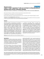
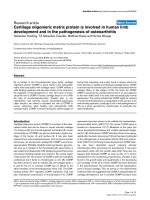
Figure 10
Diagram summarizing major events in diosgenin-induced apoptosis in human rheumatoid arthritis (RA) fibroblast-like synoviocytes (FLS)Diagram summarizing major events in diosgenin-induced apoptosis in
human rheumatoid arthritis (RA) fibroblast-like synoviocytes (FLS). The
effect of diosgenin is associated with a loss of mitochondrial membrane
potential (∆ψm), caspase-3 activation, and DNA fragmentation. Further-
more, diosgenin causes an inhibition of human RA FLS cell growth with
apoptosis induction associated with up-regulation of cyclooxygenase-2
(COX-2). Celecoxib, a selective COX-2 inhibitor, provokes a large
decrease in diosgenin-induced apoptosis whereas IL-1β, a COX-2
inducer, strongly increases diosgenin-induced apoptosis of human RA
FLS. These new studies provide strong evidence that modulation of
COX-2 is associated with diosgenin-induced human RA FLS death. As
exogenous prostaglandin E
2
(PGE
2
) alone did not induce synoviocyte
apoptosis, the exact mechanism by which endogenous PGE
2
sensitizes
human RA FLS to cell death is still not clear.
Available online />R383
24. Cruz-Correa M, Hylind LM, Romans KE, Booker SV, Giardiello FM:
Long-term treatment with sulindac in familial adenomatous
polyposis: a prospective cohort study. Gastroenterology 2002,
122:641-645.
25. Baier A, Meineckel I, Gay S, Pap T: Apoptosis in rheumatoid
arthritis. Curr Opin Rheumatol 2003, 15:274-279.
26. Arnett FC, Edworthy SM, Bloch DA, McShane DJ, Fries JF, Cooper
NS, Healey LA, Kaplan SR, Liang MH, Luthra HS, Medsger TA,
Mitchell DM Jr, Neustadt DH, Pinals RS, Schaller JG, Sharp JT,
Wilder RL, Hunder GG: The American Rheumatism Association
1987 revised criteria for the classification of rheumatoid
arthritis. Arthritis Rheum 1988, 31:315-324.
27. Glant TT, Jacobs JJ, Molnar G, Shanbhag AS, Valyon M, Galante
JO: Bone resorption activity of particulate-stimulated
macrophages. J Bone Miner Res 1993, 8:1071-1079.
28. Bonnet C, Bertin P, Cook-Moreau J, Chable-Rabinovitch H, Treves
R, Rigaud M: Lipoxygenase products and expression of 5-
lipoxygenase and 5-lipoxygenase-activating protein in human
cultured synovial cells. Prostaglandins 1995, 50:127-135.
29. Moalic S, Liagre B, Labrousse F, Beneytout JL: Enhanced apop-
tosis in retrovirally transfected osteosarcoma cells after expo-
sure to sodium butyrate. Int J Oncol 2000, 16:695-700.
30. Smiley ST, Reers M, Mottola-Hartshorn C, Lin M, Chen A, Smith
TW, Steele GD Jr, Chen LB: Intracellular heterogeneity in mito-
chondrial membrane potentials revealed by a J-aggregate-
forming lipophilic cation JC-1. Proc Natl Acad Sci USA 1991,
88:3671-3675.
31. Duval R, Bellet V, Delebassee S, Bosgiraud C: Implication of cas-
pases during maedi-visna virus-induced apoptosis. J Gen Virol
2002, 83:3153-3161.
32. Moalic S, Liagre B, Le Bail JC, Beneytout JL: Dose-dependent
modulation of apoptosis and cyclooxygenase-2 expression in
human 1547 osteosarcoma cells by NS-398, a selective
cyclooxygenase-2 inhibitor. Int J Oncol 2001, 18:533-540.
33. Accatino L, Pizarro M, Solis N, Koenig CS: Effects of diosgenin,
a plant-derived steroid, on bile secretion and hepatocellular
cholestasis induced by estrogens in the rat. Hepatology 1998,
28:129-140.
34. Turchan J, Pocernich CB, Gairola C, Chauhan A, Schifitto G, But-
terfield DA, Buch S, Narayan O, Sinai A, Geiger J, Berger JR, Elford
H, Nath A: Oxidative stress in HIV demented patients and pro-
tection ex vivo with novel antioxidants. Neurology 2003,
60:307-314.
35. Moalic S, Liagre B, Corbiere C, Bianchi A, Dauca M, Bordji K,
Beneytout JL: A plant steroid, diosgenin, induces apoptosis,
cell cycle arrest and COX activity in osteosarcoma cells. FEBS
Lett 2001, 506:225-230.
36. Corbiere C, Liagre B, Bianchi A, Bordji K, Dauca M, Netter P,
Beneytout JL: Different contribution of apoptosis to the antipro-
liferative effects of diosgenin and other plant steroids, heco-
genin and tigogenin, on human 1547 osteosarcoma cells. Int J
Oncol 2003, 22:899-905.
37. Thornberry NA, Lazebnik Y: Caspases: enemies within. Science
1998, 281:1312-1316.
38. Callsen D, Brune B: Role of mitogen-activated protein kinases
in S-nitrosoglutathione-induced macrophage apoptosis. Bio-
chemistry 1999, 38:2279-2286.
39. Skulachev VP: Why are mitochondria involved in apoptosis?
Permeability transition pores and apoptosis as selective
mechanisms to eliminate superoxide-producing mitochondria
and cell. FEBS Lett 1996, 397:7-10.
40. Itoh K, Hase H, Kojima H, Saotome K, Nishioka K, Kobata T: Cen-
tral role of mitochondria and p53 in Fas-mediated apoptosis of
rheumatoid synovial fibroblasts. Rheumatology 2003,
43:277-285.
41. Kemick ML, Chin JE, Wuthier RE: Role of prostaglandins in dif-
ferentiation of growth plate chondrocytes. Adv Prostaglandin
Thromboxane Leukot Res 1989, 19:423-426.
42. Pelletier JP, Fernandes JC, Jovanovic DV, Reboul P, Martel-Pelle-
tier J: Chondrocyte death in experimental osteoarthritis is
mediated by MEK 1/2 and p38 pathways: role of cyclooxygen-
ase-2 and inducible nitric oxide synthase. J Rheumatol 2001,
28:2509-2519.
43. Jovanovic DV, Mineau F, Notoya K, Reboul P, Martel-Pelletier J,
Pelletier JP: Nitric oxide induced cell death in human osteoar-
thritic synoviocytes is mediated by tyrosine kinase activation
and hydrogen peroxide and/or superoxide formation. J
Rheumatol 2002, 29:2165-2175.
44. Simon LS, Weaver AL, Graham DY, Kivitz AJ, Lipsky PE, Hubbard
RC, Isakson PC, Verburg KM, Yu SS, Zhao WW, Geis GS: Anti-
inflammatory and upper gastrointestinal effects of celecoxib in
rheumatoid arthritis: a randomized controlled trial. JAMA
1999, 282:1921-1928.
45. Silverstein FE, Faich G, Goldstein JL, Simon LS, Pincus T, Whelton
A, Makuch R, Eisen G, Agrawal NM, Stenson WF, Burr AM, Zhao
WW, Kent JD, Lefkowith JB, Verburg KM, Geis GS: Gastrointes-
tinal toxicity with celecoxib vs nonsteroidal anti-inflammatory
drugs for osteoarthritis and rheumatoid arthritis: the CLASS
study: A randomized controlled trial. Celecoxib Long-Term
Arthritis Safety Study. JAMA 2000, 284:1247-1255.
46. Kusunoki N, Yamazaki R, Kawai S: Induction of apoptosis in
rheumatoid synovial fibroblasts by celecoxib, but not by other
selective cyclooxygenase 2 inhibitors. Arthritis Rheum 2002,
46:3159-3167.
47. Warner TD, Giuliano F, Vojnovic I, Bukasa A, Mitchell JA, Vane JR:
Nonsteroid drug selectivities for cyclo-oxygenase-1 rather
than cyclo-oxygenase-2 are associated with human gastroin-
testinal toxicity: a full in vitro analysis. Proc Natl Acad Sci USA
1999, 96:7563-7568.