Báo cáo y học: "Improved cartilage integration and interfacial strength after enzymatic treatment in a cartilage transplantation model" potx
Bạn đang xem bản rút gọn của tài liệu. Xem và tải ngay bản đầy đủ của tài liệu tại đây (685.42 KB, 8 trang )
Open Access
Available online />R469
Vol 6 No 5
Research article
Improved cartilage integration and interfacial strength after
enzymatic treatment in a cartilage transplantation model
Jarno van de Breevaart Bravenboer
1
, Caroline D In der Maur
2
, P Koen Bos
1
, Louw Feenstra
2
,
Jan AN Verhaar
1
, Harrie Weinans
1
and Gerjo JVM van Osch
1,2
1
Erasmus Orthopaedic Research Laboratory, Department of Orthopaedics, Erasmus University Medical Center, Rotterdam, The Netherlands
2
Department of Otorhinolaryngology, Erasmus University Medical Center, Rotterdam, The Netherlands
Corresponding author: Gerjo JVM van Osch,
Received: 18 Mar 2004 Revisions requested: 4 May 2004 Revisions received: 30 May 2004 Accepted: 23 Jun 2004 Published: 6 Aug 2004
Arthritis Res Ther 2004, 6:R469-R476 (DOI 10.1186/ar1216)
http://arthr itis-research.com/conte nt/6/5/R469
© 2004 van de Breevaart Bravenboer et al.; licensee BioMed Central Ltd. This is an Open Access article: verbatim copying and redistribution of this
article are permitted in all media for any purpose, provided this notice is preserved along with the article's original URL.
Abstract
The objective of the present study was to investigate whether
treatment of articular cartilage with hyaluronidase and
collagenase enhances histological and mechanical integration
of a cartilage graft into a defect. Discs of 3 mm diameter were
taken from 8-mm diameter bovine cartilage explants. Both discs
and annulus were either treated for 24 hours with 0.1%
hyaluronidase followed by 24 hours with 10 U/ml collagenase or
left untreated (controls). Discs and annulus were reassembled
and implanted subcutaneously in nude mice for 5 weeks.
Integration of disc with surrounding cartilage was assessed
histologically and tested biomechanically by performing a push-
out test. After 5 weeks a significant increase in viable cell counts
was seen in wound edges of the enzyme-treated group as
compared with controls. Furthermore, matrix integration
(expressed as a percentage of the total interface length that was
connected; mean ± standard error) was 83 ± 15% in the treated
samples versus 44 ± 40% in the untreated controls. In the
enzyme-treated group only, picro-Sirius Red staining revealed
collagen crossing the interface perpendicular to the wound
surface. Immunohistochemical analyses demonstrated that the
interface tissue contained cartilage-specific collagen type II.
Collagen type I was found only in a small region of fibrous tissue
at the level of the superficial layer, and collagen type III was
completely absent in both groups. A significant difference in
interfacial strength was found using the push-out test: 1.32 ±
0.15 MPa in the enzyme-treated group versus 0.84 ± 0.14 MPa
in the untreated controls. The study shows that enzyme
treatment of cartilage wounds increases histological integration
and improves biomechanical bonding strength. Enzymatic
treatment may represent a promising addition to current
techniques for articular cartilage repair.
Keywords: cartilage integration, cartilage repair, enzyme, push-out test
Introduction
Localized articular cartilage defects are a major problem for
orthopaedic surgeons. Because cartilage has poor ability
to heal because of lack of intrinsic repair capacity [1-3],
chondral defects do not heal and may increase the risk for
early osteoarthritis. A number of different treatment tech-
niques, such as subchondral penetration [4-6], osteochon-
dral transplantation and mosaïcplasty [7-9], perichondrium
covering of the defect [10,11] and autologous chondrocyte
transplantation [12,13], as well as various enzymatic treat-
ment techniques [14-17], have been tried in either clinical
or laboratory settings in an attempt to restore the articular
surface. Until now none of these techniques has resulted in
long-term, durable and a predictable repair of the articular
cartilage. Many researchers focus on the production, or
local induction, of hyaline-like cartilage; however, these
techniques are generally not directly aimed at local integra-
tion with the surrounding healthy cartilage. Variable and
suboptimal wound healing and integration may be a cause
of potential failure of otherwise promising techniques.
Injury to cartilage results in the formation of an acellular and
thus metabolically inactive zone adjacent to the wound
interface [18-20], thereby prohibiting significant matrix
deposition at the wound interface area and subsequently
limiting integration. Ideally, the biochemical composition of
the integrative matrix should equal that of native cartilage,
with high contents of collagen type II and proteoglycans,
and low amounts of collagen types I and III. Furthermore,
the biomechanical properties of the interfacial tissue
should be within the range of native cartilage in order to
prevent excessive strain [21] and mechanical failure.
Arthritis Research & Therapy Vol 6 No 5 van de Breevaart Bravenboer et al.
R470
We previously showed that enzymatic treatment with
hyaluronidase and collagenase increased cell density at the
wound edges of cartilage explants after 2 weeks of in vitro
culture [22]. This treatment method could improve cartilage
integration in chondral defects and potentially could confer
benefit in clinical applications. In the present study we used
enzymatic treatment with hyaluronidase and collagenase,
and tested how this would affect wound healing and carti-
lage integration in terms of matrix composition and biome-
chanical properties. Specifically, we applied a combination
hyaluronidase and collagenase treatment on both sides of
a cartilage explant, and tested the effect of this treatment
on cell viability at the wound edge, production of collagens
types I, II and III, collagen fibre orientation, and biomechan-
ical bonding strength.
Methods
Articular cartilage samples were harvested from the meta-
carpo-phalangeal joints of calves aged 6–12 months. Full-
thickness cartilage explants of 8 mm diameter and with a
thickness of 0.9–1.2 mm were prepared using a dermal
biopsy punch and scalpel. The explants were then ran-
domly divided into two groups. From the centre of the
explants, 3-mm cores were punched out, using a custom
built device to ensure punching in the exact middle of the
explant. Group 1 (n = 12) specimens (both outer ring and
inner core) were incubated for 24 hours in 0.1% hyaluroni-
dase type I-S from bovine testes (Sigma-Aldrich Chemie
BV, Zwijndrecht, The Netherlands) followed by 24 hours in
10 U/ml highly purified collagenase VII (Sigma-Aldrich
Chemie BV), both in Dulbecco's modified eagle's medium/
Hams' F12 with 2% foetal calf serum. Specimens from
group 2 (controls; n = 12) were incubated in Dulbecco's
modified eagle's medium/Ham's F12 culture medium
(Gibco, Grand Island, NY, USA) supplemented with 2%
foetal calf serum at 37°C for 48 hours (controls). The
choices of enzymes, enzyme concentrations and treatment
times were based on the findings from our previous in vitro
study [22]. After 48 hours the samples were washed three
times for 10 min in culture medium, and the 3-mm inner
cores were reimplanted in their accompanying 8-mm outer
rings. Constructs were then implanted in four subcutane-
ous pockets on the backs of six nude mice (BALB-C nu/nu;
Harlan, Horst, The Netherlands), for which approval was
obtained from the local animal ethical committee (DEC
no.126-01-01). Each mouse carried two enzyme-treated
constructs (group 1) and two control constructs (group 2).
After 5 weeks the mice were killed by cervical dislocation
and constructs were harvested.
Histology
From each mouse, one control and one enzyme-treated
construct were processed for histology. Constructs were
divided into two halves. One half was fixed in 4% phos-
phate-buffered formalin and embedded in paraffin, and the
other half was frozen in liquid nitrogen and stored at -80°C
for later cryosection preparation. Sections (6 µm) were cut
using a standard microtome (paraffin) or cryomicrotome
and mounted on Starfrost
®
slides (Knittel, Braunschweig,
Germany). Paraffin sections were haematoxylin and eosin
stained as well as immunostained for collagen type II. Cry-
osections were used for thionin (proteoglycan) stain, picro-
Sirius Red stain and immunohistochemical stains for pro-
collagen type I, collagen type I and collagen type III.
Evaluation of chondrocyte viability
The number of vital chondrocytes was counted in surface,
middle and deep zones in both wounded and unwounded
areas using haematoxylin and eosin coloured slides at
400× magnification using a 50 × 50 µm boxed grid.
Nuclear and cytoplasmic changes, as described by Kim
and coworkers [23], were analyzed to assess cell viability/
death. Only cells with visible nuclei were evaluated. Values
for wounded cartilage were scored from a 150-µm broad
band on both sides of the lesion, and the values for both
sides of the interface were then averaged to obtain one
value per interface. Furthermore, the cartilage was divided
into superficial (100 µm from the surface down), deep (150
µm from the bottom up) and middle (between superficial
and deep) zones, as previously described [22]. For com-
parison, values from unwounded tissue were obtained from
the middle part of the outer ring, which is more then 1 mm
from the wound edge. Chondrocyte densities are repre-
sented as vital cells/mm
2
.
For each explant the amount of viable chondrocytes was
calculated from the values obtained from two to four sec-
tions. Subsequently, the averages for the control and
enzyme-treated groups were calculated and used for statis-
tical evaluation.
Evaluation of integration
Cryosections were fixed in acetone and stained with
0.04% thionin in 0.01 M sodium acetate for 5 min. For each
sample we assessed the percentage of total interface
length that had a matrix–matrix connection using a micro-
scope with a 50 µm square grid. A clear distinction could
be made between parts with a matrix connection and parts
of the cartilage touching each other but without a clearly
connected matrix, which were scored as parts with a gap.
Interface integration percentages were obtained from
measurements of two to four different sections from each
sample, resulting in one average value for each interface.
Picro-Sirius Red stain
Cryosections were fixed in acetone and stained with 0.1%
Sirius Red F3BA (Direct Red 80; Fluka Chemie, Zwijn-
drecht, The Netherlands) in a saturated picric acid solution
for 1 hour. Brief washing in 0.1% acetic acid was followed
by rapid dehydration in 100% alcohol (three changes for 3
Available online />R471
min each), after which a xylene bath (two changes for 5 min
each) was used to prepare the slides for mounting with
Entellan (Merck, Darmstadt, Germany). Slides were ana-
lyzed using a polarized light microscope (Dialux 20; Leitz,
Wetzlar, Germany) to evaluate fibre orientation in the inter-
face area. The relative sign of birefringence was deter-
mined using the analyzer filter. For semiquantitative
analyses, samples from both groups were classified as fol-
lows: 0 = no fibres crossing; 1 = occasional fibre crossing;
and 2 = many fibres crossing.
Collagen immunostaining
Cryosections were fixed in acetone for procollagen type I,
and collagen types I and III staining. For collagen type II
staining paraffin sections were deparaffinized using xylene
and rehydrated through a graded series of ethanol, after
which they were incubated with 0.2% pronase for 30 min
to retain antigenicity. Treatment with 1% hyaluronidase
(Sigma-Aldrich Chemie BV) was used to unmask the
epitopes. Nonspecific binding was blocked using 10% nor-
mal goat serum (CLB, Amsterdam, The Netherlands) fol-
lowed by incubation with the respective antibody for 2
hours. Antibodies used were M38 and II-6B3 (both 1:100;
Developmental Studies Hybridoma Bank) for procollagen
type I and collagen type II, respectively; ab6308 (mouse
monoclonal IgG antibody, 1:500; Abcam Ltd, Cambridge,
UK) for collagen type I; and ab6310 (mouse monoclonal
IgG antibody, 1:500, Abcam Ltd) for collagen type III. All
primary antibodies were previously complexed with goat
Fab fragment against mouse conjugated with alkaline phos-
phatase (GAMAP, 1:400; Immunotech, Marseilles, France)
at 4°C overnight. After coupling, 0.1% normal mouse
serum was used for 2 hours before usage to capture the
unbound GAMAP, after which the antibody solution was
used on the slides. Sections were subsequently incubated
for 30 min with alkaline phosphatase anti-alkaline phos-
phatase (APAAP, 1:100 for procollagen I and collagen II,
1:75 for collagen types I and III; Dakopatts, Copenhagen,
Denmark). New Fuchsine substrate (Chroma, Kongen,
Germany) was used for colour development and haematox-
ylin for counterstaining, after which slides were mounted
using Vectamount (Vecto Laboratories Inc., Burlingame,
CA, USA). Negative controls were subjected to the same
protocol with omission of the primary antibody.
Mechanical testing
After harvesting of the constructs, the surrounding fibrotic
tissue was carefully removed. From each of the six mice,
one control and one enzyme-treated construct were frozen
using liquid nitrogen and stored in airtight tubes at -80°C
for later mechanical testing. Immediately before testing
constructs were slowly thawed in airtight tubes. Thickness
of the sample was measured to an accuracy of 50 µm using
calipers. Constructs were then mounted in a specially
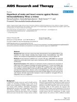
designed push-out setup (Fig. 1) on a materials testing
machine (LRX; Lloyd Instruments, Fareham, UK) equipped
with a 500 N load cell. Push-out tests were performed by
leading the push-out rod on top of the 3 mm inner core
through the specimen at 10 µm/s. During the test con-
structs were kept moist by adding a few drops of phos-
phate-buffered saline on top before starting the test, which
on average took 4–5 min. During the test both displace-
ment and load were monitored at a sample frequency of 18
Hz and the output of these values was read out and stored
on a desktop computer. For each specimen the peak load-
to-failure (maximum observed load) was used to calculate
the interface stress-to-failure (maximum load normalized to
interface area) as a representative marker of interfacial
strength. Furthermore, we also performed push-through
tests of intact cartilage for comparison (n = 4) and push-
out tests of constructs immediately after reassembly of
core and annulus (n = 8) to determine the friction compo-
nent of our setup; all this was done in a manner similar to
that described above.
Statistical analysis
Values shown are mean ± standard deviation unless other-
wise specified. Statistical analyses for both viable cell
count and mechanical testing were done using Student's t-
test for independent samples. Matrix integration scores and
results from polarized light microscopy were analyzed using
the Mann–Whitney U test. P ≤ 0.05 was considered statis-
tically significant.
Results
Histology
Cell counts in the integration area revealed significantly
more vital cells near the wound edges in the enzyme-
treated group than in the untreated group in all three layers
(Table 1), with the largest increase in the superficial layer.
Figure 1
Schematic representation of the push-out setupSchematic representation of the push-out setup. Displacement trans-
ducer and load cell are connected to the push-out rod.
Arthritis Research & Therapy Vol 6 No 5 van de Breevaart Bravenboer et al.
R472
Many cells were located in the interface region of the
enzyme-treated group (Fig. 2a,2b), but despite the appar-
ently normal average vital cell count in the 150-µm broad
band in the untreated control samples, the tissue in the
interface region was almost acellular (Fig. 2c,2d). Measure-
ment of matrix integration on thionin stained sections (Fig.
3) revealed an average matrix–matrix connection percent-
age of 83 ± 15% of wound interface length in the enzyme-
treated constructs, as compared with 44 ± 40% in the
untreated group (P < 0.05), with variability between sec-
tions of the same interface typically being less then 15%.
To assess the quality of the newly formed interface matrix
we evaluated which types of collagen were present in this
new tissue. Immunohistochemical staining revealed the
presence of limited amounts of (pro-)collagen type I in the
interfaces, which was limited to the area of ingrowth of
fibrous tissue from the top surface (Fig. 4a; four out of 10
interfaces in the treated group and three out of 10 inter-
faces in the control group). Typically, this ingrowth was
around 10% of the interface length, with Fig. 4a showing
the worst case. Furthermore, an abundance of cartilage-
specific collagen type II was found in all interfacial matrices
(Fig. 4b), whereas no collagen type III was found in any of
the interface areas (Fig. 4c). No clear differences in immu-
nohistochemical staining were observed between the two
groups.
Polarized light microscopy of picro-Sirius Red stained sec-
tions indicated that collagen fibres in the wound interface
were mainly directed perpendicular to the interface. Many
fibres were seen crossing the interface in three out of five
treated samples and in none of the control samples. Occa-
sional fibre crossing was observed in two out of five treated
samples and in three out of five control samples; in two out
of five control samples no fibre crossing was observed.
Most of the perpendicularly running fibres in the untreated
control group protruded only into the interface (Fig. 5). Sta-
tistical analyses showed a significant difference between
groups (P < 0.05).
Table 1
Effect of enzymatic treatment on cell viability in cartilage wound
edges 5 weeks after subcutaneous implantation in nude mice
Zone Unwounded Not enzyme treated Enzyme treated
Surface 1440 ± 175 1151 ± 133 2316 ± 209*
Middle 787 ± 160 866 ± 27 1097 ± 59*
Deep 646 ± 75 589 ± 16 960 ± 45*
Cartilage was treated with hyaluronidase and collagenase or left
untreated and implanted subcutaneously into nude mice for 5 weeks.
The number of vital cells were counted in a 150-mm broad band
along both sides of the wound edges, as well as in unwounded
control areas in surface, middle and deep zones. *P < 0.05 versus
unwounded areas and non-enzyme-treated wound areas.
Figure 2
HE-stained sections of enzyme-treated and untreated control con-structs 5 weeks after implantationHE-stained sections of enzyme-treated and untreated control con-
structs 5 weeks after implantation. (a) Enzyme treated construct that
shows good integration, with cells located in the interfacial region [(b)
enlargement]. (c) Untreated construct that shows a poor integration,
with no cells present in the interfacial tissue [(d) enlargement]. Magnifi-
cations: panels a and c 100×; panels b and d 200×.
Figure 3
Thionin stained sections 5 weeks after implantation in nude miceThionin stained sections 5 weeks after implantation in nude mice. (a)
Enzyme treated and (b) untreated control section. Note the clear differ-
ence in thionin staining of the interfacial tissue between the enzyme-
treated and control section. Interfaces are encircled.
Available online />R473
Mechanical testing
Mechanical assessment of the cartilage interface between
inner core and outer ring by push-out test revealed that the
interface connection was stronger in the treated group; the
enzyme-treated group exhibited a 58% increase in stress-
to-failure over the untreated controls (1.32 ± 0.15 MPa
versus 0.84 ± 0.14 MPa). Average force–displacement
curves, including standard errors, are shown in Fig. 6.
Furthermore, the push-through strength of intact articular
cartilage was 8.8 ± 0.52 MPa, with failure occurring in an
annular manner, as with the integrated constructs. Push-
out tests performed immediately after reinsertion of the
core into the annulus revealed a maximum friction stress of
22.2 ± 9.4 kPa, which is only 1.7–2.6% of the stress meas-
ured in the integrated constructs.
Discussion
In the present study we found an improvement in histologi-
cal and biomechanical integration of articular cartilage after
treatment with a combination of hyaluronidase and
collagenase, a protocol that was previously shown to
increase chondrocyte densities in wound edges in vitro
[22]. Our setup of a 3-mm disc placed in an annulus pro-
vides a reasonable representation of the in vivo situation, in
which cartilage is transplanted into a defect with wound
edges perpendicular to the surface. Because an in vitro
culture system might not provide the optimal environment
for tissue growth and repair [24], we decided to perform
our experiments in the well established nude mouse model
[25,26], creating an environment in which there is an ample
supply of nutrients.
In this setup, cellularity in nontreated wound edges
reached the levels of unwounded cartilage, and cellularity
of unwounded cartilage was increased to a level similar to
that before implantation, which is in contrast to results from
our previous in vitro study [22]. We believe that this is due
to the nutrient-rich in vivo environment. However, in this
model we confirmed [22] that the enzymatic treatment pro-
tocol enhanced the number of cells near the wound edges
as compared with nontreatment, and resulted in better
histological integration, as assessed by the percentage of
matrix connection in the interfacial area. Furthermore, the
repair tissue exhibited collagen fibres crossing the wound
edges, and the matrix in both experimental groups exhibited
cartilage specific collagen type II, limited (pro-)collagen
type I and no collagen type III. This improved integration fol-
lowing enzymatic treatment was further supported by push-
out tests, which are similar to tests described by others
[24,27].
Although enzymatic treatment significantly increased
mechanical strength to 1.32 MPa, the interfacial strength
was still almost sevenfold less than the 8.8 MPa intrinsic
failure strength values observed for intact cartilage. It
should be appreciated that the fairly simple normalization to
interface area is a rather crude method because the inter-
face stress is not uniformly distributed. Therefore, tests
using different sizes or shapes of specimens cannot readily
be compared. Because the average thickness of our sam-
ples was 1.14 ± 0.28 mm for the treated group and 1.14 ±
0.21 mm for controls, and no correlation could be found
between sample thickness and failure strength, we may
compare strength values within the present study.
Figure 4
Immunohistochemical stainings for collagens present in the interfacial area of enzyme-treated constructsImmunohistochemical stainings for collagens present in the interfacial area of enzyme-treated constructs. (a) Collagen type I, with light staining (in
red) in the area of fibrous ingrowth (circled). (b) Collagen type II, showing medium intensity staining (in red) in the entire matrix of the interfacial area.
(c) Collagen type III; staining (in red) only present in the surrounding capsule.
Arthritis Research & Therapy Vol 6 No 5 van de Breevaart Bravenboer et al.
R474
Our findings indicate a relation between interfacial strength
and cellular activity at the interface. This confirms the
results reported by DiMicco and coworkers [28], who used
fetal, calf and adult bovine cartilage; after 14 days of culture
those investigators found the highest failure stress in calf
cartilage at 77 kPa in a single lap shear test. However,
Reindel and coworkers [29] found an interface strength of
34 kPa after 3 weeks of culture, and showed that integra-
tive strength was highly dependent on the use of fetal
bovine serum in culture, which can influence cellular activ-
ity. Dependence of integration on active cell processes is
also demonstrated by lack of adhesive strength when com-
bining two lyophilized explant blocks [30]. In an 8-week
bioreactor culture of tissue engineered cartilage core con-
structs with surrounding native cartilage, Obradovic and
coworkers [24] found better mechanical integration of very
young (5 days) constructs (254 kPa) as compared with
more mature constructs (5 weeks; approximating 150 kPa).
Peretti and coworkers [26] also used lyophilized explants,
which were seeded with chondrocytes and then held
together using fibrin glue and placed subcutaneously in
nude mice for up to 6 weeks. Tensile testing showed a
clear increase of failure strength to 77 kPa, which is 10
times higher than unseeded control explants, with failure
always occurring at the interface between new tissue and
devitalized matrix. Because cellular activity is clearly an
important factor in integration, we should appreciate that in
most studies young bovine cartilage is used, which is more
cellular than human cartilage. In a previous study, however,
we did see similar effects of enzymatic treatment on cell
density in human adult articular cartilage [22]. It can be
anticipated that, because of the lower cell numbers, the
overall repair process might be slower than in the present
study but can still be stimulated using enzyme treatment.
Our findings suggest that enzymatic treatment may be a
promising technique with which to improve cartilage inte-
gration, in addition to currently developing clinical and
experimental articular cartilage repair techniques. The cell
counts along the wound edge in the control group were
comparable to those of native tissue. However, a close look
at the histological pictures (Fig. 2c,2d) shows that a thin
acellular band is still visible. In the enzyme-treated group
cell counts were even higher than those in native cartilage,
and histology did not reveal a large acellular band, as seen
in the controls (Fig. 2a,2b), thus fulfilling one of the
prerequisites for integration, namely the presence of active
Figure 5
Picro-Sirius Red stained section of the interface regions (circled)Picro-Sirius Red stained section of the interface regions (circled). (a, b)
Enzyme treated group, well integrated. (c, d) Untreated control group,
not integrated. In panels a and c show crossed polarizing filters without
analyzer filter; fibres run in parallel and perpendicular directions relative
to the interface. Note the squares around individual chondrocytes, sig-
nifying pericellular collagen shell (arrowheads). In panels b and d the
same field of view is shown as in panels a and c, but this time with the
analyzer filter in place, revealing only those fibres that run in a perpen-
dicular direction relative to the interface(circled), pointed out by the fact
that the pericellular fibres that run in the parallel direction have disap-
peared (arrowheads), as well as the lightening up of the superficial car-
tilage layer. Clearly visible are the fibres crossing through the interface
area, thus connecting both pieces of cartilage (panel b) and fibres
along the wound edge projecting into the interface area (panel d). Orig-
inal magnification: 25×.
Figure 6
Average force–displacement curves of push-out tests with standard error bars for untreated (n = 5) and enzyme-treated (n = 6) curves, respectivelyAverage force-displacement curves of push-out tests with standard
error bars for untreated (n = 5) and enzyme-treated (n = 6) curves,
respectively. Failure strength in the enzyme-treated group was signifi-
cantly higher (+58%). The failure of the curve to return to zero can be
explained by friction between pushed-out core and sample holder.
Available online />R475
chondrocytes close to the lesion site. The high cellularity at
the wound edge observed in the present study probably
resulted in the increased collagen fibre deposition across
the wound gap of adjacent cartilage surfaces, as shown in
the picro-Sirius Red slides (Fig. 5). Normally, cross-gap
deposition of collagen between native and repair tissue is
insufficient in the reparative process that occurs after full-
thickness defects [2,31,32]. The observed cross-gap dep-
osition of collagen in the present study coincides with
increased interfacial strength, as shown previously in
integration experiments using fetal, calf and adult bovine
cartilage explants [28]. Those studies showed a correlation
between increased adhesive strength and an increased
hydroxyproline incorporation in the interface area. Further-
more, inhibition of collagen cross-link formation by β-amino-
propionitrile resulted in almost complete loss of integrative
repair.
The explanation for the success of the enzymatic treatment
technique may be found by examining wound healing in
vascularized tissues. In nonvascularized articular cartilage,
proteolytic enzyme activity is either lacking or insufficient to
degrade and remove the observed acellular band in the
wounded areas, as occurs with debris and necrotic tissue
in vascularized tissues. Application of enzymes may remove
this layer, uncovering an activated area of chondrocytes
that are capable of integration. Another possible underlying
mechanism of this enzymatic treatment may be the partial
degradation of extracellular matrix surrounding the wound
edge chondrocytes, which frees chondrocytes from the
tight extracellular matrix in which they were entrapped.
Because chondrocytes have been shown to have the ability
to migrate [33], this may enable them to move to the wound
edge in need of repair. A third possible mechanism of the
enzymatic degradation of wound edge extracellular matrix
may be the stimulation of local chondrocyte proliferation,
which can be seen by looking at the cell clusters in the his-
tological images (e.g. Fig. 2). Although we did observe cell
division, the exact mechanism by which the enzymatic treat-
ment exerts its effects is still unclear. A more detailed
mechanistic study is needed to further elucidate this.
In the present study we demonstrated the potential of
hyaluronidase and collagenase treatment in a screening 'in
vivo' environment. Animal experiments with actual articular
cartilage defects are needed to determine the value of our
findings. Further studies must be undertaken to optimize
the enzymatic treatment protocol (e.g. shorter treatment
duration) and learn more about the mechanisms involved,
such as cell migration to the wound area and matrix depo-
sition, and to improve mechanical interface strength further
to the level of intact cartilage, which is still almost an order
of magnitude higher. Therefore, longer term studies are
required to judge the success of different integration
enhancing techniques against the mechanical strength of
intact cartilage, and to develop protocols that may become
clinically applicable, which in our view could be a valuable
addition to existing repair strategies.
Conclusion
The present study shows that enzymatic treatment of carti-
lage wounds increases histological integration and
improves biomechanical bonding strength. Enzymatic treat-
ment may represent a promising addition to current tech-
niques for articular cartilage repair.
Competing interests
None declared.
Acknowledgements
Monoclonal antibodies II-6B3 and M38 were obtained from the Devel-
opmental Studies Hybridoma Bank, which is maintained by the Depart-
ment of Pharmacology and Molecular Sciences, Johns Hopkins
University School of Medicine, Baltimore, Maryland, USA and the
Department of Biological Sciences, University of Iowa, Iowa City, Iowa,
USA under contract N01-HD-6-2915 from the National Institute of Child
Health and Human Development. The authors thank Nicole Kops for
immunohistochemistry, Inge van Rensen for her work on cell counting,
and Corrina de Ridder for her help in the nude mice experiments. Further
thanks go to the company T. Boer & Zn., Nieuwerkerk a/d IJssel, The
Netherlands, in particular Ton Looijen, for their kind supply of bovine
joints.
References
1. Hunter W: On the structure and diseases of articulating
cartilages. Trans R Soc Lond 1743, 42B:514-521.
2. Newman AP: Articular cartilage repair. Am J Sports Med 1998,
26:309-324.
3. Hunziker EB: Articular cartilage repair: basic science and clini-
cal progress. A review of the current status and prospects.
Osteoarthritis Cartilage 2002, 10:432-463.
4. Pridie KH: A method of resurfacing osteoarthritic knee joints. J
Bone Joint Surg Br 1959, 41:618-619.
5. Muller B, Kohn D: Indication for and performance of articular
cartilage drilling using the Pridie method [in German]. Ortho-
pade 1999, 28:4-10.
6. Insall J: The Pridie debridement operation for osteoarthritis of
the knee. Clin Orthop 1974, 101:61-67.
7. Hangody L, Kish G, Karpati Z, Udvarhelyi I, Szigeti I, Bely M: Mosa-
icplasty for the treatment of articular cartilage defects: appli-
cation in clinical practice. Orthopedics 1998, 21:751-756.
8. Hangody L, Fules P: Autologous osteochondral mosaicplasty
for the treatment of full-thickness defects of weight-bearing
joints: ten years of experimental and clinical experience. J
Bone Joint Surg Am 2003, Suppl 2:25-32.
9. Feczko P, Hangody L, Varga J, Bartha L, Dioszegi Z, Bodo G,
Kendik Z, Modis L: Experimental results of donor site filling for
autologous osteochondral mosaicplasty. Arthroscopy 2003,
19:755-761.
10. Homminga GN, Bulstra SK, Bouwmeester PS, van der Linden AJ:
Perichondral grafting for cartilage lesions of the knee. J Bone
Joint Surg Br 1990, 72:1003-1007.
11. Bouwmeester P, Kuijer R, Terwindt-Rouwenhorst E, van der
Linden T, Bulstra S: Histological and biochemical evaluation of
perichondrial transplants in human articular cartilage defects.
J Orthop Res 1999, 17:843-849.
12. Brittberg M, Lindahl A, Nilsson A, Ohlsson C, Isaksson O, Peter-
son L: Treatment of deep cartilage defects in the knee with
autologous chondrocyte transplantation. N Engl J Med 1994,
331:889-895.
13. Brittberg M, Peterson L, Sjogren-Jansson E, Tallheden T, Lindahl
A: Articular cartilage engineering with autologous
Arthritis Research & Therapy Vol 6 No 5 van de Breevaart Bravenboer et al.
R476
chondrocyte transplantation. A review of recent developments.
J Bone Joint Surg Am 2003, Suppl 3:109-115.
14. Caplan AI, Elyaderani M, Mochizuki Y, Wakitani S, Goldberg VM:
Principles of cartilage repair and regeneration. Clin Orthop
1997, 342:254-269.
15. Lee MC, Sung KL, Kurtis MS, Akeson WH, Sah RL: Adhesive
force of chondrocytes to cartilage. Effects of chondroitinase
ABC. Clin Orthop 2000, 370:286-294.
16. Hunziker EB, Kapfinger E: Removal of proteoglycans from the
surface of defects in articular cartilage transiently enhances
coverage by repair cells. J Bone Joint Surg Br 1998,
80:144-150.
17. Quinn TM, Hunziker EB: Controlled enzymatic matrix degrada-
tion for integrative cartilage repair: effects on viable cell den-
sity and proteoglycan deposition. Tissue Eng 2002, 8:799-806.
18. Hunziker E, Quinn T: Surgical removal of articular cartilage
leads to loss of chondrocytes from the wound edges. Trans
46th meeting of Orthop Res Soc, Orlando USA 2000:185.
19. Bos PK, van Osch GJ, Frenz DA, Verhaar JA, Verwoerd-Verhoef
HL: Growth factor expression in cartilage wound healing: tem-
poral and spatial immunolocalization in a rabbit auricular car-
tilage wound model. Osteoarthritis Cartilage 2001, 9:382-389.
20. Tew SR, Kwan AP, Hann A, Thomson BM, Archer CW: The reac-
tions of articular cartilage to experimental wounding: role of
apoptosis. Arthritis Rheum 2000, 43:215-225.
21. Wu JZ, Herzog W, Hasler EM: Inadequate placement of osteo-
chondral plugs may induce abnormal stress-strain distribu-
tions in articular cartilage: finite element simulations. Med Eng
Phys 2002, 24:85-97.
22. Bos PK, DeGroot J, Budde M, Verhaar JA, van Osch GJ: Specific
enzymatic treatment of bovine and human articular cartilage:
implications for integrative cartilage repair. Arthritis Rheum
2002, 46:976-985.
23. Kim HA, Song YW: Apoptotic chondrocyte death in rheumatoid
arthritis. Arthritis Rheum 1999, 42:1528-1537.
24. Obradovic B, Martin I, Padera RF, Treppo S, Freed LE, Vunjak-
Novakovic G: Integration of engineered cartilage. J Orthop Res
2001, 19:1089-1097.
25. Silverman RP, Bonasser L, Passaretti D, Randolph MA, Yaremchuk
MJ: Adhesion of tissue-engineered cartilate to native cartilage.
Plast Reconstr Surg 2000, 105:1393-1398.
26. Peretti GM, Bonassar LJ, Caruso EM, Randolph MA, Trahan CA,
Zaleske DJ: Biomechanical analysis of a chondrocyte-based
repair model of articular cartilage. Tissue Eng 1999, 5:317-326.
27. Hunter CJ, Levenston ME: Native/engineered cartilage adhe-
sion varies with scaffold material and does not correlate to
gross biochemical content. Trans 48th meeting of Orthop Rec
Soc, Dallas, USA 2002:479.
28. DiMicco MA, Waters SN, Akeson WH, Sah RL: Integrative artic-
ular cartilage repair: dependence on developmental stage and
collagen metabolism. Osteoarthritis Cartilage 2002,
10:218-225.
29. Reindel ES, Ayroso AM, Chen AC, Chun DM, Schinagl RM, Sah
RL: Integrative repair of articular cartilage in vitro: adhesive
strength of the interface region. J Orthop Res 1995,
13:751-760.
30. DiMicco MA, Sah RL: Integrative cartilage repair: adhesive
strength is correlated with collagen deposition. J Orthop Res
2001, 19:1105-1112.
31. Shapiro F, Koide S, Glimcher MJ: Cell origin and differentiation
in the repair of full-thickness defects of articular cartilage. J
Bone Joint Surg Am 1993, 75:532-553.
32. Mitchell N, Shepard N: The resurfacing of adult rabbit articular
cartilage by multiple perforations through the subchondral
bone. J Bone Joint Surg Am 1976, 58:230-233.
33. Chang C, Lauffenburger DA, Morales TI: Motile chondrocytes
from newborn calf: migration properties and synthesis of col-
lagen II. Osteoarthritis Cartilage 2003, 11:603-612.