Báo cáo y học: "The tandem CCCH zinc finger protein tristetraprolin and its relevance to cytokine mRNA turnover and arthritis" pot
Bạn đang xem bản rút gọn của tài liệu. Xem và tải ngay bản đầy đủ của tài liệu tại đây (1.98 MB, 17 trang )
248
ARE = AU-rich element; EGP = Environmental Genome Project; EST = expressed sequence tag; GM-CSF = granulocyte–macrophage colony-
stimulating factor; KO = knockout; LPS = lipopolysaccharide; MAP = mitogen-activated protein; NMR = nuclear magnetic resonance; PARN =
poly(A) exonuclease; PBS = phosphate-buffered saline; PCR = polymerase chain reaction; SNP = single-nucleotide polymorphism; TNF-α = tumor
necrosis factor-α; TNF-αR = tumor necrosis factor receptor; TTP = tristetraprolin; TZF = tandem CCCH zinc finger; UTR = untranslated region; WT =
wild-type; ZFP36L1 = zinc finger protein 36-like 1; ZFP36L2 = zinc finger protein 36-like 2.
Arthritis Research & Therapy Vol 6 No 6 Carrick et al.
Introduction
The transcript encoding the tandem CCCH zinc finger
(TZF) protein tristetraprolin (TTP) was discovered nearly
15 years ago in screens for genes that were rapidly turned
on by exposure of cultured fibroblasts to insulin, serum, or
tumor-promoting phorbol esters [1–4]. The transcript was
rapidly but transiently induced by these and other stimuli.
Because of the immediate-early response gene
characteristics of this induction, the presence of two
nearly identical putative zinc fingers in the predicted
protein sequence, and early data indicating nuclear localiza-
tion, TTP was thought likely to be a transcription factor.
Review
The tandem CCCH zinc finger protein tristetraprolin and its
relevance to cytokine mRNA turnover and arthritis
Danielle M Carrick
1
, Wi S Lai
2
and Perry J Blackshear
1,2,3
1
Office of Clinical Research, National Institute of Environmental Health Sciences, Research Triangle Park, North Carolina, USA
2
Laboratory of Neurobiology, National Institute of Environmental Health Sciences, Research Triangle Park, North Carolina, USA
3
Departments of Medicine and Biochemistry, Duke University Medical Center, Durham, North Carolina, USA
Corresponding author: Perry J Blackshear,
Published: 8 October 2004
Arthritis Res Ther 2004, 6:248-264 (DOI 10.1186/ar1441)
© 2004 BioMed Central Ltd
Abstract
Tristetraprolin (TTP) is the best-studied member of a small family of three proteins in humans that is
characterized by a tandem CCCH zinc finger (TZF) domain with highly conserved sequences and
spacing. Although initially discovered as a gene that could be induced rapidly and transiently by the
stimulation of fibroblasts with growth factors and mitogens, it is now known that TTP can bind to AU-
rich elements in mRNA, leading to the removal of the poly(A) tail from that mRNA and increased rates
of mRNA turnover. This activity was discovered after TTP-deficient mice were created and found to
have a systemic inflammatory syndrome with severe polyarticular arthritis and autoimmunity, as well as
medullary and extramedullary myeloid hyperplasia. The syndrome seemed to be due predominantly to
excess circulating tumor necrosis factor-α (TNF-α), resulting from the increased stability of the TNF-α
mRNA and subsequent higher rates of secretion of the cytokine. The myeloid hyperplasia might be
due in part to increased stability of granulocyte–macrophage colony-stimulating factor (GM-CSF).
This review highlights briefly the characteristics of the TTP-deficiency syndrome in mice and its
possible genetic modifiers, as well as recent data on the characteristics of the TTP-binding site in the
TNF-α and GM-CSF mRNAs. Recent structural data on the characteristics of the complex between
RNA and one of the TTP-related proteins are reviewed, and used to model the TTP-RNA binding
complex. We review the current knowledge of TTP sequence variants in humans and discuss the
possible contributions of the TTP-related proteins in mouse physiology and in human monocytes. The
TTP pathway of TNF-α and GM-CSF mRNA degradation is a possible novel target for anti-TNF-α
therapies for rheumatoid arthritis, and also for other conditions proven to respond to anti-TNF-α
therapy.
Keywords AU-rich elements, deadenylation, granulocyte–macrophage colony-stimulating factor, mRNA turnover,
rheumatoid arthritis, tumor necrosis factor
249
Available online />During the succeeding several years, the protein was
found to be translocated from the nucleus to the cytosol,
and also phosphorylated, by the same agents that
stimulated the accumulation of its mRNA [5,6]. Its gene,
labeled ZFP36 in humans (for zinc finger protein 36 [7]),
was found to be highly regulated by known and novel
transcription-factor-binding sites in both the classical
promoter domain and in the single intron [8,9]. Two other
predicted proteins containing the characteristic TZFs
were also identified during this time; their official gene
names are ZFP36L1 and ZFP36L2 (for zinc finger protein
36-like 1 and 2) in humans, although the encoded proteins
have numerous aliases (see [10] for a brief recent review).
Despite this progress, essentially nothing was known
about the function of this small family of proteins with the
unusual, hypothetical zinc fingers until the development of
the TTP knockout (KO) mouse [11]. These animals
appeared normal at birth but soon developed a phenotype
that included an erosive, polyarticular arthritis. A key
observation was that this arthritis, as well as most other
aspects of the phenotype, could be prevented by treating
young mice with weekly injections of an antibody against
tumor necrosis factor-α (TNF-α). This observation, and
subsequent experiments on the role of TTP in TNF-α
regulation, led to speculation that TTP might be involved in
some aspects of human arthritis. In this brief review we will
discuss the following: first, the characteristics of the TTP
KO phenotype; second, recent experiments on genetic
modifiers of this phenotype; third, recent data on the
nature of the interaction between TTP and the TNF-α
transcript; fourth, the current status of known TTP
sequence variants in humans; and fifth, potential roles of
the TTP-related proteins ZFP36L1 and ZFP36L2. This
review is not meant to be comprehensive but instead will
focus on our personal experiences with this family of
proteins over the past 14 years.
Characteristics of the TTP KO phenotype
As noted above, there seemed to be minimal, if any,
excess prenatal mortality of TTP KO mice [11]. However,
the pups soon exhibited a slow-growth phenotype, which
ranged from minimal to quite severe. This was associated
with marked secondary loss of adipose tissue in all
depots, and this cachexia was one of the key findings that
led us to suspect a link with TNF-α, formerly known as
‘cachectin’. Other aspects of the phenotype included
patchy alopecia and dermatitis, conjunctivitis, and the
‘kangaroo’ hunched posture that is characteristic of many
inflammatory syndromes in mice. However, one of the
most striking aspects of the external phenotype was the
apparent symmetrical arthritis, manifested by reddened,
swollen paw joints. This had been seen previously in other
TNF-α excess syndromes [12,13] and was a second
major clue that effective TNF-α excess might have a role in
the disease pathogenesis.
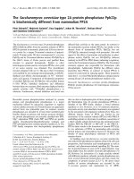
Histologically, the arthritis was severe and erosive.
Figure 1 compares interphalangeal joints from littermate
wild-type (WT) and TTP KO mice at about 7 months of
age. In this comparison there is a clear increase in the size
and inflammatory nature of the synovium, which resembles
an early pannus; however, the articular cartilage appears
relatively normal in this joint. However, there was a very
striking increase in the cellularity of the marrow
compartment in the KO mice, in which the marrow cavities
were packed with normal-appearing granulocytes, with
accompanying erosion of the surrounding bone.
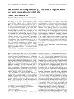
In a larger joint, in this case the ‘wrist’ of the mice, the
changes were more marked (Figs 2 and 3). The ordinarily
delicate synovium had been transformed into an exuberant
pannus, which seemed to be eroding the articular
cartilage as well as underlying bone. The smaller bones of
Figure 1
Interphalangeal joints in wild-type (a) and tristetraprolin knockout (b)
mice. Shown are matching joints from littermate mice at about 7
months of age, stained with hematoxylin and eosin. C, articular
cartilage; M, marrow; P, pannus; S, synovium; T, trabecular bone.
Scale bar, 0.5 mm. Modified from [11].
250
Arthritis Research & Therapy Vol 6 No 6 Carrick et al.
the metacarpals were often destroyed, and loss of digits
was not uncommon. As in the smaller joints described
above, there was marked proliferation of marrow
granulocytes, essentially all of which were Gr-1
+
, again
with internal erosion of both trabecular and cortical bone.
This histological picture was similar to that seen in other
models of TNF-α excess, although in most cases the
marrow granulocytosis was either not found or not
commented on [12–14]. One exception was the
syndrome induced by periodic injections of TNF-α into
mice, in which the authors reported a marked increase in
marrow granulocytes [15].
Other pathological characteristics included granulocyte
infiltration in the skin, accompanying the loss of sub-
cutaneous fat; and foci of granulocytes in the liver, spleen,
lymph nodes and other extramedullary sites. This was
accompanied by marked splenomegaly and lymph-
adenopathy in most cases. In the blood, there was an
approximately twofold increase in total white blood cell
count; most of the increase could be accounted for by
nearly a fourfold increase in Gr-1
+
granulocytes, a
threefold increase in F4/80
+
macrophages, and increases
in PK135
+
natural killer cells. There were decreases in
both B and T lymphocytes. Erythrocyte and platelet counts
were essentially normal. There were also marked
increases in myeloid progenitors in spleen and peripheral
blood, but not in bone marrow, in the KO mice.
Serologically, autoimmunity was present in the form of
high titers of antinuclear antibodies that stained in a
homogeneous pattern, as well as antibodies against both
double-stranded and single-stranded DNA. However,
rheumatoid factor titers were normal (both IgG and IgM),
as were anti-Sm antibody titers. There were some
histological abnormalities of the kidneys, but no increase
in IgG or IgM staining of the glomeruli was noted, and no
proteinuria or azotemia was found in the KO mice.
Because many aspects of the mouse phenotype
resembled previous models of chronic, systemic TNF-α
excess, we performed an experiment in which hamster
anti-mouse TNF-α monoclonal antibodies were
administered to TTP KO mice as soon as they could be
genotyped after birth [11]. Remarkably, these injections
prevented the development of essentially the entire TTP
deficiency phenotype. Specifically, the cutaneous, joint,
adipose tissue, and hematological abnormalities were
prevented. This led to the conclusion that most if not all of
the abnormalities noted in the KO mice were due to
Figure 2
Radial head histology in wild-type (a) and tristetraprolin knockout (b)
mice. This low-power view is of radial head joints from littermate mice
at about 7 months of age, with the radial head (RH) indicated. Other
abbreviations are as in the legend to Fig. 1. Modified from [11].
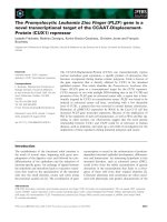
Figure 3
Higher-power view of the radial head histology for the same littermate
wild-type (a) and tristetraprolin knockout (b) mice as those shown in
Fig. 2, with the radial head at the bottom of each panel. Abbreviations
are as in the legend to Fig. 1. Modified from [11].
251
Available online />chronic ‘effective elevation’ of circulating TNF-α. This was
strongly supported by later studies in which the TTP KO
mice were interbred with mice lacking one or both types of
TNF-α receptor (see below).
The next studies were aimed at elucidating the mechanism
of this apparent TNF-α elevation. An important clue was
that the phenotype could be transferred by whole bone
marrow transplantation into Rag2
–/–
recipient mice, but
only after a rather long latent period, suggesting that the
phenotype was not transferred with lymphocyte
progenitors but instead by more slowly reconstituting cells
such as those of the macrophage/monocyte lineage [16].
We then demonstrated that KO macrophages derived from
several sources, including fetal liver, adult bone marrow,
and peritoneal cavity, all released considerably more TNF-α
than did macrophages from their WT littermates [16].
Critically, this was associated with an increase in the
steady-state levels of TNF-α mRNA in the KO cells,
implying either an increase in TNF-α gene transcription or a
decrease in TNF-α mRNA turnover rates, or both.
Subsequent studies in bone marrow-derived macro-
phages demonstrated that there was a marked decrease
in TNF-α mRNA turnover rate in KO cells stimulated with
lipopolysaccharide (LPS) and then treated with
actinomycin D to inhibit transcription, implicating TTP in
the process of TNF-α mRNA turnover [17]. To assess the
possibility that TTP could be causing this effect by first
binding directly to the transcript, we performed direct
RNA binding studies with ultraviolet crosslinking of protein
to RNA followed by immunoprecipitation with anti-TTP
antibodies, and RNA gel-shift analyses. These studies
revealed that TTP did indeed bind directly to the TNF-α
transcript, in an AU-rich region long known to be an
instability agent for this relatively unstable message, and
that the TZF domain of TTP was the RNA-binding domain
of the protein [17].
These studies showed that TTP was an AU-rich element
(ARE)-binding protein that destabilized its target mRNA, in
this case the mRNA encoding TNF-α. This led us to
postulate a TTP–TNF-α feedback loop, in which both TNF-
α and LPS stimulated the transcription of both the TNF-α
and TTP genes, with the latter protein product feeding
back to bind to the TNF-α mRNA and destabilizing it, thus
potentially reversing the effects of the initial stimulus and
the subsequent positive feedback effect that TNF-α has
on its own transcription [17].
The mechanism of the effect of TTP remains to be
determined, but an important clue came from our
evaluation of a second mRNA containing a TNF-α-like
ARE: that encoding granulocyte–macrophage colony-
stimulating factor (GM-CSF) [18]. TTP could bind to this
element as well as to the TNF-α ARE. In addition, the
transcript from KO bone marrow-derived stromal cells was
essentially completely stable after treatment of the cells
with actinomycin D, whereas the transcript was unstable
in the corresponding WT cells. This indicated that the
GM-CSF mRNA was likely to be another physiological
target for TTP. Most importantly, however, the pattern of
GM-CSF transcripts revealed by northern blotting was
extremely informative. In total cellular RNA from the WT
stromal cells, the GM-CSF mRNA existed as two species
of approximately equal abundance, differing in size by
about 200 bases. This size difference was shown to be
due to the presence or absence of a poly(A) tail. Strikingly,
almost all of the stable transcripts in the KO cells were in
the fully polyadenylated form. This meant that the absence
of TTP led to a failure of deadenylation, or removal of the
poly(A) tail, from the GM-CSF transcript. This is thought to
be the first step in vertebrate mRNA degradation [19–21],
and it seemed likely that the inability of the GM-CSF
mRNA to be deadenylated in the KO cells led directly to
the increase in its stability. Conversely, it meant that TTP
could be viewed as an mRNA-binding protein whose
destabilizing abilities were likely to be due to its ability to
promote the process of transcript deadenylation.
In subsequent studies, we developed both cell
transfection assays and cell-free deadenylation assays to
show that, indeed, TTP could promote the deadenylation
and breakdown of mRNAs containing the characteristic
ARE-binding sites [22,23]. This ability of TTP to promote
deadenylation was enhanced by increasing the
concentrations of poly(A) exonuclease (PARN), either in
intact cells or in cell-free assays, suggesting that the two
proteins could act synergistically to promote the
deadenylation of a relatively small number of transcripts
containing characteristic ARE TTP-binding sites [23]. The
exact molecular nature of this interaction is the subject of
continuing studies, but it should lead us to a molecular
understanding of how TTP promotes the deadenylation
and destabilization of a specific set of target proteins.
Now that the principal target of TTP relevant to the TTP-
deficiency phenotype is known to be the TNF-α mRNA
ARE, it is instructive to compare the TTP KO phenotype
with others in the literature that have involved TNF-α
specifically. In the first such model developed, Kollias and
colleagues created transgenic mice in which human
TNF-α was overexpressed in T lymphocytes [14]. These
animals developed many of the same phenotypes as the
TTP KO mice, including wasting and cachexia, premature
death, and universal inflammatory arthritis, all of which
could be prevented by the administration of an antibody
against TNF-α. Another directly relevant model was that
created by specifically removing the TNF-α mRNA ARE
(∆ARE) from the endogenous mouse TNF-α gene,
resulting in a markedly more stable TNF-α mRNA [13]. As
expected, this led to the chronic oversecretion of TNF-α
252
due to increased accumulation of the stabilized mRNA, in
a manner analogous to that in the TTP KO mice.
In general, the phenotype of this mouse was considerably
more severe than the TTP KO phenotype. For example,
although these mice were born alive, the KO animals
rapidly developed a marked slow-growth phenotype and
died between 5 and 12 weeks of age. Pathologically,
these mice were characterized by severe erosive arthritis
and inflammatory bowel disease, but inflammation in other
tissues was minimal. They also exhibited thymic atrophy
and blurring of thymic cortical and medullary boundaries,
also characteristic of the TTP KO phenotype. The arthritis
developed as early as 12 days of age, and resembled the
TTP KO arthritis in general character; however, the ∆ARE
mice exhibited elevated levels of mouse IgG and IgM
rheumatoid factor, not seen in the TTP KO mice. The
inflammatory bowel disease was severe and universal and
affected both the colon and the terminal ileum in some
cases. The hemizygous ∆ARE mice had a less severe
phenotype, but most aspects of the same syndrome were
seen with a delayed onset relative to the homozygous
mice [13].
In a striking difference from the TTP KO mice, both the
∆ARE homozygous and hemizygous mice developed a
severe form of Crohn’s-like colitis, a syndrome that has not
been seen in the TTP KO mice so far [13]. We have
recently shown that TTP protein is relatively highly
expressed in normal mouse colon [24], and its absence
from this tissue might be expected to have deleterious
effects on this organ. Much remains to be determined
about the specific cell types that express TTP in this
tissue, which may not be directly relevant to the
development of the colitis syndrome. Nonetheless, this
remains an interesting difference between the ∆ARE mice
and the TTP KO mice that is yet to be explained.
Genetic modifiers of the TTP-deficiency
syndrome
We and others have evaluated the TTP-deficiency
phenotype further by interbreeding the mice with other
potentially informative genotypes. To confirm the data from
the TNF-α antibody injection experiments, we interbred
the TTP-deficient mice with mice deficient in either or both
TNF-α receptors 1 and 2 (TNF-αR1 and TNF-αR2) [25].
As predicted from the antibody experiments, TTP KO mice
that were also deficient in both types of TNF-α receptor
seemed essentially normal, with none of the cutaneous or
joint manifestations of the TTP-deficiency syndrome. Most
of the protective effect was due to the TNF-αR1
deficiency; the TTP/TNF-αR1 double KO mice were also
essentially normal. Interestingly, the TTP/TNF-αR2 double
KO mice were even more severely affected than the
parental TTP KO strain, with very severe arthritis and
growth retardation in early life leading to premature death
in most cases. This supported the concept, developed in
other studies, that TNF-α acting through TNF-αR2 can
exert a protective effect on the TNF-α-induced
inflammatory response in some situations. In any case,
these studies confirmed the findings of the TNF-α
antibody injection study; it seemed clear that most
aspects of the TTP-deficiency phenotype were thus due to
chronic, elevated levels of TNF-α.
However, observation of the normal-appearing TTP/TNF-
αR1/TNF-αR2 triple KO mice for longer than about 1 year
revealed that they were not quite normal. As these animals
aged, there was the development of myeloid hyperplasia,
particularly intramedullary, which eventually resembled that
seen in the original TTP KO mice [25]. This finding,
coupled with the known effect of TTP deficiency to
stabilize the GM-CSF mRNA and lead to enhanced
secretion of this cytokine [18], suggested the possibility
that the myeloid hyperplasia seen in the aging triple KO
mice might be due to chronic increases in the
concentration of either circulating or local GM-CSF. We
are currently attempting to address this possibility by
interbreeding the TTP KO mice, and the TTP/TNF-αR1/
TNF-αR2 triple KO mice, with mice deficient in GM-CSF
[26]. The first mice of these genotypes are approaching
an age at which they can be analyzed, and should yield
valuable information: first, the extent to which the myeloid
hyperplasia of the original TTP KO mice was due to GM-
CSF; second, the extent to which the late-onset myeloid
hyperplasia of the triple KO mice is due to GM-CSF; and
third, if the myeloid hyperplasia is prevented, these
‘quadruple KO’ mice might possibly yield valuable insights
into any remaining physiologically relevant TTP target
transcripts.
Besides interbreeding with TNF-αR1, TNF-αR2 and GM-
CSF KO mice, several studies have been performed with
other potentially informative genetic backgrounds. These
studies are arduous because of the marked subfertility of
the TTP KO mice, necessitating that the line be
maintained by hemizygotes. Recently, for example, Phillips
and colleagues [27] generated mice deficient in both TTP
and another TNF-α synthesis regulator, TIA-1. TIA-1 is
thought to inhibit TNF-α production primarily by interfering
with the translation of existing transcripts. As expected,
the TTP/TIA-1 double KO mice developed more severe
arthritis than either genotype alone. An unexpected result
of this study was that macrophages derived from the
TTP/TIA-1 double KO mice secreted less TNF-α protein
than cells from either single KO alone, leading the authors
to speculate that the TNF-α secretory apparatus might
somehow be interfered with in these macrophages. They
also found that bone marrow cells from the TTP KO mice,
particularly in combination with the TIA-1 KO, secreted
considerable TNF-α in response to LPS, in contrast to the
minimal secretion from marrow cells from either WT mice
Arthritis Research & Therapy Vol 6 No 6 Carrick et al.
253
or TIA-1 KO mice. This enhanced secretion was also seen
with isolated neutrophils, which accumulate markedly in
the marrow and elsewhere in the TTP KO mice. The
authors speculated that these neutrophils might represent
the primary source of ‘arthritogenic’ TNF-α in this situation.
Other interbreeding experiments are either under way or
have not yet been published. For example, J Rivera-Nieves
has crossed the TTP-deficient mice with the TNF-α∆ARE
mice alluded to above (personal communication). This is a
potentially interesting cross because the TNF-α∆ARE
mice develop a severe, Crohn’s-like colitis, whereas the
TTP-deficient mice have not been observed to do so, at
least on the C57Bl6 background. Another interesting
cross is the TTP-deficient mice interbred with a mouse in
which TNF-α cannot be secreted because its biosynthetic
cleavage is prevented, leading to the accumulation of cell-
associated TNF-α but an absence of secreted TNF-α
(G Kollias, personal communication). A third current study
involves crossing the TTP KO mice with mice deficient in
the p38 mitogen-activated protein (MAP) kinase target
MK2, which seems to be involved in TNF-α mRNA
translation and TTP post-translational modification (see
below; M Gaestel, personal communication). Other
studies with potentially interesting genetic modifiers are
continuing, and the next several years should yield several
interesting insights into regulatory molecules and
pathways. In addition to these crosses into specific
knockout mice, we are crossing the TTP deficiency
genotype into other genetic backgrounds in the hope of
identifying other modifiers.
Recent data on the TTP-binding site in the
TNF-
αα
and GM-CSF mRNAs
Since the pioneering work of Shaw and Kamen [28], it has
been clear that certain AREs in mRNAs can confer
instability on those transcripts. These have been
categorized more recently into classes based on the
presence or absence of AU substructures, such as the
AUUUA pentamer. The TNF-α and GM-CSF AREs, so far
the only definite physiological targets for TTP, fall into the
type 2 ARE of Chen and colleagues [29], or the type 2a of
Willusz and colleagues [21], in which there are multiple
copies of tandem and overlapping AUUUA pentamers.
Several recent studies have characterized a more specific
TTP-binding site. In the first, Worthington and colleagues
used the SELEX procedure to identify the nonamer 5′-
UUAUUUAUU-3′ as the optimal site from a random
collection of oligonucleotides [30]. Using a synthetic TZF
domain peptide derived from the human TTP, known as
TTP73, our group independently showed that this same
nonamer sequence was optimal for TTP binding on gel-
shift analysis, which could be accomplished with binding
affinities in the low nanomolar range [31]. In a
heteronuclear single-quantum coherence nuclear
magnetic resonance (NMR) analysis, we found that
binding of the TTP peptide to target RNAs caused a major
conformational shift in the resonances of the first zinc
finger in the peptide, while simultaneously permitting the
demonstration of second zinc finger resonances that had
been apparently unstructured in the absence of RNA.
Using progressive shortening of a longer, TNF-α ARE-
based mRNA target, we found that the characteristic NMR
resonance shift occurred in an identical manner down to
the 5′-UUAUUUAUU-3′ nonamer; however, loss of a
single base in an octamer showed a deterioration of the
NMR resonance pattern, and still shorter oligonucleotides
were ineffective. These data suggested that the nonamer
5′-UUAUUUAUU-3′ was the minimal complete binding site
for the TTP peptide. This study also demonstrated that in
longer AREs containing several repeats of this sequence,
as are found in the TNF-α and GM-CSF AREs, several
tandem molecules of the TTP73 peptide could bind to a
single RNA strand, leaving open the possibility of multiple
occupancy of the longer ARE by mature TTP protein
molecules. The effect of these multiple tandem nonamer
repeats on the effectiveness of TTP in promoting mRNA
deadenylation is the subject of a continuing study.
Very recently, Hudson and colleagues determined the
structure of the analogous TZF domain peptide from the
TTP-related protein ZFP36L2 (TIS11D) in complex with
this RNA nonamer [32]. This novel structure confirmed the
conformational change in the peptide after RNA binding,
and revealed many interesting aspects of the interaction
between the TZF domain and RNA. For example, each of
the two zinc fingers formed very similar structures, each of
which contacted an RNA ‘half-site’ 5′-UAUU-3′, with the
binding apparently mediated by electrostatic and
hydrogen-bonding interactions. The binding is further
influenced by ‘stacking’ of the conserved aromatic amino
acid side chains with the RNA bases. The ‘lead-in’ motifs
R(K)YKTEL of each finger also participate through
hydrogen-bonding interactions with the 5′-most bases on
each half site. Importantly for future informatics analyses,
the 5′-most U of the 5′-UUAUUUAUU-3′ nonamer was
disordered in the structure, which might allow base
substitutions at this position in physiologically relevant
RNA-binding sites, as well as less conservation in the
protein sequence at the protein face near this base. The
structure of this RNA-binding domain was thought to be
unlike any previously published structure.
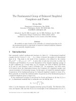
We have taken the coordinates of the ZFP36L2 TZF
domain complex and modeled the structure of the
interaction of the human TTP TZF peptide with the RNA
nonamer, using the Swiss-Model programs [33,34]. A
surface depiction of this model is shown in Fig. 4. It should
be emphasized that this is a model based on the
published ZFP36L2 TZF domain structure [32], but the
two sequences are so closely related that it seems likely
Available online />254
to be a fairly close approximation of the final TTP TZF
domain structure in complex with its RNA target.
One of the striking aspects of this model is the residues
within the TTP TZF domain peptide that are identical to
those in the ZFP36L2 peptide, which are shown in dark
blue in Fig. 4. It is apparent that essentially all of the amino
acid residues that are in direct contact with the RNA are
identical to those in the ZFP36L2 peptide, whereas those
residues that are progressively less well conserved
(ranging in order of conservation from aquamarine [best
conserved] through green, yellow, and orange [least
conserved]) are far removed from the RNA contact
surfaces. This model suggests that the RNA contact
domain of the TTP peptide is likely to be identical in
structure to that of ZFP36L2 (TIS11D), and that the
differing residues on other faces of the peptide might be
involved in potential specificity determinants, such as
protein–protein interactions. The one potentially interfering
interaction in this model is between the 5′-most U residue
and the green amino acid in the upper left corner;
however, from the data of Hudson and colleagues [32],
this initial U is unstructured in the complex and thus can
assume other conformations than that pictured, to prevent
interfering interactions with the peptide.
Other aspects of this model fit some of our experimental
observations. For example, it is possible to mutate the
middle A (U5) to C while retaining some TTP binding and
deadenylating abilities (WSL and PJB, unpublished data).
This fits with the apparent position of U5 near a ‘hole’ in
the middle of the structure shown in Fig. 4, which
represents part of the inter-finger linker region.
Conversely, mutations of either of the two A residues in
this sequence (A3 and A7) were not well tolerated, and
they are critical in the structure to stacking interactions
with hydrophobic residues in the peptide. Mutations within
these critical hydrophobic amino acids were also not well
tolerated in terms of RNA binding and promoting mRNA
deadenylation [35]. It will be interesting in the coming
years to extend these analyses to other residues in the
TZF domain, particularly in the highly conserved inter-
finger linker region, which contains two basic residues
thought to be critical for the nuclear import of the peptide
[36].
Given that these studies have all agreed that the central
optimal binding motif is 5′-UUAUUUAUU-3′, it is of
interest to compare this sequence with the naturally
occurring physiological targets of TTP found in the TNF-α
and GM-CSF mRNAs. Figure 5 shows the central ARE
sequences from all known mammalian orthologs of the
TNF-α and GM-CSF mRNAs [37]. It should be noted that
the extreme conservation of these motifs falls apart in both
the 5′ and 3′ direction in both TNF-α and GM-CSF
mRNAs. By far the most popular mammalian pattern in the
TNF-α mRNA is exemplified by the human sequence and
those of nine other mammals, in which there are five
overlapping versions of the nonamer-binding site. Within
the five overlapping nonamers are several non-overlapping
patterns. It will be of great interest to determine how many
intact TZF peptide and intact TTP protein molecules can
bind to this sequence, because it might be expected that
the overlapping nonamers might prevent binding to some
of the neighboring nonamers by steric hindrance. It is also
of interest that several other mammals have different
patterns and total numbers of these nonamers (Fig. 5a);
again, it will be of interest to determine whether there are
species differences in the number of TTP molecules that
can occupy these domains, perhaps resulting in species
differences in TTP effects on mRNA turnover rates.
The situation is somewhat different with the GM-CSF
transcript, because the common mammalian pattern is to
have three densely overlapping nonamers (Fig. 5b). The
pig is an outlier in this case, with three more widely
spaced nonamers. However, GM-CSF also has a
completely conserved 5′-UAUUUAU-3′ heptamer, with
miscellaneous bases in the 1 and 9 positions in the
Arthritis Research & Therapy Vol 6 No 6 Carrick et al.
Figure 4
Proposed structure of the human tristetraprolin (TTP) tandem zinc
finger domain in complex with the TTP-binding site 5′-UUAUUUAUU-
3′. This proposed structure was modeled on the original nuclear
magnetic resonance structure described by [32], using their pdb
coordinates and the Swiss-Model program. The RNA oligonucleotide
is shown in magenta, with the 5′ and 3′ ends indicated, along with the
key residues A3, U5, and A7. The peptide is shown as a surface
structure, with the buried zinc residues highlighted and the amino-
terminal (N-term.) and carboxy-terminal (C-term.) ends of the peptide
shown by arrows. The dark blue residues represent amino acids that
are identical between human TTP and the ZFP36L2 (TIS11D) protein
used in the original structure. The other colors represent progressively
greater amino acid differences between the two proteins, ranging from
minimally different (aquamarine, upper right), through green, yellow,
and orange, with orange representing the most marked amino acid
differences.
255
nonamer. On the basis of the structural features of the
TZF–RNA complex, it seems possible that some variability
in positions 1 and 9 can also be tolerated, and that this
more 5′ sequence in the GM-CSF mRNA might represent
a physiologically relevant TTP-binding site.
From the point of view of identifying potential targets for
these proteins by bioinformatics approaches, it seems that
multiple copies of the canonical nonamer are optimal but
that some base and length differences might be tolerable.
For example, Brewer and colleagues [38] have recently
shown that changing the ‘inter-A’ sequence from UUU to
UU or UUUU changes the dissociation constant (K
d
) of
the TTP73 synthetic peptide bound to synthetic RNA
oligonucleotides from 3.2 nM to 18 or 6.4 nM,
respectively, both close enough to the WT K
d
to make
them possibly physiologically significant. However, just
because an mRNA sequence meets these criteria does
not necessarily make it a physiologically relevant target.
There are many such potential TTP target AREs in the
database [39], and TTP can be shown to bind to and
deadenylate a variety of ARE-containing mRNAs in
overexpression studies. In our view, the key criterion for
demonstrating that a particular mRNA is a genuine target
of TTP or one of its family members is a demonstration
that the stability of the transcript is increased in cells
deficient in the protein. At the time of this writing, only the
TNF-α and GM-CSF transcripts have met this criterion
and can therefore be considered true and validated
physiological TTP targets.
Regulation of TTP
TTP was initially discovered because of the rapid and
marked inducibility of its mRNA in fibroblasts in response
to insulin, phorbol esters, and serum. This rapid and
profound induction is clearly one of the means by which
cellular TTP levels are regulated. As with many genes of
the ‘immediate early response’ type, TTP mRNA is quite
unstable, and in many cell types it returns to basal levels
only a few hours after stimulation, despite the continued
presence of the original stimulus in the culture medium.
Considerable work has been done on the TTP promoter
and its mitogen- and cytokine-responsive enhancer
elements, and it is clear that there are apparent
Available online />Figure 5
Mammalian tumour necrosis factor-α (TNF) and granulocyte/macrophage colony-stimulating factor (GM-CSF) AU-rich elements (AREs). (a) The
central ARE region of the TNF mRNA 3′ untranslated region from all mammalian species for which this region of the mRNA has been deposited in
GenBank. In most cases these were derived from EST sequences; note that the horse sequence has not been completed at the 3′ end. The
overlines indicate the nine-base tristetraprolin (TTP)-binding site 5′-UUAUUUAUU-3′. Sequences from the various mammals are divided into groups
based on the pattern of these nonamers, with the top group of 10 mammals being the most common group. (b) A similar approach was used to
align the central ARE from the GM-CSF transcript, after alignment using the program ClustalW. The asterisks below the alignment represent base
identity at that position; note that gaps were used to optimize the alignment. The overlines again represent the nonamer TTP-binding site. These
data are modified from [37].
Horse GAUUAUUUAUUAUUUAUUUAUUAUUUAUUUAUUU
Beluga GAUUAUUUAUUAUUUAUUUAUUAUUUAUUUAUUUAC
Baboon GAUUAUUUAUUAUUUAUUUAUUAUUUAUUUAUUUAC
Cow GAUUAUUUAUUAUUUAUUUAUUAUUUAUUUAUUUAC
Goat GAUUAUUUAUUAUUUAUUUAUUAUUUAUUUAUUUAC
Mouse GAUUAUUUAUUAUUUAUUUAUUAUUUAUUUAUUUGC
Sheep GAUUAUUUAUUAUUUAUUUAUUAUUUAUUUAUUUAC
Human GAUUAUUUAUUAUUUAUUUAUUAUUUAUUUAUUUAC
Dolphin GAUUAUUUAUUAUUUAUUUAUUAUUUAUUUAUUUAC
Zebu GAUUAUUUAUUAUUUAUUUAUUAUUUAUUUAUUUAC
Rabbit GAUUAUUUAUUAUUUAUUUAAUAUUUAUUUAUUUGC
Pig CAUUAUUAUUUAUUUAUUUAUUUAUUAUUUAUUUAC
Woodchuck AAUUAUUUAUUACUUAUUUAUUAUUUAUUUAUUUAC
______
Rat GACUAUUUAUUUAUUAUUUAUUAUUUAUUUAUUUGC
HUMAN UAUUUAUAUAUUUAUAUUUUUAAAAUAUUUAUUUAUUUAUUUAUUUAAGUUCAUAUUCCA
HORSE UAUUUAUAUAUUUAUGUAUUUUAA-UAUUUAUUUAUUUAUUUAUUUAAGCUCAUACUCCA
MOUSE UAUUUAUAUAUUUAUAUUUUUUAAAUAUUUAUUUAUUUAUUUAUUUAA
WOODCHUCK UAUUUAUAUAUUUAUACUUUUAAAAUAUUUAUUUAUUUAUUUAUUG
COW UAUUUAUAUAUUUAUGUAUUUUAA-UAUUUAUUUAUUUAUUUAUUUAAACUCAUACCCCA
DOG UAUUUAUAUAUUUAUGUAUUUUAA-UAUUUAUUUAUUUAUUUAUUUAAGAUCAUACUCUG
PIG UAUUAACCUAUUUAUGUAUUUUAA-UAUUUAUUUAUUUAUUUAUCUAUUUAUUUAUUUA
**** * ******* *** ** *******************
(b) GM-CSF
(a) TNF
256
transcription-factor-binding sites within the promoter and
single intron that are involved in this rapid and transient
transcriptional response, but the binding proteins remain
to be identified [40].
In contrast to this rapid but transient induction of the TTP
mRNA in response to various stimuli, the protein seems to
be surprisingly stable. In a recent study from our laboratory
in which a TTP antiserum was used to probe western
blots from mouse RAW264.7 macrophages after
stimulation with LPS [41], the protein was almost
undetectable in normally growing, unstimulated cells. This
was compatible with parallel studies in normal mouse
tissues, in which extraordinary amounts of tissue protein
were needed in western blots to be able to detect
immunoreactive protein. However, within 30–60 min of
stimulation with LPS, readily detectable protein began
appearing in the cytosol, reaching a peak value after about
2–4 hours. Surprisingly, despite the relative lability of the
mRNA in most cell types, the protein remained very stable
in the cells for many hours thereafter; it was still much
higher than baseline levels after 6 hours, and was still
readily detectable after 24 hours. Therefore, de novo
biosynthesis of protein is clearly a major mode of TTP
regulation, with manyfold increases occurring within a few
hours after stimulation with LPS. Nonetheless, the reversal
of this process seems to be rather slow.
Another interesting aspect of the protein accumulation
experiments is that, as the protein was being synthesized
and accumulated in the cellular cytoplasm, its apparent
molecular mass on SDS gels continued to increase in an
incremental manner, compatible with a stoichiometric
increase in its phosphorylation. This occurred more slowly
than the generally accepted model of protein
phosphorylation, in which a previously synthesized protein
is acted upon within a few minutes by a newly activated
protein kinase. Possible mechanisms for this slow but
profound increase in phosphorylation include the
following: first, the protein was initially in the wrong cellular
compartment to serve as a kinase substrate, and only
became accessible after a lag of 1–2 hours; second, the
protein was protected from phosphorylation by binding
proteins or even intramolecular folding events; third, the
protein was phosphorylated by one or more protein
kinases that were either slowly activated by LPS or whose
biosynthesis was also stimulated by LPS; fourth, the
phosphorylation of the protein was increased by the
activity of constitutively active cellular protein kinases in
the setting of phosphatases that were either inactivated or
destroyed in response to stimulation with LPS; or fifth,
some combination of these events.
Elucidation of these mechanisms is of increasing interest
because the effect of protein phosphorylation on TTP
behavior has begun to be elucidated in a flurry of recent
studies. In our initial report on TTP phosphorylation [5], we
demonstrated that both major bands of phosphorylated
TTP expressed in intact fibroblasts, with or without
stimulation with fetal calf serum, contained only phospho-
serine. We identified a single major serine phosphorylation
site in that study (S220 in the mouse, corresponding to
S228 in the human protein) that was a substrate for MAP
kinases both in intact cells and in cell-free systems. In both
cases — that is, in intact cells and in cell-free assays — this
phosphorylation event changed the migration behavior of
the protein in SDS gels, compatible with a stoichiometric
increase in phosphorylation. However, because nothing
was known about TTP’s physiological function at that
point, we were unable to conclude anything about the
effect of this phosphorylation event on protein behavior
except to say that the absence of this phosphorylation site
did not seem to affect TTP’s ability to translocate from the
nucleus to the cytosol in response to stimuli such as
serum.
Several more recent studies have added to our knowledge
of TTP phosphorylation and its effects on protein behavior.
In one study we found that TTP could serve as a substrate
for the p38 protein kinase and that global dephos-
phorylation of TTP in a cell-free system with alkaline
phosphatase seemed to increase its binding affinity for an
ARE-containing RNA substrate [42]. Clark and colleagues
[43] demonstrated that TTP could serve as a substrate for
the MAP kinase-activated protein kinase 2 (MK2),
although these authors did not detect direct phosphory-
lation of TTP by the p38 kinase. Johnson and colleagues
[44] demonstrated that TTP could bind to the cellular
protein 14-3-3, and that this binding could be influenced
by the phosphorylation status of the protein, specifically at
serine 178 in the mouse protein (corresponding to serine
186 of the human protein). They also demonstrated that
the binding to 14-3-3 protein promoted the localization of
TTP to the cellular cytoplasm. Very recently, Chrestensen
and colleagues [45] identified the same serine 178 as a
major phosphorylation site for MK2; they also identified
serine 52 as another major site and showed that these
two phosphorylations created functional binding sites for
14-3-3. Finally, Stoecklin and colleagues [46] showed that
phosphorylation of TTP by MK2 on serines 52 and 178
led to 14-3-3 binding, which in turn led to the exclusion of
TTP from arsenite-induced stress granules. Although the
14-3-3 interaction still permitted binding of TTP to RNA, it
inhibited the TTP-dependent degradation of ARE-
containing RNA substrates.
Taken together, these data suggest a model in which TTP
activity against ARE-containing mRNAs can be modulated
by the activation of p38 and the MK2-induced
phosphorylation of TTP, leading in turn to 14-3-3
association and cytoplasmic sequestration as well as the
inhibition of mRNA degradation. The relevance of direct
Arthritis Research & Therapy Vol 6 No 6 Carrick et al.
257
p38 phosphorylation is unclear, although some of the
minor sites identified by Christensen and colleagues [45]
might have been sites for the p38 kinase. It will be
interesting to determine how this model works in intact
organisms; this should now be capable of investigation,
given the availability of MK2-deficient mice [47]. In fact, as
pointed out by Mahtani and colleagues [43], mouse
spleen cells from MK2-deficient mice seemed to express
normal levels of TNF-α mRNA, which exhibited apparently
normal stability after LPS stimulation [47].
The studies indicating association of TTP with 14-3-3
protein highlight another possible mode of regulation of
TTP activity, namely association and dissociation from
binding proteins. In addition to 14-3-3, previous studies
have identified interactions of TTP with TIA-1 [48] and the
nucleoporin CAN/Nup214 [49]; in the latter study, the
interaction again seemed to regulate the intracellular
localization of TTP. Functional studies have suggested
interactions with the nuclear export protein CRM1 [50],
although direct binding interactions were not demon-
strated in that study. Chen and colleagues [51]
demonstrated an association of TTP with components of
the exosome, suggesting a role for this structure in the
degradation of deadenylated ARE-containing mRNAs.
Finally, a recent two-hybrid analysis by our group
(Blackshear PJ, unpublished data) identified a large
number of previously unidentified potential interacting
proteins, each of which needs to be painstakingly
validated by other interaction methods. We expect to see
the identification of many such interacting proteins in the
immediate future, and many of these binding events will
undoubtedly have effects on the cellular physiology of the
protein.
Polymorphisms in the human TTP gene (
ZFP36
)
It is possible that severe mutations in TTP coding
sequences could prevent or decrease the expression of
mature transcripts, interfere with splicing of the single
intron, or lead to frame-shift or stop-codon mutations.
These could have major effects on TTP’s ability to bind to
TNF-α transcripts and destabilize them. Linkage studies
with less severe TTP polymorphisms could provide insight
to the treatment and/or diagnosis of human disorders
associated with excess TNF-α, such as rheumatoid
arthritis or Crohn’s disease. Although such linkage studies
are yet to be completed, the first step in performing them
is to identify polymorphisms. We have taken this first step
and identified several single-nucleotide polymorphisms
(SNPs) in the human TTP gene, ZFP36 [52].
As part of the NIEHS Environmental Genome Project
(EGP), ZFP36 was resequenced in the genomic DNA
from 92 anonymous subjects (Coriell collection and
Coriell Polymorphism Discovery collection; see reference
[52] for further details). Resequencing identified 10
polymorphisms and expressed sequence tag (EST)
searches identified an additional four potential SNPs in
ZFP36. These are summarized in Table 1, and a
schematic depiction of the SNP positions on the human
gene is shown in Fig. 6.
Polymorphisms in the promoter region and single intron of
TTP are of particular interest because both of these
regions are necessary for the proper regulation of
expression [8,9]. Four polymorphisms were identified in
the promoter region. The polymorphism ZFP36*2 at base
359 in the promoter was the most common SNP
identified; it was present at 47% in the EGP subjects. The
other three promoter SNPs were ZFP36*1 (found in 1.8%
of the EGP subjects), ZFP36*3 (3.1%), and ZFP36*4
(0.6%). Two intronic polymorphisms were also found in
this group. ZFP36*5 and ZFP36*6 were present in the 92
EGP subjects at frequencies of 0.5% each.
Six polymorphisms were identified in the protein-coding
domains by resequencing; all were in exon 2. Three of the
SNPs were identified by resequencing the EGP subject
DNA. These SNPs were present at frequencies of 0.6%
for ZFP36*7, 6.2% for ZFP36*8, and 4.2% for ZFP36*9.
Of these three SNPs, only ZFP36*7 resulted in an amino
acid change from proline to serine. Because this
represented a non-conservative change, the frequency of
ZFP36*7 was determined in 422 North Carolina subjects
of varying ethnicities; it was found in 2.4% of this
population [52].
Three other potential protein-coding-domain poly-
morphisms were identified by EST searches. One of the
SNPs, ZFP36*11, was found in 4.2% of the ESTs
examined. This SNP resulted in a non-conservative amino
acid change from glycine to cysteine. Although this SNP is
within the TZF RNA-binding domain (in the 18 amino acid
‘linker’ between the two zinc fingers), it had no detectable
effect on the RNA-binding ability of TTP in a cell-free gel-
shift assay. The other two SNPs, ZFP36*12 (present in
4.1% of ESTs examined) and ZFP36*13 (11%), did not
produce an amino acid change.
An ARE in the 3′ untranslated region (UTR) of TTP confers
instability on the mRNA, as determined by deletion studies
(WSL and PJB, unpublished data). This region was also
examined for polymorphisms. Two polymorphisms,
ZFP36*10 and ZFP36*15, were identified within the 3′
UTR. ZFP36*10 was identified by resequencing the EGP
subject DNA. This polymorphism was the second most
frequently occurring polymorphism (frequency 7.6% in
EGP subjects). ZFP36*10 lies within a region of the
transcript that is highly conserved among mammals and
therefore might be significant in terms of altering TTP
mRNA stability. The other 3′ UTR polymorphism,
ZFP36*15, was identified only by EST searches (present
Available online />258
in 7.3% of the ESTs examined). Both of these SNPs are
potentially interesting in that they lie with a region of the
TTP mRNA 3′ UTR that probably contributes to the
stability of this message.
Since the completion of the initial resequencing study, we
have embarked on a more extensive study in subjects with
various potentially related diseases. These include
subjects with TNF-associated periodic inflammatory
Arthritis Research & Therapy Vol 6 No 6 Carrick et al.
Table 1
Polymorphisms in
ZFP36
encoding human tristetraprolin
Variant allele
frequency
Amino in EGP Variant allele
acid subjects (%) frequency
Polymorphism Location Base Change Sequence change (n = 92) in ESTs (%)
ZFP36*1 Promoter 316 C→A CCCCC(C/A)ATCCG 1.8
ZFP36*2 Promoter 359 A→G CGGTC(A/G)CGGCT 47
ZFP36*3 Promoter 490 C→A CCGGC(C/A)CCGGC 3.1
ZFP36*4 Promoter 492 C→T GGCCC(C/T)GGCCC 0.6
ZFP36*5 Intron 1226 G→A GGGAA(G/A)CCGGG 0.5
ZFP36*6 Intron 1256 C→G TAAGG(C/G)CTCGG 0.5
ZFP36*7 PCD (ex.2) 1525 C→T CGGGA(C/T)CCTGG P37→S 0.6
a
0/127
ZFP36*8 PCD (ex.2) 1725 C→T TCGCG(C/T)TACAA R103→R 6.2 2/127 (1.6%)
ZFP36*9 PCD (ex.2) 2235 T→C CCCTC(T/C)GTACA S273→S 4.2 1/69 (1.4%)
ZFP36*10 3′ UTR 2980 Del TT TTTTT(delTT)GTAAT 7.6 62/249 (26%)
(7T→5T)
ZFP36*11 PCD (ex.2) 1807 G→T GCCTG(G/T)GCGAG G131→C 5/118 (4.2%)
ZFP36*12 PCD (ex.2) 2112 C→T GCCTT(C/T)TCTGC F232→F 4/97 (4.1%)
ZFP36*13 PCD (ex.2) 2184 C→A AGGGC)CA_ACTCC A256→A 10/94 (11%)
ZFP36*15 PCD (ex.2) 3059 A→C TGCCT(C/T)CCGCT 17/232 (7.3%)
The base numbers correspond to the following GenBank accession numbers: ZFP36 gene, M92844; tristetraprolin cDNA, NM_003407.1;
tristetraprolin protein, NP_003398.1. Polymorphism numbering is consistent with that in [52]. Base refers to the base number in the genomic
sequence M92844. The polymorphic changes are indicated as follows: A (original base or amino acid)→G (polymorphic base or amino acid), or
(A/G) in the sequence column. EGP, Environmental Genome Project; ex. 2, exon 2; PCD, protein coding domain; 3′-UTR, 3′-untranslated region.
Table modified from [52].
a
This SNP was also found in 2.4% of subjects from Durham, North Carolina (see the text). The change labeled ‘Del TT’
refers to the removal of two T residues, changing the normal seven consecutive T residues to five in the variant sequence.
Figure 6
Schematic representation of the human tristetraprolin (TTP) gene (ZFP36) and its polymorphisms. The two exons of ZFP36 are shown as boxes,
whereas the flanking regions and intron are indicated by a thin line. Open boxes represent untranslated regions, solid filled boxes represent protein-
coding regions, and the hatched region represents the tandem zinc finger domain. The positions of the polymorphisms listed in Table 1 are
indicated by arrowheads. Kb, kilobases. The data are modified from [52].
*11
*10
*4 *5 *12 *13
*2
*1 *3 *6 *7 *8 *9
*1
5
0 0.5 1 1.5 2 2.5 3 3.5
Kb
259
syndrome (TRAPS); bronchial hyper-responsiveness to
inhaled endotoxin; rheumatoid arthritis, both responsive
and resistant to anti-TNF-α therapies; various subgroups
of juvenile rheumatoid arthritis; psoriatic arthritis; and
multiple sclerosis. If these subjects are added to the
original 92 members of the EGP participants, then we will
have resequenced the gene from a total of 507 subjects.
Although analysis of these samples is not yet complete,
we have identified an additional 20 potential SNPs,
making the total number discovered so far about 35.
Current efforts include assembling the known SNPs into
haplotypes for association studies, determining SNP and
haplotype frequency in a large number of normal subjects,
and a biochemical examination of the SNPs and
haplotypes for effects on the biosynthesis, activity and
stability of the TTP protein itself.
It is of interest in this regard that long-term follow-up of the
TTP hemizygous mice has shown that several mice older
than 1 year have developed the full-blown TTP deficiency
phenotype, apparently in a stochastic and unpredictable
manner. This suggests that TTP hemizygosity in humans
might be compatible with a completely normal life, but that
in some cases the relative TTP insufficiency might lead to
disease, perhaps in response to some environmental
perturbation. We expect that a full-blown, autosomal
recessive TTP deficiency syndrome would be severe and
fatal in childhood in humans, but so far there are no known
instances of such a condition in humans.
TTP and related CCCH proteins in humans
As noted above, there are now known to be three
members of the TZF protein family in humans, and
extensive blasting of the human genome and EST
collections has not yielded any further members, despite
the presence of a group of sequences of closely related
fourth members in fish and frogs [53]. Much less is known
about the physiological roles of these proteins in
mammalian systems. As shown in Fig. 7a, all three
members of the family can bind readily to a TNF-α ARE
probe, as demonstrated by RNA gel-shift analysis. In
addition, all three family members can promote the
deadenylation of ARE-containing polyadenylated RNA
probes, both in intact cell transfection systems and in cell-
free deadenylation assays (Fig. 7b) [23,54]. This occurs
whether or not the proteins are used to ‘effectively
activate’ endogenous deadenylating activities in HEK-293
cell extracts, or co-transfected PARN in the same cells
(Fig. 7b). This and other types of evidence suggest that all
three proteins have similar roles to TTP in the physiology
of some cell types; that is, they are capable of binding to
specific ARE sequences in certain transcripts and
promoting their deadenylation and degradation. Many
questions remain, including the following. First, in what
cell types does each protein function as an mRNA
destabilizing factor, and in what physiological or
pathological situations? Second, how are these inter-
actions regulated, by biosynthetic and post-translational
events, as well as interactions with other cellular proteins?
Third, what are the mRNA targets for each family member
in normal physiology?
For Zfp36L1 in mouse, we have recently shown that its
complete deficiency leads to universal intrauterine death
of the KO embryos, usually at about embryonic day 9–11
[55]. In most cases there was failure of chorioallantoic
fusion, which undoubtedly leads to the death of the
embryos. In the minority of cases in which fusion occurred,
there seemed to be secondary failure of the placenta,
leading to poor perfusion of the embryo, runting,
widespread apoptosis, neural tube defects, and death.
The presumed stabilized transcripts that lead to these
abnormalities are not known, but they are the subject of
continuing study in the laboratory. This embryonic lethal
phenotype means, however, that elucidation of the
physiological function of the Zfp36L1 protein in cells and
tissues from adult mice will require conditional KO
strategies.
For Zfp36L2 in the mouse, our attempts to create a
conventional KO mouse led to a mouse in which a
transcript is produced that contains a significant portion of
the single intron as well as the complete second exon,
apparently driven by the endogenous promoter (despite
the presence of the neo gene in between) [56]. The result
is that the tissues and cells of this mouse produce a
protein that lacks the amino-terminal 29 amino acids and
is expressed at a variable fraction of endogenous protein
levels in different cells and tissues. Despite this
unsatisfactory situation, a mouse was produced that has a
very specific phenotype: complete female infertility of the
KO mice, apparently due to arrest of the embryo after the
two-cell stage. This seems to be a maternal effect,
because KO embryos from heterozygous mothers seem to
develop entirely normally. This is an unusual phenotype
and seems to implicate Zfp36L2, and particularly its amino
terminus, in maternal aspects of the earliest stages of
embryonic development. The development of more
conventional and complete KO mice is currently under way.
As part of our evaluation of ZFP36 polymorphisms in
humans, we have evaluated the transcript levels for all
three family members in one human cell type, purified and
cultured monocytes prepared from normal subjects and
subjected to stimulation with LPS. It is difficult to
compare expression levels of different proteins or
transcripts by immunological and northern blotting
procedures because of differences in antibody or probe
affinity and for other reasons. However, with the use of
real-time polymerase chain reaction (PCR) it is possible
to make quantitative comparisons between different
transcripts by using primer and probe sets that are
Available online />260
carefully matched for PCR amplification efficiencies and
fluorescence intensities. Using this technique, we have
compared the expression profiles and levels of transcripts
encoding TTP, ZFP36L1, and ZFP36L2 in normal human
monocytes stimulated with LPS, to determine the
approximate percentages of expression of each transcript
in the control and stimulated states.
Purified human monocytes (from Dr Keith Hull and Dr Dan
Kastner, National Institutes of Health) were treated for
various times up to 24 hours with LPS (1 ng/µl) or
phosphate-buffered saline (PBS) as a control. The cells
were harvested, and the RNA was extracted, treated with
DNAse, and reverse transcribed to cDNA; then 5 µl of the
cDNA was subjected to Taqman analysis (Applied
Biosystems 7900 instrument) using primers and
fluorescently-labeled probes (Applied Biosystems Assays
On Demand) specific for each of the genes of interest.
The primer/probe sets were previously validated to detect
only the gene of interest and to have similar PCR
amplification efficiencies and fluorescence intensities, as
determined by experiments showing that equivalent C
t
values were obtained with the same cDNA copy number
for each gene and primer/probe set; this allowed
comparisons to be made of the relative expression levels
of each of the transcripts.
For the data shown in Fig. 8, RNA from LPS-stimulated or
PBS-stimulated monocytes from five healthy human
subjects was analyzed for expression of TTP, ZFP36L1,
and ZFP36L2 transcript levels, along with four internal
control transcripts (18S rRNA, PSMB6, HNRPL, and
PSMD7). The ∆∆Ct method of analysis [57] was used to
determine changes in gene expression. The method of
normalizing the gene expression data is based on
research in [58], in which the geometric mean of several
internal controls was used to normalize the gene
expression data. The internal controls in the present study
Arthritis Research & Therapy Vol 6 No 6 Carrick et al.
Figure 7
Effects of tristetraprolin (TTP)-related tandem CCCH zinc finger (TZF)
proteins to bind AU-rich element (ARE)-containing probes and to
promote their deadenylation. HEK-293 cells were maintained, and
transient transfection of 1.2 × 10
6
cells with expression plasmid
constructs in calcium phosphate precipitates was performed, as
described [22]. To each plate of HEK-293 cells was added 0.2 µg of
the TZF protein expression constructs CMV.hTTP.tag (hTTP), a human
TTP (hTTP) zinc finger mutant (C124R), CMV.cMG1.tag (cMG1),
CMV.mTis11D.tag (mTis11D), 0.1 µg of human poly(A) exonuclease
(hPARN) expression plasmid CMV.hPARN.flag (hPARN), or plasmid
DNA alone (BS+). The zinc finger protein expression constructs were
transfected either with vector alone or together with CMV.hPARN.flag;
vector DNA (BS+) was added to each transfection to make the total
amount of co-transfected DNA 5 µg per plate. Cytosolic extracts were
prepared and used in deadenylation assays as described [23].
(a) Extracts (10 µg of protein per sample) were incubated with probes
ARE or ARE-A50 at 37°C for 60 min in the presence (+) or absence
(–) of 20 mM EDTA, as indicated. The samples were processed as
described previously [23]. The arrow indicates the migration position
of the ARE probe (lanes 1–6) and the deadenylated product of probe
ARE-A50 (lanes 9, 11, 12, 14, 16 and 17). (b) The extracts used in
lanes 7–13 of (a) were incubated with the ARE-A50 probe and used
in a gel-shift assay. Lane 7 (P′) was loaded with probe alone (digested
with RNase T1). The migration positions of the zinc finger
protein–RNA complexes are indicated by the bracket to the right of
the gel, and the position of the free probe (FP) is also indicated. The
bands present in the gel in lane 1 represent endogenous HEK-293
cell proteins shifting the probe; note that this pattern is identical in
lane 3, representing a zinc finger mutant of TTP, and in lane 6,
representing hPARN alone.
261
were selected on the basis of the previous identification of
these genes as being stably expressed in adult and fetal
tissue [59] and the absence of evidence from previous
work that these genes were affected by LPS. Furthermore,
to minimize plate-to-plate variability between real-time
PCR assays, the signal from LPS-treated RNA was
normalized to the signal from PBS-treated RNA from the
same subject (at the same time points), assayed together
on the same plate.
Initially, all data were expressed as a fraction of maximal
expression, set at 1.0. Stimulation of human monocytes
with LPS caused a rapid increase in the accumulation of
TTP mRNA, reaching about 16-fold that in the control by
60 min, then rapidly declined again (Fig. 8a). This is similar
to the profile seen in mouse macrophages in response to
LPS [17].
Analysis of ZFP36L1 transcript expression patterns in the
monocytes after stimulation with LPS showed a similar
pattern to that of TTP mRNA, but the peak value was not
reached until 90 min, and the return to baseline was
slightly slower (Fig. 8b). In addition, ZFP36L1 transcripts
did not decrease to basal levels, even after 24 hours.
Although the temporal sequence of transcript
accumulation was similar to that of TTP, the maximal
increase at 90 min was only about 4.4-fold the control,
unstimulated values, compared with the 16-fold increase
with TTP.
Analysis of ZFP36L2 mRNA levels after LPS stimulation in
the same samples exhibited an expression profile similar to
that of TTP mRNA (Fig. 8c). Again, the overall fold
increase in expression was much less than that of TTP,
with transcript levels increasing to less than threefold that
of the control at the maximal time point (Fig. 8c).
Although the temporal expression patterns of each of the
TTP-related transcripts were similar after stimulation with
LPS, the mRNA levels relative to one another were
different (Fig. 9). At baseline (t = 0), all three transcripts
were present at similar levels, each accounting for about
one-third of the total ‘TTP transcript equivalents’ in
unstimulated human monocytes (Fig. 9). However, 1 hour
after LPS stimulation, TTP mRNA levels increased to
about threefold those of ZFP36L1 transcripts, and to
about sevenfold those of ZFP36L2 transcripts. Thus, after
stimulation for 1 hour with LPS in normal human mono-
cytes, TTP accounted for about 69% of the total TTP
equivalents, ZFP36L1 21%, and ZFP36L2 10% of the
total TTP-related transcripts.
These data have important implications for potential
treatments directed at TTP specifically as an approach to
anti-TNF-α treatment. In resting, unstimulated monocytes,
each of the family members may contribute approximately
equally to the turnover of the ARE-containing target
transcripts, whereas the TTP effect may become
predominant after stimulation of the innate immune
system. Interfering with TTP itself, for example by
completely inhibiting its biosynthesis, might have less
effect than expected because of partial compensation by
other family members expressed in the same cell. From
the TTP KO mouse experiments, there was no apparent
compensatory increase in the expression of the other two
family members in cells and tissues from the KO mice
[11]. Nonetheless, their presence at approximately equal
molar concentrations suggests that they might well
contribute to normal rates of mRNA deadenylation and
stability in physiological circumstances. Many other
factors could modify this conclusion, including major
Available online />Figure 8
Tristetraprolin (TTP), ZFP36L1, and ZFP36L2 expression patterns in
human monocytes stimulated with lipopolysaccharide (LPS). Purified
monocytes from healthy human subjects (n = 5) were stimulated with
LPS (or phosphate-buffered saline as control). Total cellular RNA from
the monocytes was converted to cDNA and analyzed by real-time
polymerase chain reaction for (a) TTP, (b) ZFP36L1, and (c) ZFP36L2
expression levels. Resulting Ct values were normalized to the
geometric mean of four internal control transcripts and then to
corresponding samples from PBS-treated cultures at the same time
points, then converted to 2
–∆∆Ct
. The normalized values were then
expressed as a fraction of the mean value at which maximum
expression occurred (t = 1 hour for TTP and ZFP36L2; t = 1.5 hours
for ZFP36L1). These were then expressed as means ± s.e.m.
Time (hours) after LPS stimulation
0
0.2
0.4
0.6
0.8
1
1.2
0 2 4 6 8 10 12
0
0.2
0.4
0.6
0.8
1
1.2
ZFP36L1 mRNA
0
0.2
0.4
0.6
0.8
1
1.2
ZFP36L2 mRNA
(c)
(b)
TTP mRNA
(a)
0 2 4 6 8 10 12
0 2 4 6 8 10 12
262
differences in the translation of the transcripts, differences
in subcellular localization, differences in post-translational
modification, and differences in associated binding
proteins.
Nonetheless, these findings might help to explain why the
phenotype of the ∆ARE mice is so much more severe than
that of the TTP KO mice [13]. In the homozygous ∆ARE
mice there would be no target ARE in the TNF-α transcript
for any of the three family members to bind to, whereas in
the TTP KO mice the other two family members could bind
to the TNF-α ARE and decrease its stability. As the other
family members are knocked out, presumably by using
conditional KO strategies, it may be possible to isolate
macrophages deficient in one, two, and three family
members to determine the potential additive effects of
each member on TNF-α transcript stability. It will also be
important to explore the expression of the various family
members in diseased tissues of humans and mice, to
determine whether certain cell types in, for example, the
inflamed joints of rheumatoid arthritis might be
overexpressing local TNF-α and other inflammatory
cytokines because of a relative local insufficiency of TTP
or its family members.
Conclusions
Although more than 6 years elapsed between the cloning
of the cDNA for TTP and the discovery of its role in
regulating TNF-α expression, the years since that
connection was made have yielded many insights into the
functions of this fascinating family of proteins. This work
by many groups has culminated most recently in the
striking structure of the TZF domain in complex with the
nine-base ARE target [32]. Can this interaction represent
a novel target for anti-TNF-α therapies? It has been
difficult to pursue studies of this type so far, because one
would ideally be searching for small molecules that would
mimic or potentiate the binding of TTP to its TNF-α ARE
target, resulting in mRNA destabilization and decreased
TNF-α secretion. Such molecular targets might also be
difficult to use therapeutically, because agents that acted
like TTP, or increased TTP’s ability to destabilize TNF-α
mRNA, might be expected to have similar effects on the
GM-CSF mRNA in other cell types, perhaps with
deleterious effects on GM-CSF functions. Conversely,
inhibitors of this interaction might be expected to increase
GM-CSF secretion, perhaps a beneficial response in
neutropenic states, but at the same time have the
potentially harmful side effect of inhibiting TNF-α mRNA
degradation. Nonetheless, we view these approaches as
potentially useful, particularly with the development of
convenient, low-volume fluorescence assays for the
binding of the TZF domain to RNA substrates [31,38];
these could conveniently be adapted to high-throughput
formats.
An alternative approach would be to try to identify small
molecules that specifically penetrate macrophages and
monocytes and stimulate TTP biosynthesis. It is clear that
transcription of the TTP gene ZFP36 is regulated
differently from that of the other two family members, and
it might be possible to uncover compounds that stimulate
its biosynthesis, specifically in macrophages, that would
Arthritis Research & Therapy Vol 6 No 6 Carrick et al.
Figure 9
Relative basal and stimulated levels of tristetraprolin (TTP), ZFP36L1,
and ZFP36L2 transcripts in cultured human monocytes. The real-time
polymerase chain reaction (PCR) values were normalized to the
geometric mean of four internal transcript controls and against PBS-
treated cells at corresponding time points, then converted to 2
–∆∆Ct
to
obtain expression level values for the TTP, ZFP36L1, and ZFP36L2
transcripts, shown in (a) as means ± s.e.m. from cultures derived from
five individual subjects. These expression levels from the three
transcripts can be compared with each other within subjects, because
the primer/probe sets for each gene yielded equivalent PCR
amplification efficiencies and fluorescence intensities, and the data
were normalized to control for plate-to-plate variations. The basal levels
are similar for each of the three genes in resting monocytes, shown as
0 hours after lipopolysaccharide (LPS) in (a) and t = 0 in (b). LPS
treatment for 1 hour stimulated the expression of all three genes;
however, after 1 hour of exposure to LPS, TTP levels were increased
most markedly (a and b, right panel). In unstimulated cells, mean levels
of all three transcripts representing TTP ‘equivalents’ were
approximately equal (b, left panel). However, after 1 hour of stimulation
with LPS, TTP represented about 69% of TTP equivalents, compared
with 21% for ZFP36L1 and 10% for ZFP36L2.
0
2
4
6
8
10
12
14
16
ZFP36L1 ZFP36L2 TTP
Relative mRNA levels
0 hrs post LPS
1 hr post LPS
(a)
(b)
1 hr post LPS
ZFP36L1
21%
ZFP36L
2
10%
TTP
69%
Basal Levels (t=0)
ZFP36L1
38%
ZFP36L2
26%
TTP
36%
263
not affect either TNF-α biosynthesis or that of the other
two family members. Such a molecule might be a useful
prototype or lead compound for a novel approach to anti-
TNF-α therapies, which have proven to be so useful in the
treatment of rheumatoid arthritis and Crohn’s disease in
the past several years [60].
Competing interests
The author(s) declare that they have no competing interests.
Acknowledgements
We are grateful to Joe Krahn for help with the peptide modeling, to
Peter Wright for communicating the TIS11d structure pdb file before
publication, to Keith Hull and Dan Kastner for the human monocytes,
and to many other colleagues and collaborators.
References
1. Lai WS, Stumpo DJ, Blackshear PJ: Rapid insulin-stimulated
accumulation of an mRNA encoding a proline-rich protein. J
Biol Chem 1990, 265:16556-16563.
2. Ma Q, Herschman HR: A corrected sequence for the predicted
protein from the mitogen-inducible TIS11 primary response
gene. Oncogene 1991, 6:1277-1278.
3. Varnum BC, Lim RW, Sukhatme VP, Herschman HR: Nucleotide
sequence of a cDNA encoding TIS11, a message induced in
Swiss 3T3 cells by the tumor promoter tetradecanoyl phorbol
acetate. Oncogene 1989, 4:119-120.
4. DuBois RN, McLane MW, Ryder K, Lau LF, Nathans D: A growth
factor-inducible nuclear protein with a novel cysteine/histi-
dine repetitive sequence. J Biol Chem 1990, 265:19185-
19191.
5. Taylor GA, Thompson MJ, Lai WS, Blackshear PJ: Phosphoryla-
tion of tristetraprolin, a potential zinc finger transcription
factor, by mitogen stimulation in intact cells and by mitogen-
activated protein kinase in vitro. J Biol Chem 1995, 270:
13341-13347.
6. Taylor GA, Thompson MJ, Lai WS, Blackshear PJ: Mitogens
stimulate the rapid nuclear to cytosolic translocation of triste-
traprolin, a potential zinc-finger transcription factor. Mol
Endocrinol 1996, 10:140-146.
7. Taylor GA, Lai WS, Oakey RJ, Seldin MF, Shows TB, Eddy RL Jr,
Blackshear PJ: The human TTP protein: sequence, alignment
with related proteins, and chromosomal localization of the
mouse and human genes. Nucleic Acids Res 1991, 19:3454.
8. Lai WS, Thompson MJ, Taylor GA, Liu Y, Blackshear PJ: Pro-
moter analysis of Zfp-36, the mitogen-inducible gene encod-
ing the zinc finger protein tristetraprolin. J Biol Chem 1995,
270:25266-25272.
9. Lai WS, Thompson MJ, Blackshear PJ: Characteristics of the
intron involvement in the mitogen-induced expression of Zfp-
36. J Biol Chem 1998, 273:506-517.
10. Blackshear PJ: Tristetraprolin and other CCCH tandem zinc-
finger proteins in the regulation of mRNA turnover. Biochem
Soc Trans 2002, 30:945-952.
11. Taylor GA, Carballo E, Lee DM, Lai WS, Thompson MJ, Patel DD,
Schenkman DI, Gilkeson GS, Broxmeyer HE, Haynes BF, et al.: A
pathogenetic role for TNF alpha in the syndrome of cachexia,
arthritis, and autoimmunity resulting from tristetraprolin (TTP)
deficiency. Immunity 1996, 4:445-454.
12. Douni E, Akassoglou K, Alexopoulou L, Georgopoulos S, Har-
alambous S, Hill S, Kassiotis G, Kontoyiannis D, Pasparakis M,
Plows D, et al.: Transgenic and knockout analyses of the role
of TNF in immune regulation and disease pathogenesis. J
Inflamm 1995, 47:27-38.
13. Kontoyiannis D, Pasparakis M, Pizarro TT, Cominelli F, Kollias G:
Impaired on/off regulation of TNF biosynthesis in mice
lacking TNF AU-rich elements: implications for joint and gut-
associated immunopathologies. Immunity 1999, 10:387-398.
14. Keffer J, Probert L, Cazlaris H, Georgopoulos S, Kaslaris E, Kious-
sis D, Kollias G: Transgenic mice expressing human tumour
necrosis factor: a predictive genetic model of arthritis. EMBO
J 1991, 10:4025-4031.
15. Ulich TR, Shin SS, J del Castillo: Haematologic effects of TNF.
Res Immunol 1993, 144:347-354.
16. Carballo E, Gilkeson GS, Blackshear PJ: Bone marrow trans-
plantation reproduces the tristetraprolin-deficiency syndrome
in recombination activating gene-2
–/–
mice. Evidence that
monocyte/macrophage progenitors may be responsible for
TNF
αα
overproduction. J Clin Invest 1997, 100:986-995.
17. Carballo E, Lai WS, Blackshear PJ: Feedback inhibition of
macrophage tumor necrosis factor-
αα
production by triste-
traprolin. Science 1998, 281:1001-1005.
18. Carballo E, Lai WS, Blackshear PJ: Evidence that tristetraprolin
is a physiological regulator of granulocyte-macrophage
colony-stimulating factor messenger RNA deadenylation and
stability. Blood 2000, 95:1891-1899.
19. Couttet P, Fromont-Racine M, Steel D, Pictet R, Grange T: Mes-
senger RNA deadenylylation precedes decapping in mam-
malian cells. Proc Natl Acad Sci USA 1997, 94:5628-5633.
20. Mitchell P, Tollervey D: mRNA stability in eukaryotes. Curr Opin
Genet Dev 2000, 10:193-198.
21. Wilusz CJ, Wormington M, Peltz SW: The cap-to-tail guide to
mRNA turnover. Nat Rev Mol Cell Biol 2001, 2:237-246.
22. Lai WS, Carballo E, Strum JR, Kennington EA, Phillips RS, Black-
shear PJ: Evidence that tristetraprolin binds to AU-rich ele-
ments and promotes the deadenylation and destabilization of
tumor necrosis factor
αα
mRNA. Mol Cell Biol 1999, 19:4311-
4323.
23. Lai WS, Kennington EA, Blackshear PJ: Tristetraprolin and its
family members can promote the cell-free deadenylation of
AU-rich element-containing mRNAs by poly(A) ribonuclease.
Mol Cell Biol 2003, 23:3798-3812.
24. Cao H, Tuttle JS, Blackshear PJ: Immunological characteriza-
tion of tristetraprolin as a low abundance, inducible, stable
cytosolic protein. J Biol Chem 2004, 279:21489-21499.
25. Carballo E, Blackshear PJ: Roles of tumor necrosis factor-
αα
receptor subtypes in the pathogenesis of the tristetraprolin-
deficiency syndrome. Blood 2001, 98:2389-2395.
26. LeVine AM, Reed JA, Kurak KE, Cianciolo E, Whitsett JA: GM-
CSF-deficient mice are susceptible to pulmonary group B
streptococcal infection. J Clin Invest 1999, 103:563-569.
27. Phillips K, Kedersha N, Shen L, Blackshear PJ, Anderson P:
Arthritis suppressor genes TIA-1 and TTP dampen the
expression of tumor necrosis factor alpha, cyclooxygenase 2,
and inflammatory arthritis. Proc Natl Acad Sci USA 2004, 101:
2011-2016.
28. Shaw G, Kamen R: A conserved AU sequence from the 3′
untranslated region of GM-CSF mRNA mediates selective
mRNA degradation. Cell 1986, 46:659-667.
29. Chen CY, Shyu AB: AU-rich elements: characterization and
importance in mRNA degradation. Trends Biochem Sci 1995,
20:465-470.
30. Worthington MT, Pelo JW, Sachedina MA, Applegate JL, Arse-
neau KO, Pizarro TT: RNA binding properties of the AU-rich
element-binding recombinant Nup475/TIS11/tristetraprolin
protein. J Biol Chem 2002, 277:48558-48564.
31. Blackshear PJ, Lai WS, Kennington EA, Brewer G, Wilson GM,
Guan X, Zhou P: Characteristics of the interaction of a syn-
thetic human tristetraprolin tandem zinc finger peptide with
AU-rich element-containing RNA substrates. J Biol Chem
2003, 278:19947-19955.
32. Hudson BP, Martinez-Yamout MA, Dyson HJ, Wright PE: Recog-
nition of the mRNA AU-rich element by the zinc finger domain
of TIS11d. Nat Struct Mol Biol 2004, 11:257-264.
33. Schwede T, Kopp J, Guex N, Peitsch MC: SWISS-MODEL: an
automated protein homology-modeling server. Nucleic Acids
Res 2003, 31:3381-3385.
34. Guex N, Peitsch MC: SWISS-MODEL and the Swiss-Pdb-
Viewer: an environment for comparative protein modeling.
Electrophoresis 1997, 18:2714-2723.
35. Lai WS, Kennington EA, Blackshear PJ: Interactions of CCCH
zinc finger proteins with mRNA: non-binding tristetraprolin
mutants exert an inhibitory effect on degradation of AU-rich
element-containing mRNAs. J Biol Chem 2002, 277:9606-
9613.
36. Murata T, Yoshino Y, Morita N, Kaneda N: Identification of
nuclear import and export signals within the structure of the
zinc finger protein TIS11. Biochem Biophys Res Commun
2002, 293:1242-1247.
Available online />264
37. Blackshear P, Phillips RS, Lai WS: Tandem CCCH zinc finger
proteins in mRNA binding. In: Zinc Finger Proteins. Edited by
Iuchi S, Kuldell N. Landes Bioscience; 2004: in press.
38. Brewer BY, Malicka J, Blackshear PJ, Wilson GM: RNA sequence
elements required for high affinity binding by the zinc finger
domain of tristetraprolin: conformational changes coupled to
the bipartite nature of AU-rich mRNA-destabilizing motifs. J
Biol Chem 2004, 279:27870-27877.
39. Bakheet T, Williams BR, Khabar KS: ARED 2.0: an update of AU-
rich element mRNA database. Nucleic Acids Res 2003, 31:
421-423.
40. Lai WS, Thompson MJ, Taylor GA, Liu Y, Blackshear PJ: Pro-
moter analysis of Zfp-36, the mitogen-inducible gene encod-
ing the zinc finger protein tristetraprolin. J Biol Chem 1995,
270:25266-25272.
41. Cao H, Tuttle JS, Blackshear PJ: Immunological characteriza-
tion of tristetraprolin as a low abundance, inducible, stable
cytosolic protein. J Biol Chem 2004, 279:21489-21499.
42. Carballo E, Cao H, Lai WS, Kennington EA, Campbell D, Blacks-
hear PJ: Decreased sensitivity of tristetraprolin-deficient cells
to p38 inhibitors suggests the involvement of tristetraprolin in
the p38 signaling pathway. J Biol Chem 2001, 276:42580-
42587.
43. Mahtani KR, Brook M, Dean JL, Sully G, Saklatvala J, Clark AR:
Mitogen-activated protein kinase p38 controls the expression
and posttranslational modification of tristetraprolin, a regula-
tor of tumor necrosis factor alpha mRNA stability. Mol Cell
Biol 2001, 21:6461-6469.
44. Johnson BA, Stehn JR, Yaffe MB, Blackwell TK: Cytoplasmic
localization of tristetraprolin involves 14-3-3-dependent and -
independent mechanisms. J Biol Chem 2002, 277:18029-
18036.
45. Chrestensen CA, Schroeder MJ, Shabanowitz J, Hunt DF, Pelo
JW, Worthington MT, Sturgill TW: MAPKAP kinase 2 phospho-
rylates tristetraprolin on in vivo sites including Ser178, a site
required for 14-3-3 binding. J Biol Chem 2004, 279:10176-
10184.
46. Stoecklin G, Stubbs T, Kedersha N, Wax S, Rigby WF, Blackwell
TK, Anderson P: MK2-induced tristetraprolin:14-3-3 complexes
prevent stress granule association and ARE-mRNA decay.
EMBO J 2004, 23:1313-1324.
47. Kotlyarov A, Neininger A, Schubert C, Eckert R, Birchmeier C,
Volk HD, Gaestel M: MAPKAP kinase 2 is essential for LPS-
induced TNF-
αα
biosynthesis. Nat Cell Biol 1999, 1:94-97.
48. Zhang T, Kruys V, Huez G, Gueydan C: AU-rich element-medi-
ated translational control: complexity and multiple activities of
trans-activating factors. Biochem Soc Trans 2002, 30:952-958.
49. Carman JA, Nadler SG: Direct association of tristetraprolin with
the nucleoporin CAN/Nup214. Biochem Biophys Res Commun
2004, 315:445-449.
50. Phillips RS, Ramos SB, Blackshear PJ: Members of the triste-
traprolin family of tandem CCCH zinc finger proteins exhibit
CRM1-dependent nucleocytoplasmic shuttling. J Biol Chem
2002, 277:11606-11613.
51. Chen CY, Gherzi R, Ong SE, Chan EL, Raijmakers R, Pruijn GJ,
Stoecklin G, Moroni C, Mann M, Karin M: AU binding proteins
recruit the exosome to degrade ARE-containing mRNAs. Cell
2001, 107:451-464.
52. Blackshear PJ, Phillips RS, Vazquez-Matias J, Mohrenweiser H:
Polymorphisms in the genes encoding members of the triste-
traprolin family of human tandem CCCH zinc finger proteins.
Prog Nucleic Acid Res Mol Biol 2003, 75:43-68.
53. Blackshear PJ: Xenopus laevis genomic biomarkers for envi-
ronmental toxicology studies. In: Biomarkers of Environmentally
Associated Disease. Edited by Wilson SH, Suk WA. Boca Raton,
FL: CRC Press, LLC; 2002: 339-353.
54. Lai WS, Carballo E, Thorn JM, Kennington EA, Blackshear PJ:
Interactions of CCCH zinc finger proteins with mRNA. Binding
of tristetraprolin-related zinc finger proteins to AU-rich ele-
ments and destabilization of mRNA. J Biol Chem 2000, 275:
17827-17837.
55. Stumpo DJ, Byrd NA, Phillips RS, Ghosh S, Maronpot RR, Cas-
tranio T, Meyers EN, Mishina Y, Blackshear PJ: Chorioallantoic
fusion defects and embryonic lethality resulting from disrup-
tion of Zfp36L1, a gene encoding a CCCH tandem zinc finger
protein of the tristetraprolin family. Mol Cell Biol 2004, 24:
6445-6455.
56. Ramos S, Stumpo DJ, Kennington EA, Phillips RS, Bock CB,
Ribeiro-Neto F Blackshear PJ: The CCCH tandem zinc finger
protein ZFP36L2 (11D) is critical for female fertility and early
embryonic development. Development 2004, 131:4883-4893.
57. Livak KJ, Schmittgen TD: Analysis of relative gene expression
data using real-time quantitative PCR and the 2
–
∆∆∆∆
CT
method.
Methods 2001, 25:402-408.
58. Vandesompele J, De Preter K, Pattyn F, Poppe B, Van Roy N, De
Paepe A, Speleman F: Accurate normalization of real-time
quantitative RT-PCR data by geometric averaging of multiple
internal control genes. Genome Biol 2002, 3:RESEARCH0034.
59. Warrington JA, Nair A, Mahadevappa M, Tsyganskaya M: Com-
parison of human adult and fetal expression and identification
of 535 housekeeping/maintenance genes. Physiol Genomics
2000, 2:143-147.
60. Feldman M, Taylor P, Paleolog E, Brennan FM, Maini RN: Anti-
TNF
αα
therapy is useful in rheumatoid arthritis and Crohn’s
disease: analysis of the mechanism of action predicts utility in
other diseases. Transplant Proc 1998, 30:4126-4127.
Arthritis Research & Therapy Vol 6 No 6 Carrick et al.