báo cáo khoa học: "Giant Merkel cell carcinoma of the eyelid: a case report and review of the literature" potx
Bạn đang xem bản rút gọn của tài liệu. Xem và tải ngay bản đầy đủ của tài liệu tại đây (1.97 MB, 5 trang )
CAS E REP O R T Open Access
Giant Merkel cell carcinoma of the eyelid: a case
report and review of the literature
Luxia Chen
1*
, Limin Zhu
1*
, Jianguo Wu
1
, Tingting Lin
1
, Baocun Sun
2*
and Yanjin He
1*
Abstract
Merkel cell carcinoma (MCC) is a rare cutaneous tumor and cases located in the eyelid have been described, but
still its rarity may lead to difficulty in diagnosis and delay in treatment. A 51-year-old female patient that presented
with large lesions in the eyelid under went surgery after the diagnosis of acute chalazion. Following respiratory
distress secondary to pulmonary metastasis, the patient’s condition deteriorated and was not fit for complete
excision treatment. Histopathological investigation of the biopsies, taken from the tumor, revealed that it was
undifferentiated small cell carcinoma. Our aim with this paper is to point out that more cases should be reported
for more effective diagnosis, histopathologica l study, clinical investigation, treatment and prognosis of this specific
neoplasm.
Keywords: Merkel cell carcinoma eyelid tumor, diagnosis, histopatholog
Background
Merkel cell carcinoma (MCC), sometimes referred to as
a neuroendocrine carcinoma of the skin, arises from the
uncontrolled growth of Merkel cells in the skin. It was
first described by Toker [1] and since then many cases
have been reported. To the best of our knowledge,
involvement of the eyelid a nd face by large MCC has
never been reported in the literature [2]. We here report
a further case of the unusual tumor in the eyelid with
histological, pictorial and immunohistochemic al studies,
which supports the hypothesis that it is derived from
Merkel cells. We consider the histopathological diagno-
sis of mass in the eyelid to be very important. And diag-
nosis and treatment approaches of this entity are
complex and require a skilled and experienced multidis-
ciplinary team.
Case Presentation
A 51-year-old white woman was referred to ophthalmol-
ogy centre at Tianjin Medical University with an enor-
mous tumor mass on her left upper eyelid that was
growing rapidly. General medical history r evealed that
the patient had been diagnosed with chalazion 3 years
ago and was being treated with removal of the chala-
zion. Ophthalmic histo ry was unrema rkable and specifi-
cally there was no previous trauma. According to the
patient and her family, the lesion first appeared on her
left upper eyelid. On examination a firm lesion of the
left eyelid measured 0.5 cm × 0.3 cm. Her physician
initially diagnosed a chalazion and the patient was trea-
ted with incision of chalazion. One year later the cystic
lesion had rec urred and occupied half of the ey elid,
measuring 1 cm × 0.6 cm, a fast-growing asymptomatic
lesion in the same location with sinuous blood vessels
covering its surface. But on her next visit three years
later the tumor lesion was even larger, with necrotic
and ulcerated areas on the surface, enlarged lymph
nodes in the left cervical part. Examination revealed a
large hard and poorly defined tumor, measuring 20 cm
× 15 cm on its basal diameter and 10 cm in height with
diffuse indurations of her left eyelid on which multiple,
extensive large ulcer, big dome-shaped nodules could be
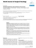
seen (Figure 1A). The clinical presentation to the
ophthalmologist and oncologist, a pate computed tomo-
graphy (CT) scan suggested a superior eyelid mass
lesion and enophthalmos (Figure 1B). Magnetic reso-
nance imaging showed no invasion in orbit, but the
results were compatible with a malignant eyelid. Further
investigation revealed systemic metastasis. A chest CT
* Correspondence: ; ;
;
1
TianJin Medical University Eye Center, 300084 TianJin P.R. China
2
Department of Pathology of TianJin Medical University, TianJin Cancer
Hospital, 300060 TianJin P.R. China
Full list of author information is available at the end of the article
Chen et al. World Journal of Surgical Oncology 2011, 9:58
/>WORLD JOURNAL OF
SURGICAL ONCOLOGY
© 2011 Chen et al; licensee BioMed Central Ltd. This i s an Open Access article distributed under the terms of the Creative Commons
Attribution License ( which permits unrestr icted use, distribution, and reproduction in
any medium, provided the original work is properly cited.
scan showed multi-metastases in the apex of lung,
metastasis mass of mediastinal lymph node and mediast-
inal lymphadenovarix (Figure 1C).
In view of the suspected diagnosis of large malignant
tumor, a biopsy was taken to confirm a provisional diag-
nosis. A biopsy was pe rformed under l ocal anaesthesia.
Histopathologic al examination of the bi opsy sample
showed a tumoral infiltration of the dermis by rounded
monomorphic cells of medium size with scant cyto-
plasm, round nuclei, and small nucleoli, clumps of a
small cell tumor, forming solid masses or small trabecu-
lar structures. The tomor cells with the mitotic index
was high (Figure 1D). The cells were arranged in large
nests, masses, and strands (Figure 2A). The formation of
glandular lumens was not observed. The t umor tissue
immunohistochemical study proved positive for cytoker-
atin 20(CK20), neuronal specific enolase (NSE) and
cytokeratin CAM5.2. The positive results are shown in
Figure 2 (2B-D). There was no immunoreactivity to pro-
tein S-100, thyroid t ranscription factor 1(TTF-1) a nd
leukocyte common antigen (LCA). Immunohistochem-
ical staining showed characteristic. All these features
above are consistent with the diagnosis of MCC. A diag-
nosis of MCC was made and the patient was referred to
the Oncology Department. The patient’s condition dete-
riorated rapidly with a midrange anaemia and she
required palliative care for disseminated MCC by her
oncologist.
Discussion
Merkel cell carcinoma is a frequently lethal skin cancer
that has a high propensity for nodal metastases and
local recurrence, has poor prognosis. Several reports
have described the association of MCC of the eyelids
[3-5]. We report the case of MCC that the patient had
been diagnosed with chalazion 3 years ago in the left
upper eyelid and was being treated with surgical treat-
ment. Although misdiagnosis of MCC pathologically as
chalazions is a p itfall, this sometimes occurs. Lesions
demonstrate a broad spectrum of clinical appearances at
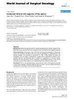
Figure 1 Phot ograph showing patient who had a red lesion of the upper eyelid, the most common localization of ocular Merkel cell
carcinoma, but the large lesion was uncommon. (A) Bottom: (lateral view) The large violaceous mass that involves the entire left eyelid and
facial surface multiple, ectensive large ulcer. The large tumor with multiple big dome-shaped nodules obscure boundary, plentiful blood vessels
in the surface. (B) CT (computed tomography) scans show a large medium to high reflectivity mass. (C) CT showed that there were tumor
metastases of mediastinal lymph node and multiple micrometastases (yellow arrow) of the lungs. (D) MCC with the mitotic index was high
(black arrows) as stained by hemotoxylin & eosin.
Chen et al. World Journal of Surgical Oncology 2011, 9:58
/>Page 2 of 5
presentation, including large ulcerated lesions, large
nodular lesions, exceeding 15 cm in diameter. Adjunc-
tive techniques, including biopsy, immunohistochemistry
and elect ron microscopy, can be he lpful in questionable
cases. In this session, speakers will present the most
current data on the clinical presentation, pathology, and
management of MCC. Representative and challenging
cases will be presented to highlight histopathological
diagnosis and treatment options.
To be exact, although MCC lacks specific clinical fea-
tures, some patients may have constitutional symptoms
with evidence of regional or distant metastasis. Heath et
al [6] reported AEIOU Features derived from 195
patientsofMCC.Thebiopsyshouldbeconsideredif
the patient presents ≥ 3 features of the above. This
study is the first to define the clinical features that may
serve as clues in the diagnosis of MCC. With this case,
the initial diagnosis was a chalazion, and no histopatho-
logic diagnoses were performed.
The histogenesis of MCC is controversial. Possible
cells of origin include the epidermal Merkel cell, a der-
mal Merkel cell equivalent, a neural-crest-derived cell of
the amine precur sor uptake. Less commonly, MCC may
simulate lymphoma, or may exhibit plasmacytoid, clear
cell, anaplastic, or spindle-cell features. Vascular or lym-
phatic invasion is not uncommon. The tumor in this
case showed multi- morphological type such as round,
small, plasmacytoid and spindle cells histology. There-
fore, this t ends to lead to misdiagnosis in some cases,
particularly if immunohistochemistry is not performed
to confirm the nature of the cells present. In this case,
the tumor tissue was positive for CK20, NSE and CAM
5.2, the patient with bad prognostic factors [7,8].CK20 is
expressed in a dotlike paranuclear or crescentic pattern.
Syn Neurofilament is also expressed in the cytoplasm of
most MCC. The above findings support the diagnosis of
primary MCC.
Diagnosis of MCC involves the following: General his-
tory, physical exam and patholo gical tests. It is a rare
type of skin cancer that is usuall y misdiagnosed.
Although MCC h as characteristic clinical features, the
diagnosis generally relies on histopathologic identifica-
tion. Innunohistochemistry is required to differentiate
MCC from other small round cell tumors; however,
Figure 2 Microscopic analysis of biopsy of Merkel cell carcinoma . (A) Photomicrograph showing that Merkel cell carcinoma tumor cells are
surrounded by intense inflammation with lymphocytes, plasma cells, and histiocytes. Proliferation of basophilic cells with round uniform nuclei,
scanty cytoplasm, patchy chromatin and inconspicuous nucleoli (black arrows). (H&E, ×400). (B) Photomicrograph showing the same tumor
stained for CK20. There is strong expression of CK20 in the cytoplasm and membrane of MCC. C Immunostaining with CAM5.2 showing
characteristic para-nuclear accentuation. (D) NSE positive suffusion expresstion was localized on in the cytoplasm and membrane. (IHC, ×400).
Chen et al. World Journal of Surgical Oncology 2011, 9:58
/>Page 3 of 5
clinical correlation may be required in differentiating
MCC from other neuroendocrine tumors that have
metastasized to the eyelids. The case we reported was
misdiagnosed as chalazion. The exact diagnosis of MCC
is made with a biopsy, for special stains are used to dis-
tinguish. Immunohistochemistry is very helpful. MCC
from other forms of cancer, such as sebaceous cyst,
small cell lung cancer (SCLC) and lymphoma, small cell
melanoma. Each of these cancers has a unique profile as
defined by special stains. CK20 and TTF-1 (positive in
SCLC) help distinguish MCC SCLC [9]. Further diag-
nostic tests are needed, for example, the imaging tests.
With this case, the differential histopathological diagno-
sisshouldbemadewith:1.Thetumorinthiscase
showed very large lesion with ulceration and mixed
epithelioid and spindle cell histology, and the above pre-
sentations may lead to misdiagnosis in some cases [10],
particularly if immunohistochemistry is not performed
to confirm t he nature of the ce lls present. In our case,
we did not see this feature. 2. In this case, the positive
assay for CK 20, NSE, CAM 5.2 and the negative one for
TTF-1and S100. In this tumor, a definition was also
supported by multi-metastases in the apex of lung and
mediastinal lymphadenovarix of pathological findings on
the plain CT chest.
Treatment is g enerally based on the stage of the dis-
ease. There are major treatments for MCC: surgical care
and m edical care [11]. MCC is chemosensitive but only
rarely chemocurable in patients with metastasis or
locally advanced tumors. Moreover, a high incidence of
toxic death occurs due to chemotherapy. Combination
chemotherapy is more effective when two or more
drugs are given at the same time because they are more
powerful in combination than either individual drug
[12]. Primary treatment of the tumor consists of exci-
sion with wide margins or micrographic surgery with or
without adjuvant radiotherapy. There is a decrease of
local recurrence after radiotherapy [13,14]. However,
this has no effect on overall survival [15]. Currently,
most eyelid MCCs are treate d without irradiation. Mer-
kel cell c arcinomas respond well to radiation therapy,
although some have recurred in the radiation field or
during radiotherapy [16]. The goal of wide surgical exci-
sion is to control local recurrence and lymph node
metastases. MCC should be r emoved with clear margins
as judged by pathology examination. It was recently
reported that sentinel lymph nodes was effective in pre-
dicting the risk of regional recurrence [17], however,
lymph node dissection does not appear to convey a sur-
vival advantag e [18]. This may be the result of the short
follow-up in most reports. There are some reports of
responses to interferon [19] and intralesional tumor
necrosis factor [20,21]. Radiation therapy, also referred
to as radiotherapy, is t he treatment of cancer with
penetrating beams of energy waves or streams of parti-
cles that ca n destroy cancer cells. Radiation therapy also
damages healthy cells in the field of radiation [22]. Cis-
platin plus etoposide, cyclophosphamide plus doxorubi-
cin plus vincristine, or cyclo phosphamide plus
epirubicin plus vincristine are the most commonly used
regimens [23]. The response rate is 70%, with a com-
plete response in 35% [24]. Interestingly, nonocular
MCC is reported to be a very aggressive tumor, lethal in
33% of patients. In contrast with the literature of MCC
at other sites, the authors found only a few patients who
died of MCC of the eyelid. This may indicate a good
prognosis for eyelid MCC. However, most MCC eyelid
studies have a limited follow-up [25]. Overall, the mor-
tality rate is less than 50% in two years, We need more
studies including longer-term follow-up.
Conclusions
In conclusion, this is t he first report of a case of MCC
with a mega lo-neoplasms, high malignance and a poor
prognosis. Although reports about MCC have appeared
successively, much still remains to be explored about
etiological factors, nosogenesis and treatment. It is
important to distinguish it from othe r tumors and early
diagnosis and therapy.
Consent
Informed consent was obtained from the patient for
publication of this case report and accompanying
images. A copy of the written consent is available for
review by the Editor-in-Chief of this journal.
List of abbreviations
(MCC): Merkel cell carcinoma; (TTF-1): Thyroid transcription factor-1; (CK20):
cytokeratin 20, (NSE): neuron specific enolase; leukocyte common antigen
(LCA) (MRI): Magnetic resonance imaging; (CT): computerized tomography.
Acknowledgements
This study was supported by Grant 09KZ102, 2010KZ101 From the Science
and technology Foundation of Health-bureau of Tianjin City, Grant from the
Tianjin Natural Science Foundation (International Cooperation, No.
09ZCZDSF04400). The authors wish to thank the patient’s family for
permission to publish the photographs.
Author details
1
TianJin Medical University Eye Center, 300084 TianJin P.R. China.
2
Department of Pathology of TianJin Medical University, TianJin Cancer
Hospital, 300060 TianJin P.R. China.
Authors’ contributions
YJH and BCS proposed the study. LXC and LMZ obtained images and
critically write the manuscript provided and reviewed pathological images.
JGW and TTL conducted a literature search. All authors read and approved
the final manuscript.
Competing interests
The authors declare that they have no competing interests.
Received: 11 January 2011 Accepted: 24 May 2011
Published: 24 May 2011
Chen et al. World Journal of Surgical Oncology 2011, 9:58
/>Page 4 of 5
References
1. Toker C: Trabecular carcinoma of the skin. Arch Dermatol 1972, , 105:
107-110.
2. Bleyen I, Wong J, Nguyen Q, Blanc JP, Hardy I: Merkel cell carcinoma of
the eyelid: a report of 2 cases. Can J Ophthalmol 2010, 45:85-86.
3. Tanahashi J, Kashima K, Daa T, Yada N, Fujiwara S, Yokoyama S: Merkel cell
carcinoma co-existent with sebaceous carcinoma of the eyelid. J Cutan
Pathol 2009, 36:983-986.
4. Rawlings NG, Brownstein S, Jordan DR: Merkel cell carcinoma
masquerading as a chalazion. Can J Ophthalmol 2007, 42:469-470.
5. Saedon H, Hubbard A: An unusual presentation of merkel cell carcinoma
of the eyelid. Orbit 2008, 27:331-333.
6. Heath M, Jaimes N, Lemos B, Mostaghimi A, Wang LC, Peñas PF, Nghiem P:
Clinical Characteristics of Merkel Cell Carcinoma at Diagnosis in 195
Patients: the AEIOU Features. Journal of the American Academy of
Dermatology 2008, 58:375-381.
7. Rund CR, Fischer EG: Perinuclear dot-like cytokeratin 20 staining in small
cell neuroendocrine carcinoma of the ovary (pulmonary-type). Appl
Immunohistochem Mol Morphol 2006, 14:244-248.
8. Bobos M, Hytiroglou P, Kostopoulos I, Karkavelas G, Papadimitriou CS:
Immunohistochemical distinction between merkel cell carcinoma and
small cell carcinoma of the lung. Am J Dermatopathol 2006, 28:99-104.
9. Llombart B, Monteagudo C, Lopez-Guerrero JA, Carda C, Jorda E,
Sanmartín O, Almenar S, Molina I, Martín JM, Llombart-Bosch A:
Clinicopathological and immunohistochemical analysis of 20 cases of
Merkel cell carcinoma in search of prognostic markers. Histopathology
2005, 46:622-634.
10. Metz KA, Jacob M, Schmidt U, Steuhl KP, Leder LD: Merkel cell carcinoma
of the eyelid: histological and immunohistochemical features with
special respect to differential diagnosis. Graefes Arch Clin Exp Ophthalmol
1998, 236:561-566.
11. Pathai S, Barlow R, Williams G, Olver J: Mohs’ micrographic surgery for
Merkel cell carcinomas of the eyelid. Orbit 2005, 24:273-275.
12. Voog E, Biron P, Martin JP, Blay JY: Chemotherapy for patients with locally
advanced or metastatic Merkel cell carcinoma. Cancer 1999, 85:2589-2595.
13. Plunkett TA, Subrumanian R, Leslie MD, Harper PG: Management of Merkel
cell carcinoma. Expert Rev Anticancer Ther 2001, 1:441-445.
14. Meeuwissen JA, Bourne RG, Kearsley JH: The importance of postoperative
radiation therapy in the treatment of Merkel cell carcinoma. Int J Radiat
Oncol Biol Phys 1995, 31
:325-331.
15. Poulsen MG, Rischin D, Porter I, Walpole E, Harvey J, Hamilton C, Keller J,
Tripcony L: Does chemotherapy improve survival in high-risk stage I and
II Merkel cell carcinoma of the skin? Int J Radiat Oncol Biol Phys 2006,
64:114-119.
16. Missotten GS, de Wolff-Rouendaal D, de Keizer RJ: Merkel cell carcinoma of
the eyelid review of the literature and report of patients with Merkel
cell carcinoma showing spontaneous regression. Ophthalmology 2008,
115:195-201.
17. Wong SL, Young YD, Geisinger KR, Shen P, Stewart JH, Sangueza O:
Intraoperative imprint cytology for evaluation of sentinel lymph nodes
from Merkel cell carcinoma. In Am Surg Edited by: Pichardo-Geisinger R,
Levine EA 2009, 75:615-619.
18. Gupta SG, Wang LC, Penas PF, Gellenthin M, Lee SJ, Nghiem P: Sentinel
lymph node biopsy for evaluation and treatment of patients with
Merkel cell carcinoma: The Dana-Farber experience and meta-analysis of
the literature. Arch Dermatol 2006, 142:685-690.
19. Durand JM, Weiller C, Richard MA, Portal I, Mongin M: Treatment of Merkel
cell tumour with interferon-alpha-2b. Br J Dermatol 1991, 124:509.
20. Pilotti S, Rilke F, Bartoli C, Grisotti A: Clinicopathologic correlations of
cutaneous neuroendocrine Merkel cell carcinoma. J Clin Oncol 1988,
6:1863-1873.
21. Güler-Nizam E, Leiter U, Metzler G, Breuninger H, Garbe C, Eigentler TK:
Clinical course and prognostic factors of Merkel cell carcinoma of the
skin. Br J Dermatol 2009, 161:90-94.
22. Garnski K, Nghiem P: Merkel cell carcinoma adjuvant therapy: Current
data support radiation but not chemotherapy. Journal of the American
Academy of Dermatology 2007, 57:166-169.
23. Feng H, Shuda M, Chang Y, Moore PS: Clonal integration of a
polyomavirus in human Merkel cell carcinoma. Science 2008,
319:1096-1100.
24. Fenig E, Brenner B, Katz A, Katz A, Rakovsky E, Hana MB, Sulkes A: The role
of radiation therapy and chemotherapy in the treatment of Merkel cell
carcinoma. Cancer 1997, 80:881-885.
25. Allen PJ, Bowne WB, Jaques DP, Brennan MF, Busam K, Coit DG: Merkel cell
carcinoma: prognosis and treatment of patients from a single institution.
J Clin Oncol 2005, 23:2300-2309.
doi:10.1186/1477-7819-9-58
Cite this article as: Chen et al.: Giant Merkel cell carcinoma of the
eyelid: a case report and review of the literature. World Journal of
Surgical Oncology 2011 9:58.
Submit your next manuscript to BioMed Central
and take full advantage of:
• Convenient online submission
• Thorough peer review
• No space constraints or color figure charges
• Immediate publication on acceptance
• Inclusion in PubMed, CAS, Scopus and Google Scholar
• Research which is freely available for redistribution
Submit your manuscript at
www.biomedcentral.com/submit
Chen et al. World Journal of Surgical Oncology 2011, 9:58
/>Page 5 of 5