báo cáo khoa học: "Clusterin confers gmcitabine resistance in pancreatic cancer" pps
Bạn đang xem bản rút gọn của tài liệu. Xem và tải ngay bản đầy đủ của tài liệu tại đây (1.22 MB, 8 trang )
RESEARCH Open Access
Clusterin confers gmcitabine resistance in
pancreatic cancer
Qingfeng Chen
1†
, Zhengkun Wang
1†
, Kejun Zhang
1†
, Xiaoyi Liu
1
, Weihong Cao
2
, Lei Zhang
3
, Shuhua Zhang
1
,
Bomin Yan
1*
, Yaoguang Wang
4
and Chunping Xia
4
Abstract
Objective: To measure clusterin expression in pancreatic cancer tissues and cell lines and to evaluate whether
clusterin confers resistance to gmcitabine in pancreatic cancer cells.
Methods: Immunohistochemistry for clusterin was performed on 50 primary pancreatic cancer tissues and 25
matched backgrounds, and clusterin expression in 5 pancreatic cancer cell lines was quantified by Western blot
and PT-PCR. The correlation between clusterin expression level and gmcitabine IC50 in pancreatic cancer cell lines
was evaluated. The effect of an antisense oligonucleotide (ASO) against clusterin(OGX-011) on gmcitabine
resistance was evaluated by MTT assays. Xenograft model was used to demonstrate tumor growth.
Results: Pancreatic cancer tissues expressed significantly higher levels of clusterin than did normal pancreatic
tissues (P < 0.01). Clusterin expression levels were correlated with gmcitabine resistance in pancreatic cancer cell
lines, and OGX-011 significantly decreased BxPc-3 cells resistance to gmcitabine (P < 0.01). In vivo systemic
administration of AS clusterin and gmcitabine significantly decreased the s.c. BxPC-3 tumor volume compared with
mismatch control ODN plus gmcitabine.
Conclusion: Our finding that clusterin expression was significantly higher in pancreatic cancer than in normal
pancreatic tissues suggests that clusterin may confer gmcitabine resistance in pancreatic cancer cells.
Introduction
Pancreatic cancer is resistant to almost all cytotoxic
drugs. Currently, gmcitabine appears to be the o nly
clinically active drug but, because of pre-existing or
acquired chemoresistance of most of the tumor cells, it
failed to significantly improve the outcome of pancreatic
carcinoma patients [1].
Clusterin, also known as t estosterone-repressed pros-
tate message-2 (TRPM-2), a polipoprotein J (ApoJ), sul-
fated glycoprotein-2 (SG P-2), and complement lysis
inhibitor(CLI), was first isolated from ram rete testes
fluid and plays important roles in various pathophysiolo-
gical processes, such as tissue remodeling, lipid trans-
port, complement regulation, and apoptosis [2]. Initial ly
clusterin has been regarded as a marker for cell death
because its expression is up-regulated in various normal
and malignant tissues undergoing apoptosis [3-5]. How-
ever, recent studies suggest a possible role for this gene
in protecting cells from death, and consistently demon-
strated that overexpression of clusterin closely correlates
with the progression of various human malignancies
[6-10] . More recent s tudies suggest that antisense oligo-
nucleotide or interfering RNAs (siRNAs) to clusterin
can enhance chemosensitivity in human cancer cells
[11-15]. Taken together, these findings indicate that
clusterin may play an important role in chemoresistance.
To extend these observations to pancreatic cancer, we
measured clusterin expression levels in pancreatic can-
cer tissue samples and cell l ines and sought to deter-
mine th e role of clu sterin in conf erring gmcitabine
resistance in pancreatic cancer cells.
Materials and methods
Cell and Tissue Collection and Preparation
Human pancreatic cancer cel l lines(PT45-P1, T3M4,
BxPc-3, Capan-1 and PancTu-1) were obtained from the
American Type Culture Collection (ATCC, Manassas,
* Correspondence:
† Contributed equally
1
Surgery, the Affiliated Hospital of Medical College, QingDao University,
QingDao.266003. R.P. China
Full list of author information is available at the end of the article
Chen et al. World Journal of Surgical Oncology 2011, 9:59
/>WORLD JOURNAL OF
SURGICAL ONCOLOGY
© 2011 Chen et al; licensee BioMed Central Ltd. T his is an Open Access article distribute d under the terms of the Creative Commons
Attribution License ( which permits unrestricted use, distribution, and reproduction in
any medium, provided the original work is properly cited.
Virginia)and maintained in Dulbecco’s modified Eagle’ s
medium with 10% fetal calf serum. Pancreas tissue sam-
ples [16] (50 tumors and 25 matched backgrounds) were
collected and part of the tissues immediately frozen in
liquid nitrogen before proce ssing, and part of the tissue
was used for immunohistochemical staining.
Immunohistochemical staining for clusterin
Serial 4-um-thick sections of the tissue array blocks
were subjected to immunohistochemical study. The sec-
tions were deparaffinized, and endogenous peroxidase
was blocked with 3% H2O2. Then the slides were
labeled with a monoclonal antibody to clusterin (clone
B-5, 1:200 dilution; Santa Cruz Biotechnology, Santa
Cruz, CA) for 1 hour. After washing with phosphate-
buffered saline, the sections were incubated with bioti-
nylated secondary antibody and then with an avidinbio-
tin streptavidin-peroxidase complex (Vectastain Elite
ABC kit; Vector Laboratorie s, Burlingame, CA). 3,3’-dia-
minobenzidine tetrahydrochloride was used as a chro-
mogen, and Mayer’s hematoxylin counterstaining was
applied. Immunohistochemical staining of clusterin was
defined as detectable immunoreaction in cytoplasm.
Clusterin expression was scored as follows: negative(-) if
no staining was seen or if weak (+) immunoreactivity
was observed in <10% of the tumor cells, and positive
(overexpression) if >10% of the tumor cells demon-
strated moderate (++) to strong (+++) staining. The
results of control staining were satisfactory.
AS Clusterin ODN
The sequences of AS clusterin ODN corresponding to
the human clusterin translation initiation site were 5’-
CAGCAGCAGAGTCTTCATCAT-3’. A 2-base clusterin
MM ODN (5’-CAGCAGCAG AGTATTTA-TCAT-3’)
was used as a control.
Treatment of Cells with ODN
Lipo fectin, a cationic lipid (Life Techno logies, Inc.), was
used to increase the ODN uptake of cells. BxPC-3 cells
were treated with various concentrations of ODN after a
preincubation for 20 min with 3 μg/ml lipofectin in
serum-free Opti-MEM (Life Techn ologies, Inc.). After
the beginning of the incubation (4 h), the medium con-
taining ODN and lipofectin was replaced with standard
culture medium as described above.
RNA Extraction and RT-PCR Analysis
The mRNA extraction and RT reaction of the tissue and
cell for synthesizing the first-strand cDNA was carried
out according to the manufacturer’s instructions. The
clusterin Primer sequences was:sense:5’-ATGATGAA-
GACTCTGCTGCT-3’,antisense:5’-TCACTCCTCCCG
GTGCTT-3’ ,GAPDH:sense:5’ -TGATGGGTGTG
AACCACGAG-3’,antisense:3’-T TGAAGTCGCAGGA
GACA ACC-5’. Fluorescent bands were visualized using
a UV-CCD video system (Epi-LightUVFA1100; AISIN
COSMOS, Tokyo, Japan) and were analyzed using
Quantity One image-analysis software (PDI, NY). The
intensity of each band r elative to the GAPDH band was
represented as t he mean ± s.d. The mean ± s.d. values
are shown in the figures. P < 0.05 was considered to be
statistically significant.
Western Blot Analysis
Samples containing equal amounts of protein (15 μg)
from lysates of the cultured PT45-P1, T3M4, BxPc-3,
Capan-1 and PancTu-1 cells and BxPc-3 tumors were
electrophoresed on an SDS-polyacrylamide gel and
transferred to a nitrocellulose filter. The filters were
blocked in PBS containing 5% nonfat milk powder at 4°
C overnight and then incubated for 1 h with a 1:400-
diluted antihuman clusterin goat polyclonal antibody
(Santa Cruz Biotechnology, Inc., Santa Cruz, CA), 1:50
0-diluted antirat b-actin mouse monoclonal antibody
(Chemicon International, Inc., Tumecula, CA), The fil-
ters were then incubated for 30 min with horseradish
peroxidase-conjugated antigoat or mouse IgG antibody
(Amersham Life Science, Arlington Heights, IL), and
specific proteins were detected using an enhanced che-
miluminescence Western blotting analysis system
(Amersham Life Science).
IC50 and MTT assay
Following the addition of 1 × 10
4
pancreatic canc er
cells into each well of a 96-well plate, 0.1 ml of med-
ium was added, containing various concentrations of
gmcitabine. The 50% inhibitory drug concentration
(IC50) was obtained by MTT assay. The result of three
repeated experiments was presented as the mean ±
standard error, and differences were analyzed using the
unpaired t.
Animal Studies
Female C57BL/6 mice at 6 -8 weeks old were o btained
from Qingdao Medical college, Qingdao University for
tumor implantation. All animals were maintained in a
sterile environment and cared for within the laboratory
animal regulations of the Ministry of Science and
Technology of the People’ s Republic of China (http://
www.most.gov.cn/kytj/kytjzcwj/200411). Full details of
the study approval by the ethics committee at the
affiliated hospital of medical college, Qingdao Univer-
sity. Each experimental group consisted of 10 mice.
Each of the tumor cell lines was trypsinized, washed
twice with PBS, and injected s.c. with 1 × 10
6
cells in
the flank as described previously [16]. After injection
for 30 days, the diameter in BxPC-3 tumors was 5~8
Chen et al. World Journal of Surgical Oncology 2011, 9:59
/>Page 2 of 8
mm. Mice bearing BxPC-3 tumors was randomly
selected for treatment with AS clusterin ODN alone,
MM control ODN alone, AS clusterin ODN plus gmci-
tabine, or MM c ontrol ODN plus gmcitabine. After
randomization, 10 mg/kg AS clusterin or MM control
ODN were injected i.p. once daily into each mouse for
28 days, and 40 uM of gmcitabine were injected i.v.
twice a week for 2 weeks. Tumor volume was mea-
suredonceweeklyandcalculatedbytheformula:1/2
(length × width × depth). Data points were reported as
average tumor volumes ± SD.
Statistical Assessment
All statistical analyses were performed using SPSS13.0
software. The results were presented as mean ± SD of
three replicate assays. Differences between various
groups were assessed using ANOVA or Dunnett t-test.
A P value of <0.05 was considered to indicate statistical
significance.
Results
Expression of clusterin in pancreatic cancer tissues
samples
Clusterin protein immunoreactivity was detected both in
matched backgrounds and pancreatic cancer cells. The
immunostaining results are presented in Table 1. Of the
50 pancreatic cancer tissues, 26 (52%)exhibited clusterin
ove rexpression in cancer cells, 4(8%) exhibited clusterin
weak expression in cancer cells(Figure 1.A-C), and no
clusterin stai ning was shown in 20 pancreatic cancertis-
sues. Of the 25 matched backgrounds tissues, Only 1 of
these showed strong immunoreactivity (++/+++), and 7
were immunoreactive in 1-10% of the tumour cells (+)
(Figure 1.D-E). A highly significant clusterin protein
immunoreactivity was shown in pancreatic cancer cells
(P < 0.05)(Table 1).
The relative c lusterin mRNA value was 0.764 ± 0.18
for tumor and 0.14 ± 0.11 for backgrounds. Levels of
clusterin was increased in tumor samples in comparison
to matched backgrounds tissues (P = 0.0136).
Relationship between clusterin expression and
gmcitabine IC50 in pancreatic cancer cell lines
The association between clusterin protein expression
and gmcitabine IC50 was examined in five pancreatic
cancer lines:PT45-P1, T3M4, BxPc-3, Capan-1 and
PancTu-1. In each of these cell lines, clusterin
expression was assayed byWestern blotting and RT-PCR
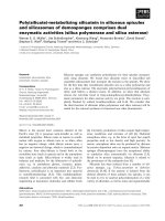
(Figure 2A a nd 2B). The highest expression of clusterin
was observed in BxPc-3 cells. The mean+SD IC50 of
gmcitabine for the PT45-P1, T3M4, BxPc-3, Capan-1
and PancTu-1 cell lines was(9.26 ± 0.0 3) × 10
-7
,(8.38 ±
0.07) × 10
-7
, (1.05 ± 0.09) × 10
-5
, (8.32 ± 0.06) × 10
-
6
and (6.04 ± 0.07) × 10
-6
M, respectively (Figure 2C).
BxPC-3 cells showed the highest resistance to gmcita-
bine. Thus, gmcitabine protein expression levels sh owed
a significant correlation with resistance to gmcitabine
(R
2
, P = 0.001).
AS ODN-mediated Inhibition of Clusterin Expression in
BxPc-3 Cells
The effect of treatment with AS clusterin ODN on clus-
terin protein expression in BxPc-3 cells, which shows the
highest level of clusterin expression was evaluated by
western blot and RT-PCR analysis. As shown in Figure
3A and 3B, daily treatment of BxPc-3 cells with AS clus-
terin ODN (100, 500, or 1000 nM) for 2 days reduced
clusterin levels by 30, 75, or 98%, respectively. In con-
trast, clusterin expression was not affected by the 2-base
MM control ODN at any of the used concentrations.
Changes in Clusterin Expression in BxPC-3 Cells after AS
Clusterin ODN and Gmcitabine Treatment
Western blot analysis was used to determine the effects of
gmcitabine treatment on clusterin protein expression in
BxPC-3 cells. As shown in Figure 4A, clusterin induction
increased in a dose-dependent manner b y gmcitabine
treatment at concentrations 0.2-20 uM. Time-course
experiments demonstrated that gmcitabine-induced clus-
terin up-regulation peaked by 24 h post-treatment and
began decreasing by 48-h post-treatment (Figure 4B). We
then examined the effects of combined treatment with AS
clusterin ODN and gmcitabine on clusterin expression in
Table 1 Immunohistochemical staining for clusterin
Clusterin
Groups n - + ++/+++ P
pancreatic cancer tissues 50 20 4 26
matched backgrounds 25 17 7 1 <0.01
Figure 1 Expression of clusterin in pancreatic cancer cells. A-C,
C lusterin protein expression was detected in the pancreatic cancer
cytoplasm. A.(+), B.(++), C.(+++). D. clusterin protein expression (+)
was detected in the matched backgrounds of the pancreatic cancer.
E. Only one patients of the matched backgrounds shown clusterin
protein expression (++/+++).
Chen et al. World Journal of Surgical Oncology 2011, 9:59
/>Page 3 of 8
BxPC-3 cells. As shown in Figure 4C, 500 nM AS clusterin
ODN combined with 10 uMgmcitabinefor48h
decreased clusterin protein levels by 60%, compared with
500 nM MM control ODN treatment.
AS Clusterin ODN Treatment Enhanced Chemosensitivity
of BxPc-3 Cells in Vitro
To determine whether treatment with AS clusterin
ODN enhances the cytotoxic effects of gmcitabine,
BxPc-3 cells were treated with 500 nM AS clusterin
ODN or MM control ODN once daily for 2 days and
then incubated with medium containing various concen-
trations of gmcitabine for 2 days. The MTT assay was
then performed to measure the number of viable cells.
As shown in Figure 5 A, AS clusterin ODN treatment
significantly enhanced chemosensitivity of gmcitabine in
a dose-dependent manner, reducing the IC
50
of gmcita-
bine by >50%.
Figure 3 Sequence-specific and dose-dependent inhibition of clusterin expression by AS clusterin ODN in BxPc-3 cells.InA, BxPc-3 cells
were treated daily with various concentrations of AS cluserin ODN or a 2-base clusterin MM ODN as a control for 2 days, total cell protein was
extracted from culture cells, and clusterin and GAPDH levels were analyzed by western blotting. In B, quantitative analysis of clusterin protein
levels after normalization to GAPDH levels in BxPc-3 cells after treatment with various concentrations of AS clusterin ODN or MM control ODN
was performed using laser densitometry. Each point represents the mean of triplicate analyses with SD. *, differs from controls (P < 0.01) by
Student’s t test.
Figure 2 Expression level of clusterin and gmcitabine IC50 of pancreatic cancer cell lines. A, RT-PCR analysis clusterin mRNA in 5
pancreatic cancer cell lines (PT45-P1, T3M4, BxPc-3, Capan-1 and PancTu-1). B, Western blot analysis of 5 pancreatic cancer cell lines (PT45-P1,
T3M4, BxPc-3, Capan-1 and PancTu-1) using a monoclonal antibody specific for the clusterin a chain. As a protein loading control, the same blot
was incubated with an anti-GAPDH monoclonal antibody. C, Correlation between clusterin expression and gmcitabine resistance. Linear
regression analysis showed a statistically significant relationship between clusterin expression and gmcitabine IC50 (R
2
, P = 0.001).
Chen et al. World Journal of Surgical Oncology 2011, 9:59
/>Page 4 of 8
To determine whether AS clusterin ODN enhances
the cytotoxic effects of gmcitabine was AS ODN dose-
dependent, BxPc-3 cells were treated with 100, 500,
1000 nM AS clusterin ODN or MM control ODN once
daily for 2 days and then incubated with medium con-
taining 5 uM concentrations of gmcitabine for 2 days.
We found AS clusterin ODN treatment significantly
enhanced chemosensitivity of gmcitabine in a dose-
dependent manner Figure 5B.
Synergistic Inhibition of Growth of BxPC-3 Cells in Vivo by
AS Clusterin ODN and Gmcitabine
The efficacy of a regim en combining AS clusterin ODN
and gmcitabine for inhibiting the growth of s.c. BxPC-3
tumors was evaluated. Mice bearing BxPC-3 tumors 5~8
mm in diameter were r andomly selected for treatment
with AS clusterin ODN alone, MM control ODN alone,
AS clusterin ODN plus gmcitabine, or MM control ODN
plus gmcitabine. Mean tumor volume was similar at the
beginning of treatment in each of these groups. Changes
in tumor volume in mice treated with MM control ODN
or AS clusterin ODN alone was similar to that of
untreated mice. MM control ODN plus gmcitabine was
similar to that with gmcitabine alone. It showed AS clus-
terin ODN alone did not have significant effect on the
tumor growth. Although gmcitabine inhibited tumor
growth, there was no s tatistical significance(data not
shown). BxPC-3 tumor growth was significantly inhibited
by t reatment wi th combined AS clusterin ODN and
gmcitabine therapy. After tumor injection (49 days), the
tumor volume in mice treated with AS clusterin ODN
plus gmcitabine was smaller than that in mice treated
Figure 4 Effects of AS clusterin ODN and/or gmcitabine treatment on clusterin expression in BxPC-3 cells.InA, cells were treated with
various concentrations of gmcitabine for 48 h, total protein was then extracted and analyzed for clusterin and GAPDH levels by western
blotting. In B, cells were treated with 10 uM gmcitabine for indicated intervals, total protein was then extracted, and clusterin and GAPDH levels
were analyzed by western blotting. In C, cells were treated daily with 500 nM AS clusterin ODN or a 2-base clusterin MM control ODN for48 h.
After a 24-h exposure to 10 uM gmcitabine, total rotein was then extracted, and clusterin and GAPDH levels were analyzed by western blotting.
Chen et al. World Journal of Surgical Oncology 2011, 9:59
/>Page 5 of 8
with AS clusterin ODN alone, MM control ODN, gmci-
tabine alone, or MM control ODN plus gmcitabine(,*P <
0.05,**P < 0.01), respectively (Figure 6).
Discussion
Resistance to anticancer agents is one of the primary
impediments to effective cancer therapy. Chemoresistance
occurs not only to clinically established therapeutic agents
but also to novel targeted therapeutics. Both intrinsic and
acquired mechanisms have been implicated in drug resis-
tance but it remains controversial which mechanisms are
responsible that lead to failure of therapy in cancer
patients [15].
The clusterin (CLU) protein was first discovered more
than 25 years ago in rat testis fluid because of its ability
to facilitate clustering of a variety of cell types in culture
[17]. Since then, homologues of the broadly expressed
CLU gene have been identified in several species and
CLU proteins have been found in most mammalian
body fluids [18]. CLU is an enigmatic molecule, impli-
cated in diverse biological processes, and has
additionally been associated with opposing functions in
regard to apoptosis. Accumulating evidence from several
studies suggests that the pro- and antiapoptotic f unc-
tions may be related to nuclear and secreted protein iso-
forms, respectively [19]. The secreted form of CLU is a
glycosylated protein of 70-80 kDa that consists of two
chains held together by five disulfide bonds, and conse-
que ntly it appears as a ~40 kDa smear on immunoblots
from reducing SDS-PAGE. Its intracellul ar pre-curser
form of 60 kDa may also exhibit an antiapoptotic func-
tion [20]. The proapoptotic CLU variant is a 50-55 kDa
protein which accumulates in the nucleus of apoptotic
cells [19]. How these different CLU protein variants are
produced from the CLU gene is poorly understood,
although it has been speculated that nuclear CLU results
from an alternativ e splice event skipping exon 2 from
the main CLU transcript otherwise translated into
secreted CLU [21].
Recent focus h as turned to clusterin (CLU) as a key
contributor to chemoresistance to anticancer agents. Its
role has been documented in prostate cancer for
Figure 5 Effect of combined treatment with AS clusterin ODN and gmcitabine BxPC-3 cell growth. A, BxPC-3 cells were treated daily with
500 nM AS clusterin ODN or MM control ODN for 2 days. After ODN treatment, the medium was replaced with medium containing various
concentrations of gmcitabine. After 48 h of incubation, the number of viable cells was determined by the MTT assay. B, BxPC-3 cells were
treated daily with 100, 500, 1000 nM AS clusterin ODN or MM control ODN for 2 days. After ODN treatment, the medium was replaced with
medium containing 5 uM concentrations of gmcitabine. After 48 h of incubation, the number of viable cells was determined by the MTT assay.
Each data point represents the mean of triplicate analyses with SD. ** and *, differs from controls (P < 0.01 and P < 0.05, respectively) by
Student’s t test.
Chen et al. World Journal of Surgical Oncology 2011, 9:59
/>Page 6 of 8
paclitaxel/docetaxel resistance as well as in renal, breast,
and lung tumor cells[22-25]. It is noteworthy that only
the cytoplasmic/secretory clusterin form (sCLU), and
not the nuclear form, is expressed in aggressive late
stage tumors, which is in line with its antiapoptotic
function [15]. Most significantly, sCLU expression is
documented to lead to broad-based resistance to other
unrelated chemotherapeutic agents such as doxorub icin,
cisplatin, etoposide, and camphothecin [15].
The current treatment of choice for metastatic pan-
creatic cance r involves single-agent gmcitabine or a
combination of gmcitabine with capecitabine or erlotinib
(a tyrosine kinase inhibitor). Only 25, 20, 13; 30% of
patients respond to this treatment and patients who do
respond initially ultimately exhibit disease progression.
Median survival for pancreatic cancer patients has
reached a plateau due to inherent and acquired resis-
tance to these agents [26]. The actual mechanisms for
pancreatic cancer to resist gmcitabine are unclear.
Xie, et al. has foun d [27] clusterin was overexpressed
in pancreatic cancer tissues and pancreatic cancer cell
lines, it was not expressed in normal pancreas. We also
shown in our study that clusterin expression is signifi-
cantly higher in pancreatic cancer tissues than in normal
pancreatic tissue. To our knowledge, there was no
report about whether overexpression enhances their
resistance to cytotoxic chemotherapy, and downregula-
tion of clusterin increases their sensitivity cytotoxic che-
motherapy. Therefore, in the present study, we
evaluated the effect of decrease in clusterin expression
in the human pancreatic cancer BxPC-3 cells using AS
ODN, and s tudy whether downregulation of clusterin
increase their sensitivity cytotoxic chemotherapy both in
vitro and in vivo.
In the present study, we showed a significant correla-
tion between clusterin expression and gmcitabine IC50
in the pancreatic cancer cell lines, we tested the hypoth-
esis that clusterin e xpression may confer gmcitabine
resistance in pancreatic cancer cells. Phosphorothioate
AS clusterin ODN corresponding to the human cluster in
translation initiation site used in this study inhibited clus-
terin expression in a dose- and sequence dependent man-
ner, even after gmcitabine treatment, which resulted in
an increase in clusterin expression. Furthermore, treat-
ment of BxPC-3(expressing hi gh levels of clusterin) with
AS clusterin ODN reduced the IC50 of gmcitabine by >
50%. These findings suggest that clusterin expression in
pancreatic cancer cells may confer a pheno type resistant
to chemotherapeutic agents;the reduction in clusterin
expression by AS clusterin ODN enhances the sensitivity
of cytotoxic chemotherapy for pancreatic cancer. Accord-
ingly, based on the findings of the present in vitro experi-
ments, we then examined whether AS clusterin ODN
therapy synergistically enhances the cytotoxic effect of
gmcitabine on the growth BxPC-3 cells in vivo .Consis-
tent with the in vitro studies, a regimen combining AS
clusterin and gmcitabine synergistically inhibited the
growth of s.c. BxPC-3 tumors in vivo. These findings sug-
gest that it might be possible t o achieve powerful cyto-
toxic effects of gmcitabine at tolerable doses by
combining with AS clusterin ODN.
In conclusion, we have shown here that clusterin
expression was significantly higher in pancreatic cancer
tumor samples than in normal pancreas tissues and that
clusterin expression was significantly correlated with
gmcitabine resistance in pancreatic canc er cell lines.
These findings may lead to the development of new
therapeutic regimens, targeting clusterin expression,
Figure 6 Effects of combined treatment with AS clusterin ODN plus gmcitabine on BxPC-3 tumor growth. Mice bearing BxPC-3 tumor
were randomly selected for treatment with AS clusterin ODN, MM control ODN, AS clusterin ODN plus gmcitabine, or MM control ODN plus
gmcitabine. After tumor cell injection (7 days), 10 mg/kg AS clusterin ODN or MM control ODN was daily injected i.p. for 28 days. gmcitabine (40
μM) was injected i.v. twice a week for 2 weeks. Tumor volume was measured once weekly and calculated by the formula: length × width ×
depth × 0.5. Each point represents the mean tumor volume in each experimental group containing eight mice with SD. *, differs from controls
(*P < 0.01) by Student’s t test.
Chen et al. World Journal of Surgical Oncology 2011, 9:59
/>Page 7 of 8
particularly in patients with gmcitabine-insensitive pan-
creatic cancers expressing high levels of clusterin.
Author details
1
Surgery, the Affiliated Hospital of Medical College, QingDao University,
QingDao.266003. R.P. China.
2
Pathology, the Affiliated Hospital of Medical
College, QingDao University, Shan Dong Province, 266003. P.R. China.
3
Molecular Biology, the Affiliated Hospital of Medical College, QingDao
University, Shan Dong Province, 266003. P.R. China.
4
Hepatobiliary surgery,
Tianjin Medical university Cancer Institute and Hospital, Tianjin, China,
300060, Huanhuxi Road, Hexi District, Tianjin; Key Laboratory of Cancer
Prevention and Therapy, Tianjin.
Received: 28 January 2011 Accepted: 24 May 2011
Published: 24 May 2011
References
1. Arlt A, Gehrz A, Müerköster S, Vorndamm J, Kruse ML, Fölsch UR, Schäfer H:
Role of NF-kappaB and Akt/PI3K in the resistance of pancreatic
carcinoma cell lines against gmcitabine-induced cell death. Oncogene
2003, 22:3243-51.
2. Rosenberg ME, Silkensen J: Clusterin: Physiologic and pathophysiologic
considerations. Int J Biochem Cell Biol 1995, 27:633-645.
3. Kyprianou N, English HF, Davison NE, Issac JT: Programmed cell death
during regression of the MCF-7 human breast cancer following estrogen
ablation. Cancer Res 1991, 51:162-166.
4. Wright PS, Cross-Doersen D, Th’ng JP, Guo XW, Crissman HA, Bradbury EM,
Montgomery LR, et al: A ribonucleotide reductase inhibitor, MDL 101,
731, induces apoptosis and elevates TRPM-2 mRNA levels in human
prostate tumor xenografts. Exp Cell Res 1996, 222:54-60.
5. Schwochau GB, Nath KA, Rosenberg ME: Clusterin protects against
oxidative stress in vitro through aggressive and nonaggressive
properties. Kidney Int 1998, 53:1647-1653.
6. Savković V, Gantzer H, Reiser U, et al: Clusterin is protective in pancreatitis
through anti-apoptotic and anti-inflammatory properties. Biochem
Biophys Res Commun 2007, 356:431-7.
7. Miyake H, Gleave M, Kamidono S, Hara I: Overexpression of clusterin in
transitional cell carcinoma of the bladder is related to disease
progression and recurrence. Urology 2002, 59:150-4.
8. Lourda M, Trougakos IP, Gonos ES: Development of resistance to
chemotherapeutic drugs in human osteosarcoma cell lines largely
depends on up-regulation of Clusterin/Apolipoprotein. J Int J Cancer
2007, 12:611-22.
9. Redondo M, Villar E, Torres-Munoz J, Tellez T, Morell M, Petito CK:
Overexpression of clusterin in human breast carcinoma. Am J Pathol
2000, 157:393-399.
10. Miyake H, Gleave ME, Arakawa S, Kamidomi S, Hara I: Introducing the
clusterin gene into human renal cell carcinoma cells enhances their
metastatic potentials. J Urol 2002, 167:2003-2008.
11. Trougakos IP, So A, Jansen B, Gleave ME, Gonos ES: Silencing expression of
the clusterin/apolipoprotein j gene in human cancer cells using small
interfering RNA induces spontaneous apoptosis, reduced growth ability,
and cell sensitization to genotoxic and oxidative stress. Cancer Res 2004,
64:1834-42.
12. Chung J, Kwak C, Jin RJ, Lee CH, Lee KH, Lee SE: Enhanced
chemosensitivity of bladder cancer cells to cisplatin by suppression of
clusterin in vitro. Cancer Lett 2004, 203:155-61.
13. Miyake H, Eto H, Hara I, So A, Li D, Gleave ME: Synergistic antitumor
activity by combined treatment with gmcitabine and antisense
oligodeoxynucleotide targeting clusterin gene in an intravesical
administration model against human bladder cancer kotcc-1 cells. J Urol
2004, 171
:2477-81.
14. Muramaki M, So A, Hayashi N, Sowery R, Miyake H, Fujisawa M, Gleave ME:
Chemosensitization of gmcitabine-resistant human bladder cancer cell
line both in vitro and in vivo using antisense oligonucleotide targeting
the anti-apoptotic gene, clusterin. BJU Int 2009, 103:384-90.
15. Djeu JY, Wei S: Clusterin and chemoresistance. Adv Cancer Res 2009,
105:77-92.
16. Zhang K, Dong Chen, Jiao X, et al: Slug enhances invasion ability of
pancreatic cancer cells through upregulation of matrix
metalloproteinase-9 and actin cytoskeleton remodeling. Laboratory
Investigation 2011, 1-13.
17. Fritz IB, Burdzy K, Setchell B, Blaschuk O: Ram rete testis fluid contains a
protein (clusterin) which influences cell-cell interactions in vitro. Biol
Reprod 1983, 28:1173-1188.
18. Trougakos IP, Gonos ES: Clusterin/apolipoprotein J in human aging and
cancer. Int J Biochem Cell Biol 2002, 34:1430-1448.
19. Shannan B, Seifert M, Leskov K, Willis J, Boothman D, Tilgen W, Reichrath J:
Challenge and promise: roles for clusterin in pathogenesis, progression
and therapy of cancer. Cell Death Differ 2006, 13:12-19.
20. Zhang H, Kim JK, Edwards CA, Xu Z, Taichman R, Wang CY: Clusterin
inhibits apoptosis by interacting with activated Bax. Nat Cell Biol 2005,
7:909-915.
21. Leskov KS, Klokov DY, Li J, Kinsella TJ, Boothman DA: Synthesis and
functional analyses of nuclear clusterin, a cell death protein. J Biol Chem
2003, 278:11590-11600.
22. Sowery RD, Hadaschik BA, So AI, Zoubeidi A, Fazli L, Hurtado-Coll A,
Gleave ME: Clusterin knockdown using the antisense oligonucleotide
OGX-011 re-sensitizes docetaxel-refractory prostate cancer PC-3 cells to
chemotherapy. BJU Int 2008, 102:389-97.
23. So A, Sinnemann S, Huntsman D, Fazli L, Gleave M: Knockdown of the
cytoprotective chaperone, clusterin, chemosensitizes human breast
cancer cells both in vitro and in vivo. Mol Cancer Ther 2005, 4:1837-49.
24. July LV, Beraldi E, So A, Fazli L, Evans K, English JC, Gleave ME: Nucleotide-
based therapies targeting clusterin chemosensitize human lung
adenocarcinoma cells both in vitro and in vivo. Mol Cancer Ther 2004,
3:223-32.
25. Zellweger T, Miyake H, July LV, Akbari M, Kiyama S, Gleave ME:
Chemosensitization of human renal cell cancer using antisense
oligonucleotides targeting the antiapoptotic gene clusterin. Neoplasia
2001, 3:360-7.
26. Sheikh R, Walsh N, Clynes M, O’Connor R, McDermott R: Challenges of
drug resistance in the management of pancreatic cancer. Expert Rev
Anticancer Ther 2010, 10:1647-61.
27. Xie MJ, Motoo Y, Su SB, Mouri H, Ohtsubo K, Matsubara F, Sawabu N:
Expression of clusterin in human pancreatic cancer. Pancreas
2002,
25:234-8.
doi:10.1186/1477-7819-9-59
Cite this article as: Chen et al.: Clusterin confers gmcitabine resistance
in pancreatic cancer. World Journal of Surgical Oncology 2011 9:59.
Submit your next manuscript to BioMed Central
and take full advantage of:
• Convenient online submission
• Thorough peer review
• No space constraints or color figure charges
• Immediate publication on acceptance
• Inclusion in PubMed, CAS, Scopus and Google Scholar
• Research which is freely available for redistribution
Submit your manuscript at
www.biomedcentral.com/submit
Chen et al. World Journal of Surgical Oncology 2011, 9:59
/>Page 8 of 8