báo cáo khoa học: "Pulmonary sclerosing hemangioma in a 21-year-old male with metastatic hereditary non-polyposis colorectal cancer: Report of a case" pps
Bạn đang xem bản rút gọn của tài liệu. Xem và tải ngay bản đầy đủ của tài liệu tại đây (1.53 MB, 6 trang )
CAS E REP O R T Open Access
Pulmonary sclerosing hemangioma in a 21-year-
old male with metastatic hereditary non-
polyposis colorectal cancer: Report of a case
Tobias S Schiergens
1*†
, Philipe N Khalil
2†
, Doris Mayr
3
, Wolfgang E Thasler
1
, Martin K Angele
1
, Rudolf A Hatz
1
,
Karl-Walter Jauch
1
and Axel Kleespies
1
Abstract
Background: Pulmonary sclerosing hemangioma (SH) is a rare tumor of the lung predominantly affecting Asian
women in their fifth decade of life. SH is thought to evolve from primitive respiratory epithelium and mostly shows
benign biological behavior; however, cases of lymph node metastases, local recurrence and multiple lesions have
been described.
Case Presentation: We report the case of a 21-year-old Caucasian male with a history of locally advanced and
metastatic rectal carcinoma (UICC IV; pT4, pN1, M1(hep)) that was eventually identified as having hereditary non-
polyposis colorectal cancer (HNPCC, Lynch syndrome). After neoadjuvant chemotherapy followed by low anterior
resection, adjuvant chemotherapy and metachronous partial hepatectomy, he was admitted for treatment of newly
diagnosed bilateral pulmonary metastases. Thoracic computed tomography showed a homogenous, sharply
marked nodule in the left lower lobe. We decided in favor of atypical resection followed by systematic
lymphadenectomy. Histopathological analysis revealed the diagnosis of SH.
Conclusions: Cases have been published with familial adenomatous polyposis (FAP) and simultaneous SH. FAP,
Gardner syndrome and Li-Fraumeni syndrome, however, had been ruled out in the present case. To the best of
our knowledge, this is the first report describing SH associated with Lynch syndrome.
Keywords: Sclerosing hemangioma Pneumocytoma, Colorectal cancer (CRC), Hereditary non-polyposis colorectal
cancer (HNPCC), Lynch syndrome, Familial adenomatous polyposis (FAP)
Background
Sclerosing hemangioma of the lung (SH), alternatively
characterized as alveolar pneumocytoma, was first
described by Liebow and Hubbel in 1956 [1] and repre-
sents a rare and, in the majority of cases, benign neo-
plasm of the lung. It predominantly affects females in
their fifth decade of life [2,3] and is more common in
Asian women. Although several theories have been pro-
posed for its histogenesis and the term implies an
endot helial derivation, an origin from immature respira-
tory epithelium is currently accepted [3-7]. Symptoms
such as atypical thoracic pain, cough, hemoptysis and
dyspnea might occur due to tumor enlargement and
compromising of surrounding tissue [3]. However, in
most patients, SH is detected incidentally during routine
chest radiographic examination because it is generally
asymptomatic [2,8]. Although SH is thought to be
benign, cases of lymph node metastases, local recurrence
and multiple lesions have been reported [2,9-11] sug-
gesting that the progression to an overtly malignant
phenotype might be po ssible. Lymph node metastases,
however, do not seem to have an impact on long-term
survival [12]. Altog ether, little is known about the asso-
ciated risk factors, prognosis and natural c ourse of SH,
and little clinical data exists from western countries.
Only a few cases have been reported affecting young
patients . There are two recent reports describing middle-
aged female patients suffering from familial adenomatous
* Correspondence:
† Contributed equally
1
Department of Surgery, University of Munich, Campus Grosshadern,
Germany
Full list of author information is available at the end of the article
Schiergens et al. World Journal of Surgical Oncology 2011, 9:62
/>WORLD JOURNAL OF
SURGICAL ONCOLOGY
© 2011 Schiergens et al; licensee BioMed Central Ltd. This is an Open Access article distributed under the terms of the Creative
Commons Attribution License ( which permits unrestricted use, distribution, and
reproduction in any medium, provided the original work is properly cited.
polyposis (FAP) and simultaneous SH that sugges t a
common tumorigenesis and report SH as a part of the
clinical phenotype of FAP [13,14]. Many hereditary syn-
dromes associated with colorectal cancer (CRC) can have
extracolonic manifestations. However, to the best of our
knowledge, we present the first case of a patient with the
diagnosis of SH and a history of Lynch syndrome.
Case Presentation
We first diagnose d a 21-year-old Caucasian male suffer-
ing from CRC in January of 2009. The patient com-
plained of having recurrent rectal bleeding for three
mont hs. He was otherwise a healthy non-smoker and in
good conditi on appro priate for his age. His medical his-
tory was uneventful. Evaluation of family history
revea led five relatives afflicted with malignant tumors at
a young age. Among them were his mother, who died at
the age of thirty-five from endometrial cancer, and the
mother’s brother, who passed aw ay at the age of forty
from CRC. The patient did not report significant weight
loss, fever or night sweats. Physical examination was
unremarkable. Carcinoembryonic antigen (CEA) and
carbohydrate antigen 19-9 (CA 19-9) were within nor-
mal range. Clinical staging diagnostics revealed a par-
tially stenosing rectal adenocarcinoma (uT4, uN+) but
no potentially metastatic lesions in the liver or lung at
that time. There was no clinical evidence of FAP or
Gardner syndrome. Li-Fraumeni syndrome was subse-
quently ruled out by sequencing of multiple TP53-exons
(3-9) after PCR amplification of genomic DNA.
With respect to locally advanced tumor growth, the
patient underwent neoadjuvant 5-fluorouracil-based che-
moradiotherapy (5-fluorouracil/folinic-acid, 50.4 Gy) fol-
lowed by low anterior resection including total
mesorectal excision in the spring of 2009. Intraoperative
sonography of the liver showed a small lesion in segment
VII, but, due to the locally advanced tumor stage (pT4,
pN2 (6/9), uM1 (hep), V1, L1, G2, R0), we decided in
favor of non-simultaneous resection of the hepatic lesion
[15]. According to revised Bethesda guidelines [16],
microsatellite instability (MSI) testing was performed by
DNA isolation and subse quent PCR amplif ication from
tissue of the primary rectal carcinoma resulting in detec-
tion of significant instability in microsatellites BAT 25,
BAT26, D17S250 and D2S12 3. This finding shaped up as
high level of MSI (MSI-H). Moreover, sequencing of the
protooncogenes KRAS and BRAF showed no mutation
(wildtype). This raised the strong suspicion of a Lynch
syndrome particularly with regard to the patient’sfamily
history, his age and the fulfillment of the Amsterdam cri-
teria [17,18]. MSI in CRC of patients under the age of
forty are estimated to be due to an underlying germline
mutation in 85.7% of the cases, a probability, which is
elevated by the presence of a BRAF-wildtype. The lat ter
can be used to distinguish sporadic MSI CRC from MSI
tumors that arise in the setting of Lynch syndrome [19].
Consecutively, t he patient underwent human genetic
counseling followed by testing for germline mut ations in
mismatch repair (MMR) genes by sequencing of their
cDNA emanating from PAX-RNA and total RNA iso-
lated from short-term lymphocyte culture. Thereby a
mutation was detecte d in MMR-gene PMS2 (exon 11).
Altogether, the diagnosis of Lynch syndrome was made.
Early restaging was perform ed during intermittent
FOLFOX chemotherapy and the patient was found to
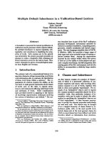
have hepatic (Figure 1a) and pulmonary lesions suspi-
cious for metastases. Thoracic computed tomography
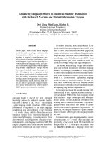
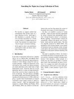
Figure 1 Diagnostic imaging: a) Magnet ic resonance imaging
(MRI) of the patient showing a colorectal liver metastasis in
segment VII of the liver (circle) prior to its resection. b) Thoracic
computed tomography exhibits a potentially metastatic, well-
circumscribed lesion of 6 mm in the left lower lobe (circle) with
homogenous contrast media enhancement. Pathological evaluation
revealed a sclerosing hemangioma of the lung.
Schiergens et al. World Journal of Surgical Oncology 2011, 9:62
/>Page 2 of 6
showed a well-circumscribed 6 mm lesion in the left
lower lobe of the lung (Figure 1b) with homogenous
contrast media enhancement as well as two smaller
lesions in the right upper lobe. There were neither signs
of infiltration of the adjacent tissue nor signs of patholo-
gically enlarged lymph nodes. We decided to first per-
form a partial hepatectomy (segment VII), which
confirmed hepatic spread of the tumor. In the light of
the patient’s young age, his early recovery and his good
general state of health, we proceeded to remove the left-
sided pulmonary lesion four weeks later. Therefore, he
underwent atypical resection of the left lower lobe
through a left anterolateral thoracotomy followed by a
systematic mediastinal and hilar lymphadenectomy [20].
The patient’s postoperative course remained uncompli-
cated and he again recovered well. Gross examination of
the specimen, however, showed a well-circumscribed
solid pulmonary tumor, 7 mm in diameter. Histological
evaluation revealed a mixed papillary, hemorrhagic and
sclerotic growth pattern of cuboidal surface cells and
polygonal stromal cells. Cuboidal surface cells were
immunopositive for thyroid transcription factor-1 (TTF-
1), epithelial membrane antigen (EMA) and pan-cyto-
keratin, whereas polygonal stromal cells were immuno-
positive for neuron-specific enolase (NSE) and S-100
protein as well as EMA. These findings are consistent
with a sclerosing hemangioma of the lung (Figure 2).
Ki-67 index was less than 5%. Both significant MSI eval-
uated by PCR amplification and loss of expression of
MMR-proteins MLH1, MSH2, MSH6 and PMS2 deter-
mined by immunohistochemistry could not be detected
in the pulmonary SH. Moreover, all lymph nodes
sampled were free of metastases.
By thoracic computed tomography, the pulmonary
lesions in the right upper lobe remained unchanged
after 3 months. According to interdisciplinary tumor
board recommendations and oncological guidelines
[21-23] we decided not to suggest further chemotherapy
or restorative proctocolectomy but to perform careful
aftercare with monitoring of the pulmonary lesions at
close intervals as well as attentive follow-up via abdom-
inal ultrasound and colonoscopy.
Furthermore, the patient’ sfamilymemberswere
referred to cancer genetics specialists for counseling
interviews and recommended germline mutation analy-
sis. During regular follow up visits CEA and CA 19-9
were within normal range. Accurate colonoscopy and
diagnostic imaging of liv er and lungs were unremark-
able, in particular pulmonary lesions of the right upper
lobe both were not identifiable any more.
Discussion
Pulmonary SH is a rare and mostly benign neoplasm of
the lung. Histologically, SH is essentially characterized
by two epithelial cell ty pes: cuboidal surface cells, which
resemble type II pneumocytes, and polygonal stromal
cells (round cells) with bland nuclei and pale cytoplasm,
which are thought to stem from primitive respiratory
epithelium [4,5]. These two cell types form four histolo-
gical patterns; papillary, which often appears to be the
predominant type, but epitheloid, sclerotic and hemor-
rhagic configurations are also found in some cases as in
the present one (Figure 2, [24]). Predominant papillary
growth patterns might make it complicated to differenti-
ate SH from a carcinoma that also exhibits a papillary
pattern. Metastatic papillary thyroid carcinoma,
mesotheli oma and bronchioloalveolar carcinoma have to
be considered accurately [11]. In this respect, however,
decreased Ki-67 labeling and low p53 expression could
help to differentiate SH from papillary thyroid carci-
noma [2]. The cuboidal surface cells of SH are typically
immunopositive for thyroid transcription factor-1 (TTF-
1), epithelial membrane antigen (EMA), surfactant pro-
tein B (SP-B), low molecular weight cytokeratin (CK-L)
as well as carcinoembryonic antigen (CEA) and negative
for neuroendocr ine markers, whereas polygonal stromal
cells (round cells) are positive for vimentin and TTF-1
and weakly positive for several neuroendocrine markers
[4,7,25]. Mitotic figures are rarely identified [2]. In the
present case, the patient’s lesion comprised mixed papil-
lary growth patterns consisting of superficial layers of
cuboidal cells that were immunopositive for TTF-1 and
EMA, as well as stromal cells positive for TTF-1 expres-
sion, and some also for neuroendocrine markers such as
neuron-specific enolase (NSE) and S-100 protein. Thus,
histological and immunohistochemical diagnosis of SH
was made, and a very low Ki-67 index of less than 5%
indicated a biologically non-active tumor [26].
In most patients, SH is detected during routine chest
radiographic examination [2,8]. Therefore, the actual pre-
valenceofSHisnotknownduetotherelativelyasymp-
tomatic nature of the disease. SH is usually diagnosed as
a single asymptomatic nodule i n the periphery of the
lung [2,8], often affecting the lower lobe [27, 28]. Radiolo-
gically, it mostly presents as a well-circumscribed lesion
with marked contrast media enhancement. Calcification
might be detected in the minority of cases. A lucent zone
around SH, the “ air m eniscus sign“ , first described in
1978 [29], is a typical radiological feature representing
trapped air around the lesion. Additionally, oth er r eports
of air spaces surrounding SH have been published [30].
However, other diagnoses must be con sidered, including
carcinoids, hamartoma, hemangioma, malignant tera-
toma, arterio-venous malformations and inflammatory
lesions. In the present case, chest radiography was nor-
mal, but thoracic computed tomography revealed a small
but well-defined lesion of the left lower lobe with homo-
geneous contrast enhancement (Figure 1b). No typical
Schiergens et al. World Journal of Surgical Oncology 2011, 9:62
/>Page 3 of 6
lucent zone was found at the periphery of the lump, and
no regional lymph node enlargement was present. Due to
the history of metastatic CRC, however, a pulmonary
spread of rectal cancer was the most probable diagnosis,
so surgical resection of the lesion was performed.
During surgical intervention, we found early stage SH.
Wedge resection in previous cases of early stage SH was
associated with excellent long-term survival and there-
fore should be the treatment of choice if an exact pre-
or intraoperative diagnosis is possible [3,31]. Otherwise,
especially in cases of uncertain intraoperative frozen sec-
tion examinations and given the uncertainty of growth,
biological behavior, local recurrence and metastatic
spread, the optimal therapeutic approach remains unde-
fined. In these cases, atypical or anatomic resection with
systematic lymphadenectomy is suggested [31]. Because
of our patient’s distinctive history, we oriented our ther-
apy toward a strong suspicion of a pulmonary metastasis
of CRC and elected to pursue a thorough surgical
approach with atypical resection followed by regional
lymphadenectomy [20].
Only a f ew cases of SH have been reported in young
patients, among them a 10-year-old, an 18-ye ar-old and
a 19-year-old Asia n female as well as a 22-year-old
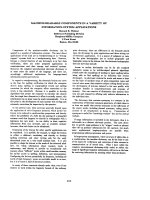
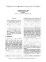
Figure 2 Histology (a, b) and immunohistochemistry (c-f) of sclerosing hemangioma of the lung: a) Well-circumscribed lesion with normal
lung tissue in the right upper corner (X), lymphoid cell infiltration (arrows) and hemorrhages (dashed arrow); hematoxylin and eosin stain (25x)
b) Mixed growth pattern of the lesion, papillary (arrows), solid (dashed arrows) and sclerotic (*); foam cells (dotted arrows); hematoxylin and
eosin stain (200x) c) Cuboidal surface cells positive for staining with pan-cytokeratin antibody (200x) d) Epithelial membrane antigen (EMA) and
e) thyroid transcription factor 1 (TTF-1) are positive in both cuboidal surface cells as well as stromal round cells (200x) f) Positive nuclear staining
for Ki67 in only few cells (Ki-67-Index < 5%) (200x).
Schiergens et al. World Journal of Surgical Oncology 2011, 9:62
/>Page 4 of 6
male, who presented with lymph node metastases imply-
ing a more malignant case of SH [12,32]. The latter
might corroborate with the monoclonality of cells within
SH, which has been described before and which suggests
a neoplastic growth pattern of the lesion [33]. With
respect to synchronous colorectal neoplasms, female
patients suffering from FAP and simultaneous SH have
been described [13,14]. In these cases, patients did not
have any extracolonic manifestations of FAP and did
notsufferfromCRCuntiltheypresentedwithSH.To
the best of our knowledge, this is the first report of SH
associated with Lynch syndrome.
Autosomal-dominant Lynch syndrome (HNPCC) is a
rare genetic disease (OMIM #609310) tha t usually shows
right-sided predominance of CRC at a young age and is
often caused by mutations of MMR-genes [34]. Although
occurrence is less frequent than CRC there is a high pre-
valence of synchronous or metachronous extracolonic
manifestations, especially endometrial cancer, which
caused the death of our patient’s mother. Other extraco-
lonic manifestations include gastric, genitourinary, ovar-
ian, small bowel, brain and sebaceous tumors [34,35].
Only one case of Muir-Torre syndrome, a variant of
Lynch syndrome with additional skin lesions, was
reported that was associated with non-small cell lung
cancer [36]. However, there are no reports of benign lung
tumors as extracolonic manifestation of Lynch syndrome.
In our patient, MSI testing of SH and immunohisto-
chemistry for MLH1, MSH2, MSH6 and PMS2 did not
reveal MSI or loss of MMR-expression in the pulmonary
nodule. On the one hand we would have judged SH as
an extracolonic manifestation of Lynch syndrome in this
specific patient if SH would have featured MSI and loss
of MMR-expression. On the other hand, one might
anticipate that high-grade MSI and loss of MMR-expres-
sion by homocygosity of a mutated PMS2 should then
have led to a more malignant growth pattern of SH.
Pulmonary SH, as in the present case (Ki-67 index
<5%), is a mostly benign and heterogeneous tumor com-
posed of different cell types and exhibits various histolo-
gical patterns [33]. Nevertheless, heterozygosity of PMS2
in the present case as exhibited by c-DNA-sequencing
might still be causally associated with the development
of this exceedingly rare tumor. Although a sporadic
coincidence of SH and Lynch syndrome could not be
ruled out in our patient, one might raise the suspicion
of a c ommon etiology being responsible for the excep-
tional concurrence of these two extremely infrequent
events in a young male Caucasian.
Conclusions
We present the first ca se of pulmonary SH in a young
Caucasian male and in a patient suffering from Lynch
syndrome.ItmightbespeculatedthatSHdidnotjust
incidentally co-occur with the patient’sCRC.Fromthis
unlikely concurrence we assume that the underlying
Lynch syndrome might have abetted the ar ising of the
patient’s SH and hypothesize a common cause for these
rare events. However, SH could not be termed as an
extracolonic manifestation of Lynch syndrome since it
obviously showed a benign behavior and did not exhibit
MSI or loss of MMR-expressio n based upon heterozyg-
osity of PMS2.
Consent
Written informed consent was obtained from the patient
for publication of this case report and a ny accompany-
ing images. A copy of the written consent is available
for review by the Editor-in-Chief of this journal.
Author details
1
Department of Surgery, University of Munich, Campus Grosshadern,
Germany.
2
Department of Surgery, University of Munich, Campus Innenstadt,
Germany.
3
Department of Pathology, University of Munich, Munich,
Germany.
Authors’ contributions
TSS, PNK and AK collected all patient’s history data with substa ntial
contribution of WET, MAK, RAH and KWJ. TSS, PNK and AK drafted the
manuscript with committed and dedicated review and discussion of WET,
MAK, RAH and KWJ. DM prepared the histopathological data and figures
including their review and evaluation. All authors contributed substantially
to the patient’s care and therapy. All authors read and approved the final
manuscript.
Competing interests
The authors declare that they have no competing interests.
Received: 14 October 2010 Accepted: 6 June 2011
Published: 6 June 2011
References
1. Liebow AA, Hubbell DS: Sclerosing hemangioma (histiocytoma,
xanthoma) of the lung. Cancer 1956, 9:53-75.
2. Iyoda A, Hiroshima K, Shiba M, Haga Y, Moriya Y, Sekine Y, et al:
Clinicopathological analysis of pulmonary sclerosing hemangioma. Ann
Thorac Surg 2004, 78:1928-1931.
3. Devouassoux-Shisheboran M, Hayashi T, Linnoila RI, Koss MN, Travis WD: A
clinicopathologic study of 100 cases of pulmonary sclerosing
hemangioma with immunohistochemical studies: TTF-1 is expressed in
both round and surface cells, suggesting an origin from primitive
respiratory epithelium. Am J Surg Pathol 2000, 24:906-916.
4. Wang E, Lin D, Wang Y, Wu G, Yuan X: Immunohistochemical and
ultrastructural markers suggest different origins for cuboidal and
polygonal cells in pulmonary sclerosing hemangioma. Hum Pathol 2004,
35:503-508.
5. Satoh Y, Tsuchiya E, Weng SY, Kitagawa T, Matsubara T, Nakagawa K, et al:
Pulmonary sclerosing hemangioma of the lung. A type II
pneumocytoma by immunohistochemical and immunoelectron
microscopic studies. Cancer 1989, 64:1310-1317.
6. Nagata N, Dairaku M, Ishida T, Sueishi K, Tanaka K: Sclerosing hemangioma
of the lung. Immunohistochemical characterization of its origin as
related to surfactant apoprotein. Cancer 1985, 55:116-123.
7. Chan AC, Chan JK: Pulmonary sclerosing hemangioma consistently
expresses thyroid transcription factor-1 (TTF-1): a new clue to its
histogenesis. Am J Surg Pathol 2000, 24:1531-1536.
8. Kim GY, Kim J, Choi YS, Kim HJ, Ahn G, Han J: Sixteen cases of sclerosing
hemangioma of the lung including unusual presentations. J Korean Med
Sci 2004, 19:352-358.
Schiergens et al. World Journal of Surgical Oncology 2011, 9:62
/>Page 5 of 6
9. Yano M, Yamakawa Y, Kiriyama M, Hara M, Murase T: Sclerosing
hemangioma with metastases to multiple nodal stations. Ann Thorac
Surg 2002, 73:981-983.
10. Noguchi M, Kodama T, Morinaga S, Shimosato Y, Saito T, Tsuboi E: Multiple
sclerosing hemangiomas of the lung. Am J Surg Pathol 1986, 10:429-435.
11. Katzenstein AL, Gmelich JT, Carrington CB: Sclerosing hemangioma of the
lung: a clinicopathologic study of 51 cases. Am J Surg Pathol 1980,
4:343-356.
12. Miyagawa-Hayashino A, Tazelaar HD, Langel DJ, Colby TV: Pulmonary
sclerosing hemangioma with lymph node metastases: report of 4 cases.
Arch Pathol Lab Med 2003, 127:321-325.
13. de Koning DB, Drenth JP, Oyen WJ, Wagenaar M, Aliredjo RP,
Nagengast FM: Pulmonary sclerosing hemangioma detected by
fluorodeoxyglucose positron emission tomography in familial
adenomatous polyposis: report of a case. Dis Colon Rectum 2007,
50:1987-1991.
14. Hosaka N, Sasaki T, Adachi K, Sato T, Tanaka T, Miura Y, et al: Pulmonary
sclerosing hemangioma associated with familial adenomatous polyposis.
Hum Pathol 2004, 35:764-768.
15. Kleespies A, Fuessl KE, Seeliger H, Eichhorn ME, Muller MH, Rentsch M, et al:
Determinants of morbidity and survival after elective non-curative
resection of stage IV colon and rectal cancer. Int J Colorectal Dis 2009,
24:1097-1109.
16. Umar A, Boland CR, Terdiman JP, Syngal S, de la CA, Ruschoff J, et al:
Revised Bethesda Guidelines for hereditary nonpolyposis colorectal
cancer (Lynch syndrome) and microsatellite instability. J Natl Cancer Inst
2004, 96:261-268.
17. Vasen HF, Mecklin JP, Khan PM, Lynch HT: The International Collaborative
Group on Hereditary Non-Polyposis Colorectal Cancer (ICG-HNPCC). Dis
Colon Rectum 1991, 34:424-425.
18. Vasen HF, Watson P, Mecklin JP, Lynch HT: New clinical criteria for
hereditary nonpolyposis colorectal cancer (HNPCC, Lynch syndrome)
proposed by the International Collaborative group on HNPCC.
Gastroenterology 1999, 116:1453-1456.
19. Schofield L, Watson N, Grieu F, Li WQ, Zeps N, Harvey J, et al: Population-
based detection of Lynch syndrome in young colorectal cancer patients
using microsatellite instability as the initial test 2. Int J Cancer 2009,
124:1097-1102.
20. Winter H, Meimarakis G, Hoffmann G, Hummel M, Ruttinger D, Zilbauer A,
et al: Does surgical resection of pulmonary metastases of head and neck
cancer improve survival? Ann Surg Oncol 2008, 15:2915-2926.
21. Schmiegel W, Pox C, Adler G, Fleig W, Folsch UR, Fruhmorgen P, et al: [S3-
Guidelines Conference “Colorectal Carcinoma
” 2004]. Z Gastroenterol
2004, 42:1129-1177.
22. Schmiegel W, Reinacher-Schick A, Arnold D, Graeven U, Heinemann V,
Porschen R, et al: [Update S3-guideline “colorectal cancer” 2008]. Z
Gastroenterol 2008, 46:799-840.
23. Vasen HF, Moslein G, Alonso A, Bernstein I, Bertario L, Blanco I, et al:
Guidelines for the clinical management of Lynch syndrome (hereditary
non-polyposis cancer). J Med Genet 2007, 44:353-362.
24. Sugio K, Yokoyama H, Kaneko S, Ishida T, Sugimachi K: Sclerosing
hemangioma of the lung: radiographic and pathological study. Ann
Thorac Surg 1992, 53:295-300.
25. Illei PB, Rosai J, Klimstra DS: Expression of thyroid transcription factor-1
and other markers in sclerosing hemangioma of the lung. Arch Pathol
Lab Med 2001, 125:1335-1339.
26. Gerdes J, Lemke H, Baisch H, Wacker HH, Schwab U, Stein H: Cell cycle
analysis of a cell proliferation-associated human nuclear antigen defined
by the monoclonal antibody Ki-67. J Immunol 1984, 133:1710-1715.
27. Gottschalk-Sabag S, Hadas-Halpern I, Glick T: Sclerosing haemangioma of
lung mimicking carcinoma diagnosed by fine needle aspiration (FNA)
cytology. Cytopathology 1995, 6:115-120.
28. Sant F, Barcelo C, Castro P, Bernardo L: Diagnosis by fine needle
aspiration cytology of sclerosing haemangioma of the lung.
Cytopathology 1995, 6:126-127.
29. Bahk YW, Shinn KS, Choi BS: The air meniscus sign in sclerosing
hemangioma of the lung. Radiology 1978, 128:27-29.
30. Hung JJ, Liu JS, Hsu WH: Sclerosing hemangioma with an air halo. J
Thorac Cardiovasc Surg 2008, 136:1365-1367.
31. Jungraithmayr W, Eggeling S, Ludwig C, Kayser G, Passlick B: Sclerosing
hemangioma of the lung: a benign tumour with potential for
malignancy? Ann Thorac Cardiovasc Surg 2006, 12:352-354.
32. Kim KH, Sul HJ, Kang DY: Sclerosing hemangioma with lymph node
metastasis. Yonsei Med J 2003, 44:150-154.
33. Niho S, Suzuki K, Yokose T, Kodama T, Nishiwaki Y, Esumi H: Monoclonality
of both pale cells and cuboidal cells of sclerosing hemangioma of the
lung. Am J Pathol 1998, 152:1065-1069.
34. Vasen HF:
Clinical description of the Lynch syndrome [hereditary
nonpolyposis colorectal cancer (HNPCC)]. Fam Cancer 2005, 4:219-225.
35. Anaya DA, Chang GJ, Rodriguez-Bigas MA: Extracolonic manifestations of
hereditary colorectal cancer syndromes. Clin Colon Rectal Surg 2008,
21:263-272.
36. Nolan L, Eccles D, Cross E, Crawford G, Beck N, Bateman A, et al: First case
report of Muir-Torre syndrome associated with non-small cell lung
cancer. Fam Cancer 2009, 8:359-362.
doi:10.1186/1477-7819-9-62
Cite this article as: Schiergens et al.: Pulmonary sclerosing hemangioma
in a 21-year-old male with metastatic hereditary non-polyposis
colorectal cancer: Report of a case. World Journal of Surgical Oncology
2011 9:62.
Submit your next manuscript to BioMed Central
and take full advantage of:
• Convenient online submission
• Thorough peer review
• No space constraints or color figure charges
• Immediate publication on acceptance
• Inclusion in PubMed, CAS, Scopus and Google Scholar
• Research which is freely available for redistribution
Submit your manuscript at
www.biomedcentral.com/submit
Schiergens et al. World Journal of Surgical Oncology 2011, 9:62
/>Page 6 of 6