báo cáo khoa học: "A rare case of xanthogranuloma of the stomach masquerading as an advanced stage tumor" ppt
Bạn đang xem bản rút gọn của tài liệu. Xem và tải ngay bản đầy đủ của tài liệu tại đây (1.23 MB, 3 trang )
CAS E REP O R T Open Access
A rare case of xanthogranuloma of the stomach
masquerading as an advanced stage tumor
Hiroyuki Kinoshita
1*
, Shunsuke Yamaguchi
1
, Yoshifumi Sakata
1
, Kazuo Arii
1
, Kazunari Mori
1
and Rieko Kodama
2
Abstract
Background: Xanthogranuloma of the stomach is an extremely rare disease, and this lesion has only been found
to coexist with early gastric cancer in 2 cases in the literature.
Case presentation: We report a case of xanthogranuloma of the stomach combined with early gastric cancer that
mimicked an advanced stage tumor. A 65-year-old female was referred to our hospital because of epigastralgia.
During a physical examination, a defined abdominal mass was palpable in the region of the left hypochondrium.
Imaging studies revealed an advanced gastric cancer, which was suspected of having infiltrated the abdominal
wall. Total gastrectomy and resection of the regional lymph node and abdominal wall were performed.
Histopathologic examination of the resected specimen demonstrated xanthogranuloma combined with early
gastric cancer.
Conclusion: Xanthogranuloma presenting as a form of SMT (submucosal tumor) of the stomach is an extremely
rare disease, and diagnosing it preoperatively is difficult. Further accumulation and investigation of this entity is
necessary.
Keywords: xanthogranuloma, early gastric cancer
Background
Xanthogranuloma was first described by Oberling in
1935 [1]. Although it is known to develop in the gall
bladder as xanthogranulomatous cholecystitis, xanthogra-
nuloma of the stomach is an extremely rare disease, and
only a few cases have been reported. Hence, we report a
case of xanthogranuloma combined with early gastric
cancer that mimicked an advanced stage tumor.
Case report
A 65-year-old female was refe rred to Naga Munic ipal
Hospital because of epigastralgia. During a physical
examination, a defined abdominal mass was pa lpable in
the region of the left hypochondrium. Neither anemia
nor jaundice was present. Blood analysis showed a white
blood cell count of 12.25 × 10
3
/μl. Her tumor marker
serum levels were within the normal limits (carcinoem-
bryonic antigen (CEA): 1.3 ng/ml, carbohydrate antigen
(CA) 19-9: 10.1 U/ml). A gastroint estinal endoscopic
examination was performed and disclosed an ulcerated
lesion in the lesser curvature of the gastric corpus at
about 7 cm from esophagogastric junction, which
squashed and isolated the gastric folds from the rest of
the stomach (Figure 1a), and an elevated lesion sim ilar to
a submucosal tumor (SMT), wh ich was suspected o f
being an advanced gastric tumor, was detected on the
anal side of the ulcerated lesion (Figure 1b). The biopsy
specimen from the ulcerated lesion indicated a moder-
ately or p oorly differentiated tubular adenocarcinoma.
Computed tomography (CT) revealed thickening of the
gastric wall and findings that seemed to indicate abdom-
inal wall invasion (Figure 1c).
Open surgery was carried out and revealed that the
tumor had infiltrated into the abdominal wall. There-
fore, total gastrectomy and resection o f the regional
lymph node and parts of the abdominal wall were per-
formed. Upon macroscopic examination, the specimens
showed an elevated and supe rficial depressed-type (IIa
+IIc type) gast ric cancer, and the adjacent tumor had
extended into the abdominal wall beyond the gastric
serosa (Figure 2). Histopathological examination of the
specimens demonstrated moderately d ifferentiated
* Correspondence:
1
Department of Surgery, Naga Municipal Hospital, 1282, Uchita, Kinokawa,
Wakayama 649-6414, Japan
Full list of author information is available at the end of the article
Kinoshita et al. World Journal of Surgical Oncology 2011, 9:67
/>WORLD JOURNAL OF
SURGICAL ONCOLOGY
© 2011 Kinoshita et al; licensee BioMed Central Ltd. This is an Open Access article distributed u nder the terms of the Creative
Commons Attribution Lice nse ( which permits unrestricted use, distribution, and
reproduction in any medium, provided the original work is properly cited.
adenocarcinoma without metastasis to the resected
lymph nodes and xanthogranuloma consisting of foamy
histiocyte s, many lymphocytes, plasma cells, and granu-
locytes which were immunohistochemically positive for
CD68 and were non reactive with CAM5.2, AE1/3 and
S-100 protein (Figure 3). The xanthogranuloma was
located near to the gastric cancer, but w as not in con-
tact with it. The patient recovered rapidly and was
discharged on postoperative day 16. She has been symp-
tom free ever since.
Discussion
Xanthogranuloma is a tumor that is macroscopically
characterized by the formation of multiple golden yellow
or bright yellow nodules, and histologically, the lesion is
predominantly composed of foamy histiocy tes mixed
with acute and chronic inflammatory cells. The patho-
genesis of xanthogranuloma has not been fully esta b-
lished, although it is thought to be a chronic lesion
associated with in fectio n, immunological disorders , lipid
transport, and lymphatic obstruction [1].
To the best of our knowledge, only seven cases of
xanthogranuloma of the stomach have been reported
[2-8], and the coexistence of this lesion with early gastric
cancer has only been reported in 2 cases. Our histopatho-
logical inspection in these cases did not support continu-
ity between the xanthogr anuloma and early gastric
cancer. Therefore, it is unclear whether early gastric
cancer participates in xanthogranuloma.
Pathologically, stromal tumors such as GIST, m yoge-
netic tumors, and neurogenic tumors account for 54
percent of all SMT, followed by heterotopic pancreas,
cyst, lipoma, carcinoid, lymphangioma, and hemangioma
[9]. There have been no previous cases of preoperatively
diagnosed xanthogranuloma as was found in the current
case.
In our case, the gastric xanthogranuloma was preopera-
tively misdiagnosed as an advance d gastric cancer. T his
occurred for the following reasons: First, a gastrointest-
inal endoscopic examination demonstrated an elevated
lesion close to the anal side of an ulcerated lesion and a
moderately or poorly differentiated adenocarcinoma was
detected by the endoscopic biopsy. Second, CT indicated
a
b
c
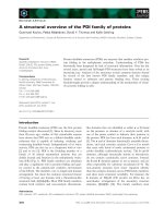
Figure 1 Gastrointestinal endoscopic examination and
Computed tomography. a. A gastrointestinal endoscopic
examination was performed and disclosed an ulcerated lesion in
the lesser curvature of the gastric corpus located at 7 cm from the
esophagogastric junction, which squashed and isolated the gastric
folds from the rest of the stomach. b. An elevated lesion that
appeared to be a submucosal tumor (SMT), which was suspected of
being an advanced gastric cancer, was detected on the anal side of
the ulcerated lesion. c. Computed tomography (CT) revealed
thickening of the gastric wall and findings indicative of abdominal
wall invasion.
a
b
Figure 2 Macroscopic examination of the specimens.a.Upon
macroscopic examination, the specimens showed an elevated and
superficial depressed-type (IIa+IIc type) gastric cancer (arrow) and an
elevated lesion similar to a submucosal tumor (arrow head). b. The
abdominal wall (arrow) was resected together with the stomach.
a
b
Figure 3 Histopatholo gical examination of the speci mens.
Histopathological examination revealed that an SMT was located in
the subserosal layer (a) and it consisted of foamy histiocytes, many
lymphocytes, plasma cells, and granulocytes (b).
Kinoshita et al. World Journal of Surgical Oncology 2011, 9:67
/>Page 2 of 3
that the elevated lesion had invaded the abdominal wall,
and a defined abdominal mass was palpable on physical
examination. Therefore, the tumor was recognized as an
advanced gastric cancer. Biopsy of the elevated lesion
should have been carried out preoperatively to obtain a
correct diagnosis in consideration of the coexistence of
the two lesions.
Conclusion
We report an extremely rare case of gastric xanthogra-
nuloma combined with early gastric cancer. W hen we
find SMT of the stomach, we should bear in mind not
only neoplastic tumors but also inflammatory tumors.
Further accumulation and investigation of gastric
xanthogranuloma cases is necessary.
Consent
Written informed consent was obtained from the patient
for publication of this case report and accompanying
images. A copy of the written consent is available for
review by the Editor-in-Chief of this journal.
Author details
1
Department of Surgery, Naga Municipal Hospital, 1282, Uchita, Kinokawa,
Wakayama 649-6414, Japan.
2
Department of Pathology, Naga Municipal
Hospital, Japan.
Authors’ contributions
HK did the literature search and writing of the manuscript. SY, YS, KA and
KM collected the clinical data. RK was responsible for the histology
consulting and pathology examination. All authors read and approved the
final manuscript.
Competing interests
The authors declare that they have no competing interests.
Received: 7 January 2011 Accepted: 2 July 2011 Published: 2 July 2011
References
1. Oberling C: Retroperitoneal xanthogranuloma. Am J Cancer 1935,
23:477-489.
2. Zafisaona G: Inflammatory fibrous histiocytoma of the stomach. Apropos
of a case of xanthogranuloma? Arch Anat Cytol Pathol 1987, 35:149-153.
3. Zhang L, Huang X, Li J: Xanthogranuloma of the stomach: a case report.
Eur J Surg Oncol 1992, 18:293-295.
4. Guarino M, Reale D, Micoli G, Tricomi P, Cristofori E: Xanthogranulomatous
gastritis: association with xanthogranulomatous cholecystitis. J Clin
Pathol 1993, 46:88-90.
5. Lespi PJ: Gastric xanthogranuloma (inflammatory malignant
fibrohistiocytoma). Case report and literature review. Acta Gastroenterol
Latinoam 1998, 28:309-310.
6. Lai HY, Chen JH, Chen CK, Chen YF, Ho YJ, Yang MD, Shen WC:
Xanthogranulomatous pseudotumor of stomach induced by perforated
peptic ulcer mimicking a stromal tumor. Eur Radiol 2006, 16:2371-2372.
7. Kubosawa H, Yano K, Oda K, Shiobara M, Ando K, Nunomura M,
Sarashina H: Xanthogranulomatous gastritis with pseudosarcomatous
changes. Pathol Int 2007, 57:291-295.
8. Aikawa M, Ishii T, Nonaka K, Nakao M, Ishikawa K, Arai S, Kita H,
Miyazawa M, Koyama I, Motosugi U, Ban S: A case of gastric
xanthogranuloma associated with early gastric cancer. Nippon Shokakibyo
Gakkai Zasshi 2009, 106:1610-1615.
9. Polkowski M: Endoscopic ultrasound and endoscopic ultrasound-guided
fine-needle biopsy for the diagnosis of malignant submucosal tumors.
Endoscopy 2005, 37:635-645.
doi:10.1186/1477-7819-9-67
Cite this article as: Kinoshita et al.: A rare case of xanthogranuloma of
the stomach masquerading as an advanced stage tumor. World Journal
of Surgical Oncology 2011 9:67.
Submit your next manuscript to BioMed Central
and take full advantage of:
• Convenient online submission
• Thorough peer review
• No space constraints or color figure charges
• Immediate publication on acceptance
• Inclusion in PubMed, CAS, Scopus and Google Scholar
• Research which is freely available for redistribution
Submit your manuscript at
www.biomedcentral.com/submit
Kinoshita et al. World Journal of Surgical Oncology 2011, 9:67
/>Page 3 of 3