báo cáo khoa học: "Multimodality treatment of brain metastases: an institutional survival analysis of 275 patients" pdf
Bạn đang xem bản rút gọn của tài liệu. Xem và tải ngay bản đầy đủ của tài liệu tại đây (412.26 KB, 9 trang )
RESEARCH Open Access
Multimodality treatment of brain metastases:
an institutional survival analysis of 275 patients
Ameer L Elaimy
1,2
, Alexander R Mackay
1,3
, Wayne T Lamoreaux
1,2
, Robert K Fairbanks
1,2
, John J Demakas
1,4
,
Barton S Cooke
1
, Benjamin J Peressini
5
, John T Holbrook
2
and Christopher M Lee
1,2*
Abstract
Background: Whole brain radiation therapy (WBRT ), surgical resection, stereotactic radiosurgery (SRS), and
combinations of the three modalities are used in the management of patients with metastatic brain tumors. We
present the previously unreported survival outcomes of 275 patients treated for newly diagnosed brain metastases
at Cancer Care Northwest and Gamma Knife of Spokane between 1998 and 2008.
Methods: The effects treatment regimen, age, Eastern Cooperative Oncology Group-Performan ce Status (ECOG-PS),
primary tumor histology, number of brain metastases, and total volume of brain metastases have on patient overall
survival were analyzed. Statistical analysis was performed using Kaplan-Meier survival curves, Andersen 95%
confidence intervals, approximate confidence intervals for log hazard-rati os, and multivariate Cox proportional
hazard models.
Results: The median clinical follow up time was 7.2 months. On multivariate analysis, survival statistically favored
patients treated with SRS alone when compared to patients treated with WBRT alone (p < 0.001), patients treated
with resection with SRS when compared to patients treated with SRS alone (p = 0.020), patients in ECOG-PS class 0
when compared to patients in ECOG-PS classes 2 (p = 0.04), 3 (p < 0.001), and 4 (p < 0.001), patients in the non-
small-cell lung cancer group when compared to patients in the combined melanoma and renal-cell carcinoma
group (p < 0.001), and patients with breast cancer when compared to patients with non-small-cell lung cancer
(p < 0.001).
Conclusions: In our analysis, patients benefited from a combined modality treatment approach and physicians
must consider patient age, performance status, and primary tumor histology when recommending specific
treatments regimens.
Keywords: Brain metastases, Survival, Treatment regimen, Age, Performance status, Primary tumor histology, Tumor
number, Tumor volume
Background
Brai n metastases are defined as cancerous lesions in the
brain that originate and spread from an extracranial pri-
mary cancer. Brain metastases occur in 20 to 40% of
patients with systemic cancer and the incidence is grow-
ing due to advances in imaging technologies and the
treatment of extracranial disease [1]. The site of metas-
tasis often depends on the nearest location of vascular
clusters. As a consequence, the most common primary
cancers that have the ability to metastasize to the brain
are cancers that develop from the lung o r breast [2].
However, metastasis to the brain originating from mela-
noma, colorectal cancer, renal-cell carcinoma, and carci-
noma of multiple other origins may also lead to the
development of one or more metastatic brain tumors
[3]. D ue to a large amount of blood flow, the cerebrum
accounts for approximately 80% of all brain m etastases,
while metastases that arise within the cerebellum and
brain stem account for the remaining 20% of metastatic
tumors [4].
Patients diagnosed with brain metastases have several
potential management options and treatment regimens
* Correspondence:
1
Gamma Knife of Spokane, 910 W 5
th
Ave, Suite 102, Spokane, WA 99204,
USA
Full list of author information is available at the end of the article
Elaimy et al. World Journal of Surgical Oncology 2011, 9:69
/>WORLD JOURNAL OF
SURGICAL ONCOLOGY
© 2011 Elaimy et al; licensee BioMed Central Ltd. This is an Open Access article distributed under the terms of the Creative Commons
Attribution License (http://creativecommons.o rg/licenses/by/2.0), which permits unrestricted use, distribution, and reproduction in
any medium, provided the original work is properly cited.
are dependent on the patient’s performance status, age,
control of primary ca ncer, presence of extracranial dis-
ease, number of brain metastases, size of brain metas-
tases, and location of brain metastases [1,5]. In general,
patients with brain metastases have a poor outlook and
survive an average of 1 to 2 months when treated with
steroid therapy alone [6]. Whole brain radiation therapy
(WBRT) has been historically a standard of care for
patients with brain metastases. WBRT takes advantage
of differences in radiobiology between tumor cells and
nervous tissue by targeting rapidly dividing tumor cells
in all areas of the brain, w hile minimizing damage to
the adjacent brain tissue [3]. Due to this favorable radia-
tion cell-kill therapeutic ratio, WBRT extends the survi-
val tim e of patient s who undergo treatment to an
average of 4 to 7 months [1]. Surgical resection followed
by WBRT has proven to be a superior treatment modal-
ity than WBRT alone or surgical resection alone for
patients with a high performance status (functionally
independent and spend no more than 50% of their day
in bed) that possess a single, surgically accessible brain
metastasis [7-9]. However, surgical resection followed by
WBRT is considered an excessive and potentially
destructive treatment modality for patients with multiple
brain metastases and has no t been investigated in a ran-
domized controlled trial [10].
Stereotactic radiosurgery (SRS) is a highly technical form
of radiation therapy that delivers a focused dose of radia-
tion to a single volume, while minimizing damage to
nearby, critical structures. The patient’s skull is immobi-
lized, allowing a controlled dose of radiation to be deliv-
ered to a specified target with sub-millimeter precision.
There are currently 4 devices utilized for SRS treatment:
Gamma Knife (GK) radiosurgery, linear accelerator
(LINAC) based treatment, a cyclotron-based proton beam,
and cyberkni fe technology [3]. Although GK remains the
“gold standard” of brain radiosurgery, published reports by
Andrews et al. [6] and Sneed et al. [11] concluded that
patient prognosis did not differ in terms of the method in
which SRS was delivered. The evidence assessing the effi-
cacy of SRS in the treatment of patients with brain metas-
tases is continuously increasing due to the fact that it is
capable of targeting any area in the brain with accuracy
and can be utilized to irradiate multiple lesions during the
same clinical treatment setting. For specific patient subsets
that have newly diagnosed brain metastases, WBRT alone,
SRS alone, SRS with WBRT, SRS with surgical resection,
or a combination of the three treatments can be the opti-
mal management approach.
We present a retrospective survival analysis of the 275
patients treated for newly diagnosed brain metastases at
Cancer Care Northwest and Gamma Knife of S pokane
between 1998 and 2008; including a comprehensive ana-
lysis o f the effects treatment regimen, age, Eastern
Cooperative Oncology Group-Performance Status
(ECOG-PS), primary tumor histology, number of brain
metastases, and total vo lume of brain metastases have
on patient survival.
Methods
We analyzed the patient population baseline characteris-
tics and survival of 275 patients treated for newly diag-
nosed brain meta stases at Ca ncer Care N orthwest and
Gamma Knife of Spokane (Deaconess Hospital, Spokane,
WA) between 1998 and 2008. After obtaining approval
from IRB Spokane (IRB 1554) and the University of
Washington Human Subjects Division (Human Subjects
Application 36306 ), the following pre-treatment factors
were recorded from the patient’s medical records: age at
first brain metastasis diagnosis, ECOG-PS at first brain
metastasis diagnosis, number of brain metastases, pri-
mary tumor histology, and total volume of brain metas-
tases at the time of SRS for patients who received SRS,
or at an imaging a ppointment prior to the patients first
treatment session for patients who did not receive SRS.
Patients were categorized by age at first brain metastasis
diagnosis (<65 years and ≥65 years), number of brain
metastases at first diagnosis (1 tumor, 2-4 tumors, >4
tumors), primary tumor histology (non-small-cell lung
cancer, small-cell lung cancer, breast cancer, melanoma,
renal-cell carcinoma, other/unknown primary), total
volume of brain metastases in cm
3
(2.0, 2.0-3.9, 4.0-5.9,
6.0-7.9, ≥8.0), and ECOG-PS class (0, 1, 2, 3, 4).
Treatment regimens were prescribed based on the
patient’ s performance status, age, control of primary
cancer, presence of extracranial disease, number of
brain metastases, size of brain metastases, location of
brain metastases, and at the discretion of the treating
physician. Of the 275 patients, 117 were treated with
WBRT alone, 65 were treated with SRS alone, 48 were
treated with WBRT with SRS, 11 were treated with sur-
gical resection with WBRT, 15 were treated with surgi-
cal resection with SRS, and 19 were treated with
surgical resection + WBRT + SRS. SRS was performed
using the Leksell
60
Co Gamma Knife (model C ). The
prescribed SRS dose to the 50% isodose line was com-
pleted in a single treatment and was based on the
patient’s tumor volume, tumor location, tumor shape,
prior radiation treatment, a nd standard Radiation Ther-
apy Oncology Group (RTOG) guidelines. The median
SRS dose was 18 Gy (13 Gy to 22 Gy). For patients
receiving WBRT, the median total dose prescribed was
30 Gy (5 Gy to 54 Gy). Length of follow-up was deter-
mined as the time interval between the date of first
treatment and the date of the most recent clinical
encounter or imaging appointment. Period of survival,
in months, was based upon the patient’s first treatment
session.
Elaimy et al. World Journal of Surgical Oncology 2011, 9:69
/>Page 2 of 9
Kaplan-Meier survival curves were utilized to compare
survival differences between the treatment groups, a ge
groups, ECOG-PS groups, tumor volume groups, pri-
mary tumor histology groups, and number of br ain
metastases groups. Andersen 95% confidence intervals
for the median survival times of the groups were con-
structed. Log-rank tests were employed to determine
statistically significant differe nces between the survival
curves of each group. Approximate confidence intervals
for the log hazard-ratio were calculated using the esti-
mate of standard error. Finally, the Cox proportional
hazard was used in a multivariate analysis of the treat-
ment groups, age groups, ECOG-PS groups, and primary
tumor histology groups. All statistical analyses were per-
formed using StatsDirect Version 2.5.7 (StatsDirect Ltd.,
Altrincham, UK) and SigmaPlot Version 11.0 (SYSTAT
Software, Inc. San Jose, CA). Statistical significance was
set at a p value < 0.05.
Results
We identified 275 patients treated at Cancer Care
Northwest and Gamma Knife of Spokane for newly
diagnosed brain metastases. The median patient age was
60 yea rs (29 years to 86 years) at the time of diagnosis.
Non-small-cell lung cancer (NSCLC) was the most com-
mon primary tumor histology. Patients possessing a sin-
gle brain metastasis were the largest tumor number
category. Of the 275 total pat ients, ECOG-PS class was
not recorded in 162 patients and total tumor volume
was not recorded in 151 patients. Table 1 shows the
number of patients according to treatment regimen, age,
ECOG-PS class, primary tumor h istology, number of
Table 1 Patient population baseline characteristics
Characteristic WBRT SRS WBRT+ SRS Surgery+ SRS Surgery + WBRT Surgery + WBRT + SRS Total
(n = 117) (n = 65) (n = 48) (n = 15) (n = 11) (n = 19) (n = 275)
Age at diagnosis, median (range) 62 (31-86) 61 (37-84) 57.5 (36-79) 57 (29-72) 60 (42-80) 60 (31-86) 60 (29-86)
<65 61 37 38 13 7 12 168
≥65 56 28 10 2 4 7 107
ECOG-PS
01251009
129191331166
2166400329
34300007
40010012
Unknown 67 35 25 11 10 14 162
Primary Tumor Histology
NSCLC 37 30 22 6 6 11 112
SCLC 18 5 1 1 2 0 27
Breast 20 8 12 0 0 2 42
Melanoma 7 7 3 4 1 3 25
Renal-cell carcinoma 5 1 1 2 0 0 9
Other 26 9 7 2 0 1 45
Unknown 4 5 2 0 2 2 15
# Brain Metastases
1 34 38 16 10 7 12 117
2-4 26 20 16 3 0 6 71
>4 9 2 7 1 0 0 19
Unknown 48 5 9 1 4 1 68
Tumor Volume (cm
3
)
<2 1 18 8 1 0 2 30
2-3.9 0 16 8 2 0 3 29
4-5.9 0 6 5 3 0 1 15
6-7.9 0 6 9 0 0 2 17
≥8 0 10 11 3 0 9 33
NA/Unknown 116 9 7 6 11 2 151
ECOG-PS = Eastern Cooperative Oncology Group-Performance Status; NSCLC = non-small-cell lung cancer; SCLC = small-cell lung cancer; SRS = stereotactic
radiosurgery; WBRT = whole brain radiation therapy
Elaimy et al. World Journal of Surgical Oncology 2011, 9:69
/>Page 3 of 9
brain metastases, and tumor volume of brain metastases.
The median patient clinical follow-up time was 7.2
months (0.20 months to 117 months).
An initi al statistica l analysis was performed using uni-
variate median survival confidence intervals and hazard
ratio confidence interval s. Within each category a refer-
ence group was selected (treatment regimen = SRS
alone, age = less than 65 years, ECOG-PS = 0, primary
tumor histology = NSCLC, number of brain metastases
= 1, tumor volume = less than 2 cm
3
)andwastested
against the other groups’ hazard ratios. Univariate
hazard ratio analysis of treatment groups indicated that
the survival of the SRS alone treatment group was statis-
tically superior (p < 0.001) to the survival of the WBRT
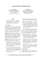
alone treatment group (95% CI, 1.37-2.53). Kaplan-
Meier survival curves i llustrating overall survival based
on treatment modality can be found in Figure 1. Uni-
variate hazard ratio analysis of age groups (95% CI,
1.14-1.98) indicated that survival statistically favored
patients <65 years of age (p = 0.002). Comparison of
univariate hazard ratios in relation to ECOG-PS class
indicated that survival statistically favored patients cate-
gorized in ECOG-PS class 0 when compared to patients
categorized in ECOG-PS c lass 2 (95% CI, 1.57-6.4) and
ECOG-PS class 3 (95% CI, 1.12-15.06), with p values o f
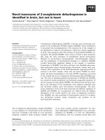
0.006 and 0.005, respe ctively. Comparison of univariate
hazard ratios in relation to primary tumor histology
indicated that survival statistically favored patients with
NSCLC when compared to patients with small-cell lung
cancer (SCLC) (95% CI, 0.94-2.61) and patients in the
other primary tumor histology group (95% CI, 1.14-
2.65), with p values of 0.04 and 0.002, respectively.
Kaplan-Meier survival curves illustrating overall survival
based on primary tumo r histology can be found in
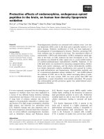
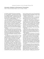
Figure 2. The analysis of the number of brain metastases
groups and tumor volume groups did not yield any sta-
tistically significant results. Kaplan-Meier survival curves
showing overall su rvival based on the nu mber of brain
metastases and volume of brain metastases are shown in
Figures 3 and 4.
The overall patient median survival time was deter-
mined to be 7.9 months. The median survival time for
patients treated with WBRT al one was 4.3 months (95%
CI, 3.30-5.38), 9.4 months (95% CI, 6.41-12.45) for
patients treated with SRS alone, 10 months (95% CI,
8.17-12.15) for patients treated with resection with
WBRT, 12 months (95% CI, 8.74-15.98) for patients
treated with WBRT with SRS, 13 months (95% CI, 9.70-
16.54) for patients t reated with resection + WBRT +
SRS, and 24 months (95% CI, 1.73-45.55) for patients
treated with resection with SRS. Patients <65 years of
age survived a median time of 11 months (95% CI, 8.42-
12.88), while patients ≥65 years of age survived a med-
ian time of 5.7 months (95% CI, 4.29-7.09). The median
survival time for patients in ECOG-PS class 0 was 22
mont hs (95% CI, 4.43-39.69) , 9.5 months (95% CI , 3.84-
15.16) for patients in ECOG-PS class 1, 6.0 months
Figure 1 Kaplan-Meier survival curve illustrating overall
survival based on treatment modality.
Figure 2 Kaplan-Meier survival curve illustrating overall
survival based on primary tumor histology.
Elaimy et al. World Journal of Surgical Oncology 2011, 9:69
/>Page 4 of 9
(95% CI, 2.64-9.26) for patients in ECOG-PS class 2, and
1.5 months (95% CI, 0.94-1.96) for patients in ECOG-PS
class 3. In regard to primary tumor histology, the med-
ian survival time for patients with NSCLC was deter-
mined to be 9.78 months (95% CI, 7.90-11.56), 9.2
months (95% CI, 4.04-14.30) for patients with breast
cancer, 8.6 months (95% CI, 3.67-13.55) for the com-
bined melanoma and renal-cell carcinoma group, 6.7
months (95% CI, 3.47-10.01) for patients with SCLC,
and 5.7 months (95% CI, 2.66-8.72) for patients classi-
fied in the other primary tumor histology group.
Further statistical analysis was conducted using multi-
variate Cox regression analysis with hazard ratio estimates
and confidence interv als (Table 2). The multivariate ana-
lyses utilized patients treated with SRS alone, patients <65
years of age, patients in ECOG-PS class 0, and patients
with NSCLC as the reference groups. Multivariate hazard
ratio analysis of treatment groups indicated that the survi-
val of patients in the SRS alone treatment group was sta-
tistically superior (p < 0.001) to the survival of the patients
in the WBRT alone treatment group (95% CI, 1 .37-2.73)
and that the survival of the resection with SRS treatment
group was statistically superior (p = 0.020) to the survival
of the SRS alone treatment group (95% CI, 0.49-0.9 4).
Comparison of multivariate hazard ratios in relation to
ECOG-PS class indicated that survival statistically favored
patients categorized in ECOG-PS cl ass 0 when compared
to patients categorized in ECOG-PS class 2 (95% CI, 1.02-
2.72), ECOG-PS class 3 (95% CI, 4.28-4.91), and ECOG-
PS class 4 (95% CI, 5.98-21.2), with p values of 0.04,
<0.001, <0.001, respectively. Multivariate hazard ratio ana-
lysis of primary tumor histology groups indicated that the
survival of patients in the breast cancer group was statisti-
cally superior (p < 0.001) to the survival of patients in the
NSCLC group (95% CI, 0.78-0.96) and that the survival of
patients in the NSCLC group was statistically superior
(p < 0.001) to the survival of patients in the combined
melanoma and renal-cell carcinoma group (95% CI, 1.06-
1.3). Multivariate hazard ratio analysis of age groups did
not yield any statistically significant results.
Discussion
Patients with metastatic brain disease have a poor prog-
nosis and curative treatment is not achievable in most
clinical situations, with 50% of patients dying from their
neurological cancer rather than their extracranial cancer
[12]. Due to this unfortunate outlook, maximizing
patient’s period of survival and comfort level is of great
importance. Although several Phase III studies have been
published assessing the efficacy of different treatment
modalities, many questions still remain unanswered and
further randomized evidence is needed not only to prove
superior treatments in comparison studies, but to identify
optimal courses of treatment in unique patient subsets
Figure 4 Kaplan-Meier survival curve illustrating overall
survival based on volume of brain metastases.
Figure 3 Kaplan-Meier survival curve illustrating overall
survival based on number of brain metastases.
Elaimy et al. World Journal of Surgical Oncology 2011, 9:69
/>Page 5 of 9
[6-9,13-17]. Our comprehensive analysis evaluates the
clinical effects treatment regimen, age, performance sta-
tus, primary tumor histology, number of brain metas-
tases, and total volume of brain metastases have on
patient survival.
Perhaps the most questionable matter in the manage-
ment of patients with brain metastases is whether the
addition of WBRT to SRS will provide patients with a
superior prognosis when compared to patients treated
with SRS alone [3]. Our study did not find statistically
significant survival differences between the SRS alone
treatment group and the SRS with WBRT treatment
group in both univariate and multivariate analysis. In
the randomized controlled trial published by Aoyama et
al. [13], the authors evaluated the clinical outcomes of
patients treated with SRS with or without WBRT and
also witnessed no significant (p = 0.4) differences in sur-
vival between the two treatment arms. However, the
patients treated with WBRT wit h SRS had a substan-
tially better 12-month brain tumor recurrence rate (p <
0.001) and underwent salvage therapy (p < 0.001) less
often than the patients treated with SRS alone, but these
increases in tumor control did not affect patient survi-
val. Several retrospective cohort studies published in the
last ten years have also reported t hat the addition of
WBRT to SRS does not resul t in superior levels of
patient survival [11,18-21].
On multivariate analysis, we found that the survival of
the SRS alone treatment arm did not statistically differ
when compared to the survival of the resection with
WBRT treatment arm. These data correlate with the
Phase III randomized trial conducted by Muacevic et al.
[17]. A total of 64 patient s with a si ngle, surgically acces-
sible brain metastasis ≤30 mm in diameter, a Karnofsky
Performance Score (KPS) ≥70, and a controlled primary
cancer were randomized into a GK radiosurgery alone
group (31 patients) and a surgery with WBRT group (33
patients). The authors reported non-significant differ-
ences in survival between the t wo treatment groups.
Rades et al. [22] retrospectively compared SRS alone and
surgery with WBRT in 260 patients classified in RPA
class 1 or 2 [5] that were diagnosed with 1 to 2 brain
metastases and also reported that the two groups did not
differ in survival. Our m ultivariate analysis also found
superior levels of survival in patients treated with resec-
tion with SRS when compared to patients treated with
SRS alone. The body of world literature lacks sufficient
studies comparing patients treated with SRS alone
against patients treated with resection with SRS. How-
ever, survival differences between patients treated with
SRS alone and patients treated with resection with SRS
was recently reported in another study by Rades et al.
[23]. The authors analyzed the clinical outcomes of 164
patients of adva nced age (≥65 years). Speci fically, 34
patients were treated with WBRT alone, 43 patients were
treated with SRS alone, 41 patients were treated with
resection + SR S, and 46 patients were treated with resec-
tion + WBRT+ SRS boost. In contrast to our re sults,
which favored the resection with SRS treatment group,
the authors reported that treatment regimen influenced
survival, with the SRS alone treatment group surviving a
greater time than the resection + SRS treatment group.
The results reported by Rades et al. [23] can be explained
when considering the risks of surgery in elderly patients.
This data permits the tr eatment of select patients who
are <65 years of age and are functionally independent
with resection in combination with SRS.
In subset analysis, patients treated with WBRT alone
at our institution exhibited the shortest period of survi-
val, with each of the other five treatment arms surviving
a substantially greater time than the WBRT alone treat-
ment arm. Although it is likely that the treatment arms
consisted of very different patient subsets with respect
to ECOG-PS class, tumor number, tumor volume, and
extent of systemic disease, both univariate and multi-
variate analysis found statistically significant differences
Table 2 Multivariate hazard ratios, confidence intervals,
and p values
Hazard Ratio
Estimate 95% CI p value**
Treatment Groups
SRS* reference
Surgery + SRS 0.68 0.49-0.94 0.020
WBRT + SRS 0.99 0.93-1.05 0.660
Surgery + WBRT + SRS 0.79 0.61-1.02 0.070
WBRT 1.94 1.37-2.73 <0.001
Surgery + WBRT 1.04 0.76-1.43 0.800
Age at diagnosis
<65* reference
≥65 1.21 0.91-1.62 0.190
ECOG-PS
0* reference
1 1.07 0.58-1.95 0.830
2 1.67 1.02-2.72 0.040
3 4.58 4.28-4.91 <0.001
4 11.26 5.98-21.2 <0.001
Primary Tumor Histology
NSCLC* reference
SCLC 1.11 0.97-1.26 0.130
Breast 0.87 0.78-0.96 <0.001
Melanoma and Renal-cell 1.17 1.06-1.3 <0.001
Other 1.41 0.95-2.1 0.080
ECOG-PS = Eastern Cooperative Oncology Group-Performance Status; NSCLC =
non-small-cell lung cancer; SCLC = small-cell lung cancer; SRS = stereotactic
radiosurgery; WBRT = whole brain radiation therapy
* Reference group against which other groups’ survival experience are
compared
** p value for test if groups’ survival experience is same as reference group
Elaimy et al. World Journal of Surgical Oncology 2011, 9:69
/>Page 6 of 9
between the hazard ratio of patients treated with WBRT
and the hazard ratio of patients treat ed with SRS alone.
No randomized controlled trials have been conducted
assessing patients treated with SRS alone compared with
patients treated with WBRT alone. However, in a recent
literature review, Li nskey et al. [12] found level 3 evi-
dence i ndicating that patients with 1 to 3 brain metas-
tases that are treated with SRS alone have superior
levels of survival when compared to patients treated
with WBRT alone.
As expected, we found that age and performance sta-
tus are both significant predictors in determining patient
prognosis, as survival statistically favored patients <65
years old in univariate analysis and patients in a lower
ECOG-PS class in both univariate and multivariate ana-
lysis. Several comparison studies have reported a survi-
val dependency on patient age and performance status.
Sanghavi et al. [24] retrospectively analyzed the out-
comes and potential prognostic factors of a total of 502
patients treated with SRS with WBRT and 1200 patients
treated with WBRT alone and found that survival was
more pronounced in patients with a higher KPS (p =
0.0001), a controlled primary cancer (p = 0.0023), the
absence of extr acranial cancer (p = 0.0001), and a lower
RPA class (p = 0.000007). Kocher et al. [25] compa red
the efficacy of SRS alone against WBRT alone in 255
patients with 1 to 3 brain metastases and reported sta-
tistically significant increases in median survival in
patients categorized in RPA class 1 (p < 0.0001) and
RPA class 2 (p < 0.04). Frazier et al. [26] retrospect ively
analyzed 237 patients treated with SRS ± WBRT and
also found that survival statistically favored patients that
were <65 years of age (p = 0.008) with KPS values >70
(p = 0.034).
The number and volume of brain metastases patients
possess at the time of diagnosis are crucial factors in
prescribing the most advantageous course of treatment
in select patient groups. When evaluating our six treat-
ment arms in univariate analysis; however, the number
and size of brain metastases di d not influence patient
survival. Tumor resection in combination with WBRT
and/or SRS in treating patients with a single brain
metastasis is recommended for those who present with
severe neurologic deficits, a ventricular obstruction, or a
tumor of a large intracranial volume (which often pro-
duces mass effect) [1]. When the patient has controlled
neurological symptoms, a tumor/s of a small intracranial
volume, a single brain metastasis, a surgically inoperable
brain metastasis, or multiple brain metastases, SRS
alone or in combination with WBRT is often the recom-
mended course of treatment [1]. Questions remain
regarding the survival dependency on the number and
size of brain m etastases patient groups possess. Studies
have shown increased survival levels in patients with a
single brain metastasis that were treated with radiosur-
gery [6,26]. However, o ther publications have reported
that tumor volume has a greater impact on patient sur-
vival than number of brain metastases and primary
tumor histology, with patients possessing small tumor
volumes surviving a greater period of time [27-30].
Further s tudy and research is needed on how the num-
ber and total volume of brain metastases affect patient
survival.
The histologic subtype of the primary tumor may be
an ess ential predictor in assessing the survival advantage
of specific patient subsets. NSCLC is known to produce
the greatest amount of metastatic brain lesions [31,32].
In univariate analysis, survival statistically favored
patients with NSCLC when compared to patients with
SCLC and patients classified in the other primary histol-
ogy group. In multivariate analysis; however, survival
statistically favored patients in the breast cancer g roup
when compared to patients in the NSCLC group.
Increases in the survival of breast cancer patients when
compared to NSCLC patients was also recently reported
in the survival analysis of 237 patients treated with
radiosurgery by Frazier et al. [26]. These results are
likely due to advances in the surgical and chemothera-
peutic care of breast cancer patients [33]. It was also
observed in multivariate analysis that survival statisti-
cally favored patients with NSCLC when compared to
the combined melanoma and renal-cell carcinoma
group. Traditionally, melanoma and renal-cell carcinoma
have been classified as “radioresistant” tumor histologies
bec ause of their negative response to standard radiation
treatment. However, several studies have reported posi-
tive outcomes when treating patients with melanoma
and renal-cell carcinoma primaries with radiosurgery
[34-40]. In a phase II trial conducted by Manon et a l.
[41], 31 patients diagnosed with melanoma, renal-cell
carcinoma, and sarcoma primary cancers with 1 to 3
brain metastases were t reated with SRS alon e. The 3
and 6 month intracranial failure rate for the ev aluated
patients was found to be 25.8 and 48.3%, respectively.
The authors concluded that delaying WBRT for patients
with melanoma, renal-cell carcinoma, and sarcoma pri-
mary cancers may be appropriate for specific subgroups
of patients, but must be approached with caution.
Conclusions
We report retrospectively on the effects treatment regi-
men, age, performance status, primary tumor histology,
number of brain metastases, and volume of brain metas-
tases have on the survival of patients diagnosed with
brain metastases. Multivariate analysis of treatment regi-
mens showed that survival statistically favored patients
treated with SRS alone and patients treated with resec-
tion with SRS when compared to patients treated with
Elaimy et al. World Journal of Surgical Oncology 2011, 9:69
/>Page 7 of 9
WBRT alone and patients treated with SRS alone,
respectively. Comparison of multivariate hazard ratios in
relation to ECOG-PS class indicated that survival stat is-
tically favored patients categorized in ECOG-PS class 0
when compared to patients categorized i n ECOG-PS
classes of 2, 3, and 4. Multivariate analysis of primary
tumor histology groups indicated that the survival of
patients in the breast cancer group was statistic ally
superior to the survival of patients in the NSCLC group
and that the survival of p atients in the NSCLC group
was statistically super ior to the survival of patients in
the combined melanoma and renal-cell carcinoma
group. In our analysis, patients benefited from a com-
bined modality treatment approach and physicians must
consider patient age, performance status, and primary
tumor histology when recommending specific treatment
regimens.
Acknowledgements
We would like to acknowledge Eric Reynolds, Rachel Garman, and Jill
Adams, as well as the entire Gamma Knife of Spokane and Cancer Care
Northwest research staff for their contributions to this manuscript. We would
also like to acknowledge that this project was funded in part by The Breast
Cancer Society in Mesa, Arizona.
Author details
1
Gamma Knife of Spokane, 910 W 5
th
Ave, Suite 102, Spokane, WA 99204,
USA.
2
Cancer Care Northwest, 910 W 5
th
Ave, Suite 102, Spokane, WA 99204,
USA.
3
MacKay & Meyer MDs, 711 S Cowley St, Suite 210, Spokane, WA 99202,
USA.
4
Spokane Brain & Spine, 801 W 5
th
Ave, Suite 210, Spokane, WA 99204,
USA.
5
DataWorks Northwest, LLC, 3952 N Magnuson St, Coeur D’Alene, ID
83815, USA.
Authors’ contributions
ALE and CML reviewed relevant literature and drafted the manuscript. BJP
conducted all statistical analyses. ARM, WTL, RKF, JJD, BSC, and JTH provided
clinical expertise and participated in drafting the manuscript. All authors
read and approved the final manuscript.
Competing interests
The authors declare that they have no competing interests.
Received: 29 April 2011 Accepted: 5 July 2011 Published: 5 July 2011
References
1. Hazard LJ, Jensen RL, Shrieve DC: Role of stereotactic radiosurgery in the
treatment of brain metastases. Am J Clin Oncol 2005, 28:403-410.
2. Hart MG, Grant R, Walker M, Dickinson H: Surgical resection and whole
brain radiation therapy versus whole brain radiation therapy alone for
single brain metastases. Cochrane Database Syst Rev 2005, CD003292.
3. Suh JH: Stereotactic radiosurgery for the management of brain
metastases. N Engl J Med 2010, 362:1119-1127.
4. Delattre JY, Krol G, Thaler HT, Posner JB: Distribution of brain metastases.
Arch Neurol 1988, 45:741-744.
5. Gaspar L, Scott C, Rotman M, Asbell S, Phillips T, Wasserman T,
McKenna WG, Byhardt R: Recursive partitioning analysis (RPA) of
prognostic factors in three Radiation Therapy Oncology Group (RTOG)
brain metastases trials. Int J Radiat Oncol Biol Phys 1997, 37:745-751.
6. Andrews DW, Scott CB, Sperduto PW, Flanders AE, Gaspar LE, Schell MC,
Werner-Wasik M, Demas W, Ryu J, Bahary JP, Souhami L, Rotman M,
Mehta MP, Curran WJ Jr: Whole brain radiation therapy with or without
stereotactic radiosurgery boost for patients with one to three brain
metastases: phase III results of the RTOG 9508 randomised trial. Lancet
2004, 363:1665-1672.
7. Patchell RA, Tibbs PA, Regine WF, Dempsey RJ, Mohiuddin M, Kryscio RJ,
Markesbery WR, Foon KA, Young B: Postoperative radiotherapy in the
treatment of single metastases to the brain: a randomized trial. JAMA
1998, 280:1485-1489.
8. Patchell RA, Tibbs PA, Walsh JW, Dempsey RJ, Maruyama Y, Kryscio RJ,
Markesbery WR, Macdonald JS, Young B: A randomized trial of surgery in
the treatment of single metastases to the brain. N Engl J Med 1990,
322:494-500.
9. Vecht CJ, Haaxma-Reiche H, Noordijk EM, Padberg GW, Voormolen JH,
Hoekstra FH, Tans JT, Lambooij N, Metsaars JA, Wattendorff AR, et al:
Treatment of single brain metastasis: radiotherapy alone or combined
with neurosurgery? Ann Neurol 1993, 33:583-590.
10. Schackert G: Surgery of brain metastases - pro and contra. Onkologie
2002, 25:480-481.
11. Sneed PK, Suh JH, Goetsch SJ, Sanghavi SN, Chappell R, Buatti JM,
Regine WF, Weltman E, King VJ, Breneman JC, Sperduto PW, Mehta MP: A
multi-institutional review of radiosurgery alone vs. radiosurgery with
whole brain radiotherapy as the initial management of brain
metastases. Int J Radiat Oncol Biol Phys 2002, 53:519-526.
12. Linskey ME, Andrews DW, Asher AL, Burri SH, Kondziolka D, Robinson PD,
Ammirati M, Cobbs CS, Gaspar LE, Loeffler JS, McDermott M, Mehta MP,
Mikkelsen T, Olson JJ, Paleologos NA, Patchell RA, Ryken TC, Kalkanis SN:
The role of stereotactic radiosurgery in the management of patients
with newly diagnosed brain metastases: a systematic review and
evidence-based clinical practice guideline. J Neurooncol 2010, 96:45-68.
13. Aoyama H, Shirato H, Tago M, Nakagawa K, Toyoda T, Hatano K, Kenjyo M,
Oya N, Hirota S, Shioura H, Kunieda E, Inomata T, Hayakawa K, Katoh N,
Kobashi G: Stereotactic radiosurgery plus whole-brain radiation therapy
vs stereotactic radiosurgery alone for treatment of brain metastases: a
randomized controlled trial. JAMA 2006, 295:2483-2491.
14. Chang EL, Wefel JS, Hess KR, Allen PK, Lang FF, Kornguth DG, Arbuckle RB,
Swint JM, Shiu AS, Maor MH, Meyers CA:
Neurocognition in patients with
brain
metastases treated with radiosurgery or radiosurgery plus whole-
brain irradiation: a randomised controlled trial. Lancet Oncol 2009,
10:1037-1044.
15. Kondziolka D, Patel A, Lunsford LD, Kassam A, Flickinger JC: Stereotactic
radiosurgery plus whole brain radiotherapy versus radiotherapy alone
for patients with multiple brain metastases. Int J Radiat Oncol Biol Phys
1999, 45:427-434.
16. Mintz AH, Kestle J, Rathbone MP, Gaspar L, Hugenholtz H, Fisher B,
Duncan G, Skingley P, Foster G, Levine M: A randomized trial to assess the
efficacy of surgery in addition to radiotherapy in patients with a single
cerebral metastasis. Cancer 1996, 78:1470-1476.
17. Muacevic A, Wowra B, Siefert A, Tonn JC, Steiger HJ, Kreth FW:
Microsurgery plus whole brain irradiation versus Gamma Knife surgery
alone for treatment of single metastases to the brain: a randomized
controlled multicentre phase III trial. J Neurooncol 2008, 87:299-307.
18. Chidel MA, Suh JH, Reddy CA, Chao ST, Lundbeck MF, Barnett GH:
Application of recursive partitioning analysis and evaluation of the use
of whole brain radiation among patients treated with stereotactic
radiosurgery for newly diagnosed brain metastases. Int J Radiat Oncol
Biol Phys 2000, 47:993-999.
19. Clarke JW, Register S, McGregor JM, Grecula JC, Mayr NA, Wang JZ, Li K,
Gupta N, Kendra KL, Olencki TE, Cavaliere R, Sarkar A, Lo SS: Stereotactic
radiosurgery with or without whole brain radiotherapy for patients with
a single radioresistant brain metastasis. Am J Clin Oncol 2010, 33:70-74.
20. Fokas E, Henzel M, Hamm K, Surber G, Kleinert G, Engenhart-Cabillic R:
Radiotherapy for brain metastases from renal cell cancer: should whole-
brain radiotherapy be added to stereotactic radiosurgery?: analysis of 88
patients. Strahlenther Onkol 2010, 186:210-217.
21. Jawahar A, Willis BK, Smith DR, Ampil F, Datta R, Nanda A: Gamma knife
radiosurgery for brain metastases: do patients benefit from adjuvant
external-beam radiotherapy? An 18-month comparative analysis.
Stereotact Funct Neurosurg 2002, 79:262-271.
22. Rades D, Bohlen G, Pluemer A, Veninga T, Hanssens P, Dunst J, Schild SE:
Stereotactic radiosurgery alone versus resection plus whole-brain
radiotherapy for 1 or 2 brain metastases in recursive partitioning
analysis class 1 and 2 patients. Cancer 2007, 109:2515-2521.
23. Rades D, Pluemer A, Veninga T, Schild SE: Comparison of different
treatment approaches for one to two brain metastases in elderly
patients. Strahlenther Onkol 2008, 184:565-571.
Elaimy et al. World Journal of Surgical Oncology 2011, 9:69
/>Page 8 of 9
24. Sanghavi SN, Miranpuri SS, Chappell R, Buatti JM, Sneed PK, Suh JH,
Regine WF, Weltman E, King VJ, Goetsch SJ, Breneman JC, Sperduto PW,
Scott C, Mabanta S, Mehta MP: Radiosurgery for patients with brain
metastases: a multi-institutional analysis, stratified by the RTOG
recursive partitioning analysis method. Int J Radiat Oncol Biol Phys 2001,
51:426-434.
25. Kocher M, Maarouf M, Bendel M, Voges J, Muller RP, Sturm V: Linac
radiosurgery versus whole brain radiotherapy for brain metastases. A
survival comparison based on the RTOG recursive partitioning analysis.
Strahlenther Onkol 2004, 180:263-267.
26. Frazier JL, Batra S, Kapor S, Vellimana A, Gandhi R, Carson KA, Shokek O,
Lim M, Kleinberg L, Rigamonti D: Stereotactic radiosurgery in the
management of brain metastases: an institutional retrospective analysis
of survival. Int J Radiat Oncol Biol Phys 2010, 76:1486-1492.
27. Bhatnagar AK, Flickinger JC, Kondziolka D, Lunsford LD: Stereotactic
radiosurgery for four or more intracranial metastases. Int J Radiat Oncol
Biol Phys 2006, 64:898-903.
28. Jawahar A, Shaya M, Campbell P, Ampil F, Willis BK, Smith D, Nanda A: Role
of stereotactic radiosurgery as a primary treatment option in the
management of newly diagnosed multiple (3-6) intracranial metastases.
Surg Neurol 2005, 64:207-212.
29. Selek U, Chang EL, Hassenbusch SJ, Shiu AS, Lang FF, Allen P, Weinberg J,
Sawaya R, Maor MH: Stereotactic radiosurgical treatment in 103 patients
for 153 cerebral melanoma metastases. Int J Radiat Oncol Biol Phys 2004,
59:1097-1106.
30. Sheehan J, Kondziolka D, Flickinger J, Lunsford LD: Radiosurgery for
patients with recurrent small cell lung carcinoma metastatic to the
brain: outcomes and prognostic factors. J Neurosurg 2005,
102(Suppl):247-254.
31. Marcou Y, Lindquist C, Adams C, Retsas S, Plowman PN: What is the
optimal therapy of brain metastases? Clin Oncol (R Coll Radiol) 2001,
13:105-111.
32. Posner JB: Management of brain metastases. Rev Neurol (Paris) 1992,
148:477-487.
33. Akyurek S, Chang EL, Mahajan A, Hassenbusch SJ, Allen PK, Mathews LA,
Shiu AS, Maor MH, Woo SY: Stereotactic radiosurgical treatment of
cerebral metastases arising from breast cancer. Am J Clin Oncol 2007,
30:310-314.
34. Adler JR, Cox RS, Kaplan I, Martin DP: Stereotactic radiosurgical treatment
of brain metastases. J Neurosurg 1992, 76:444-449.
35. Amendola BE, Wolf AL, Coy SR, Amendola M, Bloch L: Brain metastases in
renal cell carcinoma: management with gamma knife radiosurgery.
Cancer J 2000, 6:372-376.
36. Auchter RM, Lamond JP, Alexander E, Buatti JM, Chappell R, Friedman WA,
Kinsella TJ, Levin AB, Noyes WR, Schultz CJ, Loeffler JS, Mehta MP: A
multiinstitutional outcome and prognostic factor analysis of
radiosurgery for resectable single brain metastasis. Int J Radiat Oncol Biol
Phys 1996, 35:27-35.
37. Flickinger JC, Kondziolka D, Lunsford LD, Coffey RJ, Goodman ML, Shaw EG,
Hudgins WR, Weiner R, Harsh GRt, Sneed PK, et al: A multi-institutional
experience with stereotactic radiosurgery for solitary brain metastasis.
Int J Radiat Oncol Biol Phys
1994, 28:797-802.
38. Maor MH, Dubey P, Tucker SL, Shiu AS, Mathur BN, Sawaya R, Lang FF,
Hassenbusch SJ: Stereotactic radiosurgery for brain metastases: results
and prognostic factors. Int J Cancer 2000, 90:157-162.
39. Sheehan JP, Sun MH, Kondziolka D, Flickinger J, Lunsford LD: Radiosurgery
in patients with renal cell carcinoma metastasis to the brain: long-term
outcomes and prognostic factors influencing survival and local tumor
control. J Neurosurg 2003, 98:342-349.
40. Shuto T, Inomori S, Fujino H, Nagano H: Gamma knife surgery for
metastatic brain tumors from renal cell carcinoma. J Neurosurg 2006,
105:555-560.
41. Manon R, O’Neill A, Knisely J, Werner-Wasik M, Lazarus HM, Wagner H,
Gilbert M, Metha M, Eastern Cooperative Oncology Group: Phase II trial of
radiosurgery for one to three newly diagnosed brain metastases from
renal cell carcinoma, melanoma, and sarcoma: an Eastern Cooperative
Oncology Group study (E 6397). J Clin Oncol 2005, 23:8870-8876.
doi:10.1186/1477-7819-9-69
Cite this article as: Elaimy et al.: Multimodality treatment of brain
metastases: an institutional survival analysis of 275 patients. World
Journal of Surgical Oncology 2011 9:69.
Submit your next manuscript to BioMed Central
and take full advantage of:
• Convenient online submission
• Thorough peer review
• No space constraints or color figure charges
• Immediate publication on acceptance
• Inclusion in PubMed, CAS, Scopus and Google Scholar
• Research which is freely available for redistribution
Submit your manuscript at
www.biomedcentral.com/submit
Elaimy et al. World Journal of Surgical Oncology 2011, 9:69
/>Page 9 of 9