báo cáo khoa học: "Hepatic actinomycosis mimicking an isolated tumor recurrence" ppsx
Bạn đang xem bản rút gọn của tài liệu. Xem và tải ngay bản đầy đủ của tài liệu tại đây (795.54 KB, 3 trang )
CAS E REP O R T Open Access
Hepatic actinomycosis mimicking an isolated
tumor recurrence
Michael G Wayne
*
, Rahul Narang, Arif Chauhdry and Justin Steele
Abstract
Actinomyces species has been described as an opportunistic pathogen, particularly in the oral cavity; however, in
rare cases these bacteria can cause actinomycosis which is characterized by formation of abscesses in the mouth,
lungs, or gastrointestinal tract.
Actinomycosis was commonly present in the pre-antibiotic era; however, it has a low prevalence now days. It has
been recognized since 150 years ago, but because of its variable clinical presentati on and indolent course, its
recognition is difficult and patients are often misdiagnosed. Here we present a case of primary hepatic
actinomycosis presenting as a metastatic liver tumor.
Case report
This is the case of a 65-year-old male, who originally
presented on August 18, 2008 with obstructive jaundice.
His past medical history includes diabetes and hyperten-
sion. He underwent ERCP with stenting of the bile duct.
The patient also had a spiral CT and an endoscopic
ultrasound of the pancreas. These tests helped to deter-
mine resectability. He underwent a pancreaticoduode-
nectomy (Whipple procedure) on Sept. 8, 2008 for a
pancreatic head adenocarcinoma. He had a standard
whipple performed, with removal of the gallbl adder and
distal stomach, as well as the head of the pancreas and
duodenum. There were no intraoperative events and no
gallstones were spilled. He received adjuvant chemother-
apy for 7 months. The chemotherap y was gemzar, oxali-
platinum and tarceva. The patient had routine follow-up
and surveillance for recurrence every 4 months the first
year and every 6 months for the second year. This
included a CT scan and CA 19-9 level. Over the course
of one year he was admitted to the hospital on several
occasions for low-grade fever, for which the diagnosis
was not established, and at times treated as an outpati-
ent for multiple urinary tract infections. During these
episodes the patie nt always had a normal white blood
cell count and normal neutrophils/lymphocytes on dif-
ferential. At no time did he complain of any abdominal
pain or any difficulty tolerating food. All CT scans
during this first year were negative for recurrent malig-
nant disease.
At approximately 18 months in follow-up, the
repeated computed tomography (CT) scan showed a
non-specificabnormalityintherightlobeoftheliver,
suspicious for a mass. A magnetic res onance imaging
(MRI) and positron emission tomography (PET) scan
were then performed, which was positive for a mass,
suspicious for an isolated tumor recurrence between
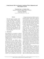
segments5and6intheliver.BoththeCTandMRI
showed a large liver lesion present in the inferior aspect
of segment 6 measuring 7 cm AP, 5 cm transverse, and
4.5 cm cranioc audal (Figure 1). This mass demonstrated
heterogeneous thick peripheral rim enhancement with
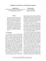
central hypo-enhancement and likely necrosis. The PET
scan showed an abnormal robust focus of metabolic
activity in inferior lateral right hepatic lobe, c oncerning
for metastatic disease, SUV 11.6 (Figure 2).
Laboratory examination of the patient’sblooddemon-
strated: white blood cell count 6.1 (reference range 4.5-
10.8 K/uL), alkaline phosphatase level 141 (reference
range 38-126 U/L), and AST 36 (reference range 15-46
U/L), ALT 50 (reference range 13-69 U/L). The total
and direct bilirubin levels were normal. The CA 19-9
was 84, which was increased from 39 on the previous
sample from 3 months prior. Physical exam of the abdo-
men was unremarkable. No masses were palpated, the
liver was not enlarged, and the abdomen was non-ten-
der. There was no cervical, umbilical, or inguinal nodes
appreciated.
* Correspondence:
Pancreas Center at Beth Israel Medical Center, NY 37 Union Square West, NY
10003, USA
Wayne et al. World Journal of Surgical Oncology 2011, 9:70
/>WORLD JOURNAL OF
SURGICAL ONCOLOGY
© 2011 Wayne et al; licensee BioMed Central Ltd. This is an Open Access articl e distribut ed under the terms of the Cre ative Commons
Attribution License (http://creativecom mons.org/licenses/by/2.0), w hich permits unrestricted use, distribution, and repro duction in
any medium, provided the original work is properly cited.
FNA biopsy was discussed at the GI tumor board, but
in the setting of a rising CA19-9 and suspicious CT,
MRI, and PET scan, the decision was made to proceed
to surgery without a biopsy. The tumor board felt that
this was recurrent pancreatic cancer and because of this
decided the small risk of seedingtheneedletractby
doing an FNA was not warranted in this case.
Patient underwent a resection of segment 5 and 6 of
the liver without any complications. Final pathology of
the specimen returned as inflammatory granulation tis-
sue and granules consistent with hepatic actinomycosis.
This was confirmed on tis sue cultures. Special staining of
this specimen showed gram positive filamentous bacteria.
Infectious disease con sult was called to review the
case. They selected doxcycline iv , based on culture and
sensitivity report, which would be changed to po doxcy-
cline on discharge. The patient had an uneventful recov-
ery and was discharged home on post-operative day 5.
He was discharged home on oral doxycycline for a 6
month treatment course. 30-day follow-up shows no
recurrence of this lesion and he continues to remain
afebrile.
Discussion
The genus Actinomyces species are a slowing growing,
gram-positive, non-spore-forming bacteria, that thrive in
microareophilic and anaerobic conditions [1]. There are
13 different species of actinomycosis of which 6 are
associated with human disease (arachina propionica,
bovis, israelli, naseslundii, odontolyticus and viscosus).
The most common pathogen encountered is Actino-
myces israelii, which gives rise to chronic suppurative
infection [2,3].
They tend to be associated with infections of the cer-
vicofacial and oral region. A rare cause of abdominal
infecti on, the pathogenesis is presumed to be caused by
hematogeneous spread via the portal vein from either a
mucosal injury or other abdominal focus of injury and/
or infection. The non-specific symptoms of fever, weight
loss and abdominal pain make diagnosis of this condi-
tion a challenge. Despite advanced imaging tec hniques
and difficulty in obtaining in positive cultures, the rate
of preoperative diagnosis is less than 10% [4].
According to the literature, actinomycosis of the
abdomen and pelvis accounts for 10-20% of reported
cases. Typically, these patients have a history of recent
or remote bowel surgery ( eg, perforated appendicitis,
perforated colonic diverticulitis, spilled gallstones dur-
ing cholecystectomy) or ingestion of foreign bodies (eg,
chicken or fish bones), during which actinomyce tes are
introduced into the deep tissues. Diagnosis is usually
established postoperatively, following exploratory lapar-
otomy for a suspected malignancy. Pelvic actinomyco-
sis most commonly ascends from the uterus in
association with intrauterine contraceptive devices
(IUCDs). Female genital Actinomyces infection i s rela-
tively rare, although strongly related to long-lasting
intrauterine contraceptive device (IUD) application. An
infective pathway has been postulated extending
upward from the female perineum to the vagina and
cervix.Thetraumaticeffectofthedeviceandaprior
infection may contribute to the Actinomyces infection
in the female genitalia.
Figure 1 Liver lesion present in the inferior aspect of segment
6 (arrow). This lesion appears to be an isolated tumor recurrence
18 months after Whipple procedure.
Figure 2 PET scan showed abnormal robust focus of metabolic
activity in inferior lateral aspect of right hepatic lobe
concerning for metastatic disease (arrow).
Wayne et al. World Journal of Surgical Oncology 2011, 9:70
/>Page 2 of 3
Risk factors associate d with this condition include
abdominal wall trauma, gastrointestinal foreign body,
previous abdo minal/pe lvic surgery, gastrointestinal tract
lesions and immunosuppressi on [5]. Since gastrointest-
inal foreign bodies are a risk factor it is of no surprise
tha t recen t literature has ci ted an increased risk for this
infection with biliary and pancreatic stent placement [6]
Liver involvement has been reported in 15% of the
cases, with majority of them resulting from translocation
from either an intra-abdominal or intrathoracic site [7].
Primary liver involvement has only been reported in 5%
of the cases [7].
Primary hepatic actinomycosis typically presents with
an indolent course with symptoms typically present for
greater than two weeks [8]. Immunocomptetent adults
with variable age distribution and a male prevalence has
been observed [8].
Difficult task for the management of actinomycosis is
to reach the diagnosis before surgical approach is taken.
In our patient the p atient presents with isolated hepat ic
mass on MRI and PET scan str ongly suspicious for
recurrence o f tumor after one year of tumor free survi-
val. Because imaging studies frequently reveal single or
multiple lesions, actinomycosis is often misdiagnosed as
a pri mary or metastatic tumor. Radiographicallly, lesions
frequently present as a single hypodense mass/abscess
68.4%) [9].
For diagnosis, macroscopic, microscopic and histo-
chemical examinations of surgical specimens are
required, but the definitive diagnosis is based on tis-
sue culture. The infection tends to lead a chronic and
suppurative infection leading to fibrosis with draining
sinuses that are pale yellow and often referred to as
“sulfur granules” [6]. The wall of these mass are often
“ wooden” in consistency [2]. Our patient presented
with one year history of indolent course with nonspe-
cific and undiagnosed causes of fevers and hospital
admissions, treated w ith antibiotics. He never had an
immunosuppressive work-up but his white blood cell
count and total lymphocyte count were always
normal.
Treatment of this infection consists of intravenous
penicillin-G for four weeks and then oral penicillin-V
for 12 months [10,11]. Prolonged treatment is rec om-
mended because of the incidence of recurrence.
Although no true surgical intervention guidelines has
been established, it has been used for treatment with
patients who present with extensive necrotic tissue or
large abscesses that cannot be adequately drained
[12,13]. Our patient’s actinomycoses was sensitive to
both penicillin-G and doxcycline. Do xycycline was the
antibiotic selected by our infectious disease doctor. No
specific reason given for his choice.
Conclusions
Our conclusion from this case report is t hat primary
hepatic actinomycosis can present in patients as a soli-
tary hepatic mass after gastrointestinal malignancy. In
our case it was mimi cking a recurrence of the tumor. It
isarareandoftenoverlookedetiologyforalivermass.
An indolent course with unexplainable fevers, occasion-
ally requiring hospital admissions, in patients with his-
tory of GI malignancies, should make physicians aware
of this possible diagnosis. Nevertheless, the variable clin-
ical presentation of this disease and its low incidence
makes it difficult to recognize.
Consent
Written informed consent was obtained from the patient
for p ublication of this Case report and any accompany-
ing images. A copy of the written consent is available
for review by the Editor-in-Chief of this journal
Authors’ contributions
MW-main author and primary surgeon for patient; RN-assisted in writing
case report; AC-assisted in gathering data; JS-assistant surgeon and helped
edit the article; All authors read and approved the final manuscript.
Competing interests
The authors declare that they have no competing interests.
Received: 29 November 2010 Accepted: 11 July 2011
Published: 11 July 2011
References
1. Bowden GHW: Actinomycosis in:Baron’s Medical Microbiology. In Univ of
Texas Medical Branch Edited by: Baron S et al. , 4 1996.
2. Brook I: Actinomycosis: diagnosis and management. South Med J 2008,
101:1019-23.
3. Jermini I, et al: An unusual case of hepatic abscess. Praxis 1994, 20:1781-4.
4. Brown JR: Human actinomycosis: CT features. J Comput Assist Tomogr
1986, 10:335-337.
5. Lee JD, Kim PG, Jo HJ, et al: A case of primary hepatic Actinomycosis.
J Korean Med Sci 1993, 8:385-9.
6. Kanellopoulou T, et al: Primary Hepatic Actinomycosis. American J Med Sci
2010, 4:362-65.
7. Gulglielmi A, et al: Primary hepatic actinomycosis: a clinical case report
and review of literature. Ann Ital Chir 1991, 62:185-9.
8. Lai AT, et al: Hepatic actinomycosis presenting as a liver tumor: a case
report and literature review. Asian J Surg 2004, 27:345-7.
9. Christodoulou N, Papadakis I, Velegrakis M: Actinomycotic live abscess.
Case report and review of the literature. Chir Ital 2004, 56(1):141-146.
10. Felekouras E, Menenakos C, Griniatsos J, Deladetsima I, Kalaxanisi N,
Nikiteas N, Papalambros E, Kordossis T, Bastounis E: Liver resection in cases
of isolated hepatic actinomycosis: case report and review of the
literature. Scand J Infect Dis 2004, 36(6-7):535-538.
11. Chen LW, Chang LC, Shie SS, CHien RN: Solitary actinomycotic abscesses
of the liver: report of two cases. Int J Clin Pract 2006, 60(1):104-107.
12. Islam T, Athar MN, Athar MK, Usman MH, Misbah B: Hepatic actinomycosis
with infiltration of the diaphragm and right lung: a case report. Can
Respir J 2005, 12(6):336-337.
13. Lall T, et al: Isolated hepatic actinomycosis: A case report. J Med Case
Report 2010, 4
:45.
doi:10.1186/1477-7819-9-70
Cite this article as: Wayne et al.: Hepatic actinomycosis mimicking an
isolated tumor recurrence. World Journal of Surgical Oncology 2011 9:70.
Wayne et al. World Journal of Surgical Oncology 2011, 9:70
/>Page 3 of 3