báo cáo khoa học: "Sentinel lymph node biopsy for high-risk cutaneous squamous cell carcinoma: clinical experience and review of literature" ppsx
Bạn đang xem bản rút gọn của tài liệu. Xem và tải ngay bản đầy đủ của tài liệu tại đây (1.33 MB, 7 trang )
REVIEW Open Access
Sentinel lymph node biopsy for high-risk
cutaneous squamous cell carcinoma: clinical
experience and review of literature
Steve Kwon
1
, Zhao Ming Dong
2
and Peter C Wu
1,3*
Abstract
High-risk cutaneous squamous cell carcinoma (SCC) is associated with an increased risk of metastases. The role of
sentinel lymph node (SLN) biopsy in these patients remains unclear. To address this uncertainty, we collected
clinical data on six patients with clinical N0 high-risk SCC that underwent SLN biopsy between 1999 and 2006 and
performed a literature review of SLN procedures for SCC to study the utility of SLN biopsy. There were no positive
SLN identified among six cases and there was one local and one distant recurrence on follow-up. Literature review
identified 130 reported cases of SLN biopsy for SCC. The SLN positivity rate was 14.1%, 10.1%, and 18.6%; false
negative rate was 15.4%, 0%, and 22.2%; and the negative predictive value was 97.8%, 100%, and 95.2% for all sites,
head/neck, and truncal/extremity sites, respectively. SLN biopsy remains an investigational staging tool in clinically
node-negative high-risk SCC patients. The higher false negative rate and lower negative predictive value among
SCC of the trunk/extremity compared to SCC of the head/neck sites suggests a more cautious approach when
treating patients with the former. Given the paucity of long-term follow up, an emphasis is placed upon the need
for close surveillance regardless of SLN status.
Keywords: sentinel lymph node, squamous cell carcinoma, cutaneous, staging
Introduction
Cutaneous squamous cell carcinoma (SCC) is overall the
second most common skin cancer with approximately
200,000 new cases diagnosed each year in the U.S. and
accounts for nearly 25% of annual skin cancer deaths
[1-4]. Fortunately, the majority of cases is associated
with a favorable prognosis and is often curable by surgi-
cal or local destructive therapy. However, a small subset
of SCC tumors can be characterized by aggressive biolo -
gic behavior with an increased risk of locoregional
recurrence and distant metastases. Numerous studies
have identified high-risk factors in SCC patients [5-7]
associated with a worse prognosis includin g large size,
rapid growth rate, irregular borders, moderate/poor dif-
ferentiation, perineural invasion, recurrent lesions, sites
of prior radiotherapy or chronic inflammation, immuno-
compromised states, and genetic disorders including
albinism and xeroderma pigmentosum. In terms of size
and location, SCC tumors are considered high-risk
when measuring greater than 2 cm on the trunk and
extremities; > 1 cm on the cheeks, forehead, scalp and
neck; and > 0.6 cm on the “ mask areas” of the face, gen-
itals, hands and feet. More recent studies have suggested
that tumor thickness (Clark’ s level IV), desmoplastic
growth, and development of nodal metastases are the
strongest predictors for survival resembling cutaneous
melanoma [8,9]. Patients with cutaneous SCC associated
with high-risk tumor features reportedly have a higher
rates of local recurrence ranging between 10-47.2%, and
rates of regional and distant metastases between 11-
47.3% [5,10].
Prognosis is generally poor in patients who develop
nodal metastases with an expected 5-year survival of 26-
34% and a 10-year survival rate of only 16%, underscor-
ing the importance of early detection and treatment
[5,10]. Recognizing that SCC typically spreads first to
regional lymph nodes prior to the development of dis-
tant metastases [10-12], there may be a beneficial role
to identify subclinical nodal metastasis for prognostic
* Correspondence:
1
Department of Surgery, University of Washington, Seattle, WA, USA
Full list of author information is available at the end of the article
Kwon et al. World Journal of Surgical Oncology 2011, 9:80
/>WORLD JOURNAL OF
SURGICAL ONCOLOGY
© 2011 Kwon et al; licensee BioMed Central Ltd. This is an Open Access arti cle distributed under the terms of the Creative Commons
Attribution License ( which permits unre stricted use, distri bution, and reproduction in
any medium, provided the original work is pro perly cited.
staging and guide further therapy including therapeutic
lymph node dissection and adjuvant radiation. Cur-
rently, there is no consensus agreement on the standard
of care staging pract ice for patients with high-ri sk cuta-
neous SCC.
Sentinel lymph node (SLN) biopsy has been widely
accepted as a minimally invasive and highly accurate
technique for detecting occult nodal metastases in breast
cancer and cutaneous melanoma and has been validated
as an independent prognostic factor for survival [13-17].
The utility of SLN biopsy for the staging of cutaneous
SCC remains unproven and there is a lack of evidence-
based practice guidelines. We contribute our institu-
tional experience with SLN biopsy in patients diagnosed
with high-risk cutaneous SCC and perform a review of
current medical literature to define the predictive value
and role of SLN biopsy in the management of occult
nodal metastases from cutaneous SCC.
Materials and methods
We reviewed our cumulative experience with SLN biopsy
in patients diagnosed with high-risk cutaneous SCC
undergoing surgical treatment between 1/1/1999 and 12/
31/2006 at the VA Puget Sound Health Care System and
theUniversityofWashington Medical Center. Institu-
tional review board approval was obtained from both
institutions to conduct this retrospective study. Data
were collected based upon retrospective review of the
medical record and institutional tumor registry. A total
of 6 patie nts were identified with clinically node-negative
cutaneous squamous cell carcinoma associated with at
least two high-risk features as shown in Table 1. The
diagnosis of SCC was verified on histological examination
and all patients had no clinical evidence of nodal metas-
tases on physical examination or imaging studies.
All patients underwent preoperative lymphoscintigra-
phy using technetium-labeled sulfur colloid. Skin
landmarks were marked to assist intraoperative SLN
localization. Lymphazur in 1% isosu lfan blue was injected
intradermally surrounding the primary tumor site at the
beginning of the procedure in 4 of 6 SCC patients. Two
patients with cutaneous SCC lesions of the head and face
did not undergo i ntraoperative blue dye injection. A
small skin incision was made overlying the SLN location
as determined by preoperative lymphoscintigraphy and
intraoperative hand-held gamma probe guidance. All
SLNs and any additional palpable nodes were harvested
for pathologic examination. Surgi cal excision of the pri-
mary tumor was performed in 5 patients with a mini-
mum 1 cm wide margin. One patient with a recurrent
SCC of the temple was excised with a 0.4 cm narrow
margin due to anatomic constraints. Submitted candidate
sentinel lymph nodes were step-sectioned with the
microtome at intervals of 150 micrometers (um) and
examined under light microscopy with conventional
H&E staining. Three patients underwent additional
immunohistochemical staining using a pancytoke ratin
marker.
We conducted a literature review of sentinel lymph
node procedures performed for th e primary diagnosis of
cutaneous S CC. The Medline, Ovid and Cochrane
Library databases were searched using the following
terms: sentinel lymph node, squamous cell carcinoma,
cutaneous. All publications available in English were
reviewed and data recorded including: number of cuta-
neous SCC cases, SLN results, adjuvant treatments, and
follow up status. Using these cumulative results, we
evaluated the utility of S LN biopsy to predict nodal dis-
ease/recurrence and excluded those studies without fol-
low up informati on for this analysis. We calculated the
probability of sentinel lymph node positivity, based
upon the total number of patients undergoing successful
SLN biopsy for all sites, head/neck, and truncal/extre-
mity sites. The accuracy of SLN could not be assessed
Table 1 Patient characteristics, sentinel lymph node results, and followup status
Patient Age Sex Primary
Site
High Risk
Features*
SLN
region
SLN # SLN
status
Excision
Margins
Adjuvant
Therapy
Follow up Time
(mos)
Recurrence
1 51 M forearm a, c axilla 1 neg neg no 1.3 no
2 76 M chest wall a, c axilla 2 neg neg no 2.6 no
3 75 M temporal a, c, e, f parotid 1 neg 4 mm no 15.5 yes, local
4 89 F temporal a, e, g parotid 3 neg neg no 11.8 no
5 67 M upper arm d, e axilla 2 neg neg no 8.5 no
6 73 M perineum a, e, f inguinal 2 neg neg no 12.8 yes, distant
* High risk features defined below:
a = size ≥20 mm (trunk/extremities), size ≥10 mm (head), size ≥6 mm (face, genitalia, hand/feet).
b = poorly defined borders
c = recurrent lesion
d = immunosuppression
e = moderate or poorly differentiated
f = rapidly growing
g = perineural involvement
Kwon et al. World Journal of Surgical Oncology 2011, 9:80
/>Page 2 of 7
since completion lymph node dissection (LND) was not
routinely performed following negative SLN biopsy. Pre-
vious studies in melanoma have also applied SLN failure
rate, which is defined as the percentage of recurrences
in the SLN-negative biopsied nodal basins, t o estimate
the overall rate of SLN biopsy failure to detect regional
spread of the disease [14]. We also calculated the SLN
failure rate for high-risk cutaneous SCC. The false nega-
tive rate, as defined in previous studies [18,19] as the
rate of nodal recurrences to the number of false nega-
tive and true positive SLN cases, was also calculated
along with the negative predictive value.
Results
Six patients (5:1, M:F) with high-risk cutaneous SCC
underwent SLN biopsy (mean age = 72 years, range 51-
89 years). All patients had at least two previously
described high-risk factors, two patients had 3 high-risk
factors, and one patient had 4 high-risk factors. One
patient developed a cutaneous SCC of the extremity
during immunosuppression fo llowing successful heart
transplantation. Mean tumor size in this case series was
3.2 cm (range: 1.3- 7 cm) and were located on the extre-
mities (n = 2), head/face (n = 2), chest wall (n = 1) and
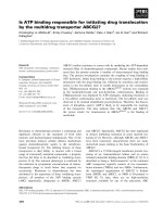
perineum (n = 1, Figure 1). Three patients were referred
for recurrent SCC tumors that had been previously trea-
ted within one year prior to the SLN procedure. Preo-
perative lymphoscintigraphy was performed in all 6
patients and identified 10 suspected SLNs. Intraopera-
tive blue dye injection was used in 4 patients with extre-
mity, truncal and perineal lesions. SLN exploration
identified a combined total of 11 SLNs (median: 1.7
nodes per patient; range 1-3) as shown in Table 1.
Upon pathologic examination with conventional H&E
staining, there was no evidence of metastatic carcinoma
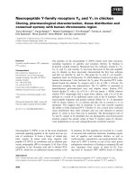
in any of the submitted lymph nodes. Immunostaining
was performed with pancytokeratin in three cases
which showed no evidence of micrometastatic disease
(Figure 2). The re were no surgical complications follow-
ing wide excision and SLN biopsy.
None of the patients received further adjuvant therapy
and no completion LNDs were performed following
negative SLN biopsy. Four patients are alive without evi-
dence of disease progression after a median follow up of
10.1 months (range 1.3 - 15.5 months). One patient
with a high-risk recurrent SCC of the right temple
developed a second local recurrence 15.2 months fol-
lowing narrow-margin excision with negative SLN
biopsy. A second patient with a high-risk large and deep
perineal SCC developed metastatic lesions in the lung
and vertebral bone 6.6 months after undergoing negative
wide margin excision and negative SLN biopsy.
A review of the literature identified a total of 161
worldwide patients in 14 case series including this study
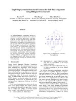
Figure 1 (A) High-risk invasive perineal squamous cell
carcinoma (B) Blue-stained inguinal sentinel lymph node.
Figure 2 Wide excision of perineal squamous cell carcinoma:
H&E staining at 2X (A) and 40X (B). Sentinel lymph node biopsy:
H&E at 10X (C) and immunostaining with pancytokeratin at 10X (D)
showing no evidence of occult metastasis.
Kwon et al. World Journal of Surgical Oncology 2011, 9:80
/>Page 3 of 7
[9,10,20-30], and 5 case reports [31-35] describing the
use of SLN biopsy in patients with cutaneous SCC.
Three case series [27-29] and one case report [31] were
excluded since these patients were later combined into
larger institutional case series resulting in a total of 130
evaluable cases (Table 2). All of the studies, except
Hatta et al. [30] clearly designated cutaneous SCC cases
with at least one high-risk feature. SLNs were success-
fully identified in 128 cases (98.5%). The probability of
SLN positivity for all sites, head/neck, and truncal/extre-
mity sites was found to be 14.1%, 10.1% and 18.6%,
respectively. An evaluation of SLN outcomes from all
available studies was performed (Table 3). Three studies
[20,22,30] did not provide follow up status after SLN
biopsy and only three studies [9,21,34] had a median
follow up exceeding 2 years. A total of 100 SCC patients
in 12 studies who underwent SLN biopsy had useful fol-
low up information. Despite this limitation, an analysis
of all documented recurrences showed an overall nega-
tive predictive value (NPV) of 97.8% for SLN status in
high-risk patient s. Among the head and neck cases (n =
51), the NPV for SLN biopsy was 100%, i.e. t here were
no regional noda l recurrences in any patient found to
have a negative SLN. On the other hand, SLN biopsy
for patients with high-risk l esions of the trunk a nd
extremities (N = 49) had a noticeably lower NPV of
95.2%. Two patients in this high-risk group developed
recurrent nodal d isease despite undergoin g a negative
SLN biopsy. Also of note, there were two patients who
relapsed with distant metastases despite a negative SLN
biopsy (not included for NPV calculation).
TheSLNfailureratewas2.2%.Therewerenofalse-
negative SLN among the g roup of head/neck SCC
tumors, while two patients with truncal/extremity SCC
developed nodal recurren ces despite negative SLN
biopsy resulting in a SLN failure rate of 4.8%. The false
negative rate was found to be 15.4% for all cases and
22.2% for the truncal/extremity group.
Discussion
Though metastases from SCC of the skin are uncom-
mon with a cumulative incidence between 2-6%, high-
risk skin lesions are reported to have metastatic rates
exceeding 30% [2]. It has been shown that regional
nodal involvement increases both the risk of recurrence
and mortality [9]. Metastases from cutaneous SCC tend
to spread first to regional nodal basins and generally
appear within the first 2 years of follow up [36]. Aggres-
sive surgical treatment has been shown to benefit
selected patients with locoregionally confined advanced
SCC and long term survivors have been reported follow-
ing radical salvage resection and therapeutic LND,
Table 2 Summary of studies reporting SLN procedures for cutaneous squamous cell carcinoma
Author, year # SCC
cases
Location SLN results and histological
methods
Adjuvant Treatment Disease
Recurrence
Stadelmann, 1997 [36] 1 Extremity 1/1 (100%), H&E LND LR (n = 1, +SLN)
Weisberg, 2000 [37] 1 Head 0/1 (0%), H&E and IHC XRT none
Altinyollar, 2002 [23] 20 Head 3/18 (17%), H&E LND N/A
Reschly, 2003 [10] 9 Head, Truncal/
Extremity
4/9 (44%), H&E and IHC LND (n = 3), XRT (n =
1)
LR (n = 1, +SLN),
DR (n = 1, +SLN)
Michl, 2003 [24] 9 Head, Truncal/
Extremity
2/9 (22%), H&E and IHC LND + CTX/XRT (n = 2) DR (n = 1, +SLN)
Eastman, 2004 [25] 6 Extremity 4/6 (67%). H&E and IHC N/A N/A
Ozcelik, 2004 [38] 1 Extremity 0/1 (0%), H&E none none
Wagner, 2004 [26] 12 Head, Truncal/
Extremity
2/12 (17%), H&E XRT (n = 2) none
Hatta, 2005 [34] 4 Extremity 0/4 (0%), H&E and IHC N/A N/A
Perez-Naranjo, 2005
[39]
1 Extremity 0/1 (0%), N/A none none
Nouri, 2006 [27] 15 Head 1/15 (6.7%), H&E and IHC LND (n = 4) none
Mullen, 2006 [9] 14 Truncal/Extremity 0/14 (0%), H&E + IHC none LR (n = 2, -SLN)
NR (n = 1, -SLN)
Sahn, 2007 [29] 9 Head, Truncal/
Extremity
0/9 (0%), H&E and some IHC XRT (n = 3) NR (n = 1, -SLN)
DR (n = 1, -SLN)
Renzi, 2007 [30] 22 Head, Truncal/
Extremity
1/22 (5%), H&E and IHC LND (n = 1) DR (n = 1, +SLN)
Kwon, 2010 6 Head, Truncal/
Extremity
0/6 (0%), H&E and some IHC. none LR (n = 1, -SLN)
DR (n = 1, -SLN)
H&E = hematoxylin and eosin, IHC = immunohistochemistry
LND = lymph node dissection, XRT = radiation therapy, CTX = chemotherapy
LR = local recurrence, NR = nodal recurrence, DR = dist ant metastases, N/A = not available
Kwon et al. World Journal of Surgical Oncology 2011, 9:80
/>Page 4 of 7
though complication and mortality rates were reported
in one study to be as high as 42% and 11%, respectively
[6,9]. The role fo r elective LND in high- risk SCC
remains undefined with most studies limited to head
and neck primary sites. For these reasons, SLN biopsy is
an unproven and yet theoretically appealing surgical
technique to accurately stage high-risk SCCs with mini-
mal morbidity, identify early occult nodal disease and
select patients that might benefit from therapeutic LND
or other adjuvant therapy
The optimal management of clinical N0 patients with
cutaneous SCC remains unclear. It appears that the
overall SLN positivity r ate (14.1%) for high-risk SCC is
comparable to studies of high-risk melanoma which
ranges from 13.9% - 29.4% [18]. SLN failure rate, false
negative rate and NPV for SCC also resemble rates
described in numerous melanoma studies. The standar-
dized use of serial sectioning and immunostaining has
significantly improved staging results of occult lymph
node metastases in melanoma patients with one group
reporting improved SLN positivity rates from 17.2 to
34% [37]. Ho wever, the benefit of routine immunostain-
ing with cyt okeratin markers for SCC patients has not
been established. Given the distinct morphologic
appearance of SCC characterized by very large and clus-
tered cells [10], routine immunohistochemistry may not
provide additional benefit. In fact, none of the studies
reporting a positive SLN (Table 2) described a case
where cytokeratin markers identified micrometastases
not readily apparent on conventional H&E staining.
Regional node involvement of SCC is associated with
an increased risk of recurrence and decreased survival.
LND is recommended for patients with regi onal lymph
node disease, though there are no significant studies
that have shown whether this impacts overall survival in
SCC patie nts. In a larger series of patients from the M.
D. Andersen Cancer Center [9], 52% of patients who
underwent LND for SCC regional nodal disease (n = 23)
had disease recurrence and 75% of these patients later
developed distant metastases. Unfortunately, there are
no published prospective studies comparing LND with
close observation in patients with clinical N0 high-risk
SCC. Further studies on the utility of SLN biopsy as
well as survival benefit from undergoing an elective
LND after a positive SLN biopsy are needed.
We found, compared to head/neck sites, there were
increased false negative rate and lower NPV for high-risk
SCC of the trunk and extremities. This may have been sec-
ondary to differences in important prognostic factors for
metastasis such as tumor thickness, immunosuppresion,
desmoplasia, and increased horizontal size [38]. This was
not evaluable given that many studies lacked these infor-
mation. We cannot rule out the possibility that there may
be inherent tumor biology differences b etween the two
sites, and suggest a more cautious approach when treating
patients with high-risk SCC of the trunk and extremities.
In addition, considering the relatively short follow up in
the majority of studies, the calculated NPV of SLN biopsy
may in fact be overestimated.Consideringtherarityof
this tumor and lack of long-term follow up in the majority
of studies, including our study, a clear emphasis is placed
upon the need for close surveillance regardless of the SLN
status. This study and review of literature highlights the
potential limitations of SLN biopsy for SCC and the criti-
cal importance of careful long-term follow-up in these
high-risk patients.
Though cytokeratin immunostaining may not directly
impact the sensitivity or specificity of SLN status, recent
studies have suggested that other pathologic markers
can provide additional insight into tumor biology and
cancer prognosis. A prospective study of non-well-differ-
entiated SCC and matched controls confirmed that
tumor thickness is the strongest prognostic risk factor
in these SCCs [39]. This study also identified the poten-
tial value of Ki-67 expression to predict recurrence. Ki-
67 is a cell-cycle protein that is upregulated during cel-
lular proliferation and has been shown to corr elate with
the differentiation status of skin cancers. There is
ongoing research to identify novel tumor biomarkers to
define cancer prognosis and promote individualized
therapies.
Conclusions
We conclude that SLN biopsy remains an investiga tional
staging tool in clinically node-negative high-risk cutaneous
squamous cell carcinoma patients. It is obvious that larger,
prospective studies with longer follow-up times are needed
Table 3 Cumulative results of sentinel lymph node (SLN)
biopsy for high-risk cutaneous squamous cell carcinoma
All
sites
Head/
Neck
Truncal/
Extremity
# total cases 130 71 59
# total cases with identified
SLN
128 69 59
# cases with SLN follow up 100 51 49
# cases with +SLN 18 7 11
# cases with +SLN and follow
up
11 4 7
# local recurrences (LR) 5 1 4
# nodal recurrences (NR) 2 0 2
# distant recurrences (DR) 5 0 5
Rate of SLN positivity 14.1% 10.1% 18.6%
SLN failure rate* 2.2% 0% 4.8%
SLN negative predictive value 97.8% 100.0% 95.2%
SLN false negative rate† 15.4% 0% 22.2%
*defined as the percentage of recurrences in the SLN-negative biopsied nodal basin s
†defined as the rate of nodal recurrences to the number of false negative and
true positive SLN cases
Kwon et al. World Journal of Surgical Oncology 2011, 9:80
/>Page 5 of 7
to establish the efficacy of SLN biopsy and define the opti-
mal treatment of occult nodal metastasis for high-risk
cutaneous SCC. It is unlikely that a large randomized con-
trolled trial can be accomplished considering the relative
low incidence of high-risk SCC and long accrual period
that would be required. An alternative approach would be
to contribute a nd analyze large prospective databases to
define the role and limitations of SLN biopsy in this
unique subset of SCC patients. Meanwhile, it is incumbent
upon treating physicians and teams to closely follow these
high-risk patients at greater risk for recurrence whether
they undergo SLN biopsy or not.
Abbreviations list
CTX: chemotherapy; DFS: disease-free survival; DR: distant recurrence; H&E:
hematoxylin and eosin; IHC: immunohistochemistry; LND: lymph node
dissection; LR: local recurrence; N/A: not available; NPV: negative predictive
value; NR: nodal recurrence; SCC: squamous cell carcinoma; SLN: sentinel
lymph node.
Acknowledgements
The authors wish to thank Drs. Noel Weiss and Thomas Lumley for their
helpful review of the epidemiological and analytical methods.
Author details
1
Department of Surgery, University of Washington, Seattle, WA, USA.
2
Department of Pathology, VA Puget Sound Health Care System, Seattle, WA,
USA.
3
Department of Surgery, VA Puget Sound Health Care System, Seattle,
WA, USA.
Authors’ contributions
SK did the data collection and data analysis, reviewed the literature, and
wrote the manuscript. ZD provided the pathology figures and legends. PW
wrote the manuscript and supervised the work. All authors read and
approved the final manuscript.
Competing interests
The authors declare that they have no competing interests.
Received: 3 March 2011 Accepted: 19 July 2011 Published: 19 July 2011
References
1. Alam M, Ratner D: Cutaneous squamous-cell carcinoma. N Engl J Med
2001, 344:975-983.
2. Rudolph R, Zelac DE: Squamous cell carcinoma of the skin. Plast Reconstr
Surg 2004, 114:82e-94e.
3. Strom SS, Yamamura Y: Epidemiology of nonmelanoma skin cancer. Clin
Plast Surg 1997, 24:627-636.
4. Preston DS, Stern RS: Nonmelanoma cancers of the skin. N Engl J Med
1992, 327:1649-1662.
5. Rowe DE, Carroll RJ, Day CL Jr: Prognostic factors for local recurrence,
metastasis, and survival rates in squamous cell carcinoma of the skin,
ear, and lip. Implications for treatment modality selection. J Am Acad
Dermatol 1992, 26:976-990.
6. North JH Jr, Spellman JE, Driscoll D, Velez A, Kraybill WG, Petrelli NJ:
Advanced cutaneous squamous cell carcinoma of the trunk and
extremity: analysis of prognostic factors. J Surg Oncol 1997, 64:212-217.
7. Veness MJ: Time to rethink TNM staging in cutaneous SCC. Lancet Oncol
2008, 9:702-703.
8. Brantsch KD, Meisner C, Schonfisch B, Trilling B, Wehner-Caroli J, Rocken M,
Breuninger H: Analysis of risk factors determining prognosis of
cutaneous squamous-cell carcinoma: a prospective study. Lancet Oncol
2008, 9:713-720.
9. Mullen JT, Feng L, Xing Y, Mansfield PF, Gershenwald JE, Lee JE, Ross MI,
Cormier JN: Invasive squamous cell carcinoma of the skin: defining a
high-risk group. Ann Surg Oncol 2006, 13:902-909.
10. Reschly MJ, Messina JL, Zaulyanov LL, Cruse W, Fenske NA: Utility of
sentinel lymphadenectomy in the management of patients with high-
risk cutaneous squamous cell carcinoma. Dermatol Surg 2003, 29:135-140.
11. Martinez JC, Cook JL: High-risk cutaneous squamous cell carcinoma
without palpable lymphadenopathy: is there a therapeutic role for
elective neck dissection? Dermatol Surg 2007, 33:410-420.
12. Garcia-Zuazaga J, Olbricht SM: Cutaneous squamous cell carcinoma. Adv
Dermatol 2008, 24:33-57.
13. Morton DL, Thompson JF, Essner R, Elashoff R, Stern SL, Nieweg OE,
Roses DF, Karakousis CP, Mozzillo N, Reintgen D, et al: Validation of the
accuracy of intraoperative lymphatic mapping and sentinel
lymphadenectomy for early-stage melanoma: a multicenter trial.
Multicenter Selective Lymphadenectomy Trial Group. Ann Surg 1999,
230:453-463, discussion 463-455.
14. Gershenwald JE, Thompson W, Mansfield PF, Lee JE, Colome MI, Tseng CH,
Lee JJ, Balch CM, Reintgen DS, Ross MI: Multi-institutional melanoma
lymphatic mapping experience: the prognostic value of sentinel lymph
node status in 612 stage I or II melanoma patients. J
Clin Oncol 1999,
17:976-983.
15. McMasters KM, Tuttle TM, Carlson DJ, Brown CM, Noyes RD, Glaser RL,
Vennekotter DJ, Turk PS, Tate PS, Sardi A, et al: Sentinel lymph node
biopsy for breast cancer: a suitable alternative to routine axillary
dissection in multi-institutional practice when optimal technique is used.
J Clin Oncol 2000, 18:2560-2566.
16. Krag D, Weaver D, Ashikaga T, Moffat F, Klimberg VS, Shriver C, Feldman S,
Kusminsky R, Gadd M, Kuhn J, et al: The sentinel node in breast cancer–a
multicenter validation study. N Engl J Med 1998, 339:941-946.
17. Morton DL, Thompson JF, Cochran AJ, Mozzillo N, Elashoff R, Essner R,
Nieweg OE, Roses DF, Hoekstra HJ, Karakousis CP, et al: Sentinel-node
biopsy or nodal observation in melanoma. N Engl J Med 2006,
355:1307-1317.
18. van Akkooi AC, Verhoef C, Eggermont AM: Importance of tumor load in
the sentinel node in melanoma: clinical dilemmas. Nat Rev Clin Oncol
2010, 7:446-454.
19. Nowecki ZI, Rutkowski P, Nasierowska-Guttmejer A, Ruka W: Survival
analysis and clinicopathological factors associated with false-negative
sentinel lymph node biopsy findings in patients with cutaneous
melanoma. Ann Surg Oncol 2006, 13:1655-1663.
20. Altinyollar H, Berberoglu U, Celen O: Lymphatic mapping and sentinel
lymph node biopsy in squamous cell carcinoma of the lower lip. Eur J
Surg Oncol 2002, 28:72-74.
21. Michl C, Starz H, Bachter D, Balda BR: Sentinel lymphonodectomy in
nonmelanoma skin malignancies. Br J Dermatol 2003, 149:763-769.
22. Eastman AL, Erdman WA, Lindberg GM, Hunt JL, Purdue GF, Fleming JB:
Sentinel lymph node biopsy identifies occult nodal metastases in
patients with Marjolin’s ulcer. J Burn Care Rehabil 2004, 25:241-245.
23. Wagner JD, Evdokimow DZ, Weisberger E, Moore D, Chuang TY, Wenck S,
Coleman JJ: Sentinel node biopsy for high-risk nonmelanoma cutaneous
malignancy. Arch Dermatol 2004, 140:75-79.
24. Civantos FJ, Moffat FL, Goodwin WJ: Lymphatic mapping and sentinel
lymphadenectomy for 106 head and neck lesions: contrasts between
oral cavity and cutaneous malignancy. Laryngoscope 2006, 112:1-15.
25. Sahn RE, Lang PG: Sentinel lymph node biopsy for high-risk nonmelanoma
skin cancers. Dermatol Surg 2007, 33:786-792, discussion 792-783.
26. Renzi C, Caggiati A, Mannooranparampil TJ, Passarelli F, Tartaglione G,
Pennasilico GM, Cecconi S, Potenza C, Pasquini P: Sentinel lymph node
biopsy for high risk cutaneous squamous cell carcinoma: case series and
review of the literature. Eur
J Surg Oncol 2007, 33:364-369.
27. Nouri K, Rivas MP, Pedroso F, Bhatia R, Civantos F: Sentinel lymph node
biopsy for high-risk cutaneous squamous cell carcinoma of the head
and neck. Arch Dermatol 2004, 140:1284.
28. Tartaglione G, Potenza C, Caggiati A, Maggiore M, Gabrielli F, Migliano E,
Pagan M, Concolino F, Ruatti P: Lymphatic mapping and sentinel node
identification in squamous cell carcinoma and melanoma of the head
and neck. Tumori 2002, 88:S39-41.
29. Tartaglione G, Potenza C, Caggiati A, Gabrielli F, Russo A, Pagan M: Sentinel
node radiolocalisation and predictive value in lip squamous cell
carcinoma. Radiol Med 2003, 106:256-261.
30. Hatta N, Morita R, Yamada M, Takehara K, Ichiyanagi K, Yokoyama K:
Implications of popliteal lymph node detected by sentinel lymph node
biopsy. Dermatol Surg 2005, 31:327-330.
Kwon et al. World Journal of Surgical Oncology 2011, 9:80
/>Page 6 of 7
31. Yamada M, Hatta N, Sogo K, Komura K, Hamaguchi Y, Takehara K:
Management of squamous cell carcinoma in a patient with recessive-
type epidermolysis bullosa dystrophica. Dermatol Surg 2004, 30:1424-1429.
32. Stadelmann WK, Javaheri S, Cruse CW, Reintgen DS: The use of selective
lymphadenectomy in squamous cell carcinoma of the wrist: a case
report. J Hand Surg Am 1997, 22:726-731.
33. Weisberg NK, Bertagnolli MM, Becker DS: Combined sentinel
lymphadenectomy and mohs micrographic surgery for high-risk
cutaneous squamous cell carcinoma. J Am Acad Dermatol 2000,
43:483-488.
34. Ozcelik D, Tatlidede S, Hacikerim S, Ugurlu K, Atay M: The use of sentinel
lymph node biopsy in squamous cell carcinoma of the foot: a case
report. J Foot Ankle Surg 2004, 43:60-63.
35. Perez-Naranjo L, Herrera-Saval A, Garcia-Bravo B, Perez-Bernal AM,
Camacho F: Sentinel lymph node biopsy in recessive dystrophic
epidermolysis bullosa and squamous cell carcinoma. Arch Dermatol 2005,
141:110-111.
36. Marks R: Squamous cell carcinoma. Lancet 1996, 347:735-738.
37. Cook MG, Green MA, Anderson B, Eggermont AM, Ruiter DJ, Spatz A,
Kissin MW, Powell BW: The development of optimal pathological
assessment of sentinel lymph nodes for melanoma. J Pathol 2003,
200:314-319.
38. Brantsch KD, Meisner C, Schonfisch B, Trilling B, Wehner-Caroli J, Rocken M,
Breuninger H: Analysis of risk factors determining prognosis of
cutaneous squamous-cell carcinoma: a prospective study. Lancet Oncol
2008, 9:713-720.
39. Jensen V, Prasad AR, Smith A, Raju M, Wendel CS, Schmelz M, Leyva W,
Warneke J, Krouse RS: Prognostic criteria for squamous cell cancer of the
skin. J Surg Res 2010, 159:509-516.
doi:10.1186/1477-7819-9-80
Cite this article as: Kwon et al.: Sentinel lymph node biopsy for high-
risk cutaneous squamous cell carcinoma: clinical experience and review
of literature. World Journal of Surgical Oncology 2011 9:80.
Submit your next manuscript to BioMed Central
and take full advantage of:
• Convenient online submission
• Thorough peer review
• No space constraints or color figure charges
• Immediate publication on acceptance
• Inclusion in PubMed, CAS, Scopus and Google Scholar
• Research which is freely available for redistribution
Submit your manuscript at
www.biomedcentral.com/submit
Kwon et al. World Journal of Surgical Oncology 2011, 9:80
/>Page 7 of 7