Báo cáo khoa học: " Surgical management of mediastinal liposarcoma extending from hypopharynx to carina: Case report" pptx
Bạn đang xem bản rút gọn của tài liệu. Xem và tải ngay bản đầy đủ của tài liệu tại đây (571.1 KB, 2 trang )
CAS E REP O R T Open Access
Surgical management of mediastinal liposarcoma
extending from hypopharynx to carina: Case
report
Thomas L Gethin-Jones, Nathaniel R Evans III, Christopher R Morse
*
Abstract
We describe the complete resection of a giant, well-differentiated mediastinal liposarcoma extending retropharynx
to envelop the aortic arch, trachea and esophagus following preoperative radiotherapy.
Background
Lipo sarc omas represent only 1% of all malignancies and
are commonly found in the lower limbs and retroperito-
neum [1]. Rarely are liposarcomas foun d in t he medias-
tinum and, of all primary mediastinal sarcomas only 9%
are liposarcomas [2]. Several reports suggest radiation
and chemotherapy without surgical resection are ineffec-
tive treatments for mediastinal liposarcoma despite often
daunting preoperative imaging [1,3]. In this case we
repo rt on the surgical resection of a large primary med-
iastinal liposarcoma by sternotomy.
Case presentation
A 70-year-old male with no histor y of radiotherapy pre-
sented with gradual swelling of the neck and dyspnea of
7 to 8 months duration. Magnetic resonance imaging
(MRI) and computed tomography (CT) scans of the
neck and chest revealed a large mass extending from
the hypopharynx to the carina (Figures 1 &2), causing
sig nifi cant displacement of the larynx, trachea, and eso-
phagus as well as encasing the aortic arch. Fine needle
aspiration (FNA) biopsy returned well-differentiated
liposarcoma. Improvement of symptoms came with 10
cycles of neoadjuvant radiotherapy prior to surgical
resection.
The patient was intubated while s pontaneously venti-
lating and with rigid bronchoscopy available. Initial
bronchoscopy revealed compression of the right main-
stem bronchus. Passage of an upper gastrointestinal
endoscope proved difficult with compression of the eso-
phagus. Through an initial collar incision and with rota-
tion of the carotid sheaths laterally, a well encapsulated
11 × 4 centimeter mass was dissected from behind the
hypopharynx. As it extended far into the mediastinum, a
sternotomy was performed and the left and right pleural
spaces opened. The liposarcoma surrounded the aortic
arch, and separated the trachea from esophagus. The
tumor was dissected from under the brachiocephalic
artery and rotated down from the neck. Laterally, a
plane was identified along the esopha gus and trachea,
but the lesion was too large to move between the tra-
chea and esophagus. Consequently, a lobulated portion
of the mass was divided and removed through the right
chest. A final component was dissected off the distal
arch of the aorta to complete the resection (Figure 3).
* Correspondence:
Division of Thoracic Surgery, Massachusetts General Hospital, Blake 1570, 55
Fruit St, Boston, MA 02114, USA
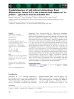
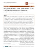
Figure 1 Axia l CT image of t he mediastinal liposarcoma.(a)
indicates the position of the esophagus, (b) indicates the trachea,
and (c) demonstrates the arch vessels.
Gethin-Jones et al. World Journal of Surgical Oncology 2010, 8:13
/>WORLD JOURNAL OF
SURGICAL ONCOLOGY
© 2010 Gethin-Jones et al; licensee BioMed Central Ltd. This is an Open Access article distributed under the terms of the Creative
Commons Attribution License ( which permits unrestricted use, distribution, and
reproduction in any medium , provided the original work is properly cited.
Postoperatively the patient was extubated and was dis-
charged to home on postoperative day eight. He
received postoperative radiation for a total of 60 Gy.
Discussion
In the literature, less than 150 cases of primary mediast-
inal liposarcomas have been reported [1,4] and because
of their rarity, there is no consistent approach to man-
agement. Warranting further study, radiology and che-
motherapy alone seem to be insufficient forms of
treatment but are possibly effective as induction or adju-
vant therapies [1,2,5]. When determining if surgical
intervention is feasible, radiographic films, given the
complex anatomy of the mediastinum, can be daunti ng.
However, given the often encapsulated nature of the
lesion s, complete resection is often possible and debulk-
ing can lead to symptomatic relief and often a long-
term solution in well-differentiated tumors.
Conclusions
Despite the complex nature of the anatomy surrounding
mediastinal liposarcomas, surgical intervention is not
unreasonable and t hought to be the most effective form
of treatment [1,3] especi ally in this particular case of an
encapsulated, well-differentiated mediastinal
liposarcoma.
Consent
Written informed consent was obtained from the patient
for publication of this case report and a ny accompany-
ing images. A copy of the written consent is available
for review by the Editor-in-Chief of this journal.
Authors’ contributions
TLG-J helped draft the manuscript. CRM and NRE reviewed and edited the
manuscript. All authors read and approved the final manuscript.
Competing interests
The authors declare that they have no competing interests.
Received: 13 November 2009 Accepted: 2 March 2010
Published: 2 March 2010
References
1. Barbetakis N, Samanidis G, Samanidou E, Kirodimos E, Kiziridou A,
Bischiniotis T, Tsilikas C: Primary mediastinal liposarcoma: a case report. J
of Medical Case Reports 2007, 1:161.
2. Burt M, Ihde JK, Hajdu SI, Smith JW, Bains MS, Downey R, Martini N,
Rusch VW, Ginsberg RJ: Primary sarcomas of the mediastinum: results of
therapy. J Thorac Cardiovasc Surg 1998, 115(3):671-80.
3. Ohta Y, Murata T, Tamura M, Sato H, Kurumaya H, Katayanagi K: Surgical
resection of recurrent bilateral mediastinal liposarcoma through the
clamshell approach. Ann Thorac Surg 2004, 77:1837-1839.
4. Vega AR, Muthuswamy MR: Primary mediastinal liposarcoma: case report
and review of the literature. Chest 2006, 130(4):334S.
5. Munden RF, Nesbitt JC, Kemp BL, Chasen MH, Whitman GJ: Primary
liposarcoma of the mediastinum. AJR Am J Roentgenol 2000, 175:1340.
doi:10.1186/1477-7819-8-13
Cite this article as: Gethin-Jones et al.: Surgical management of
mediastinal liposarcoma ex tending from hypopharynx to carina: Case
report. World Journal of Surgical Oncology 2010 8:13.
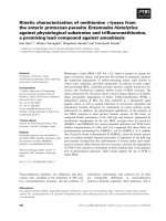
Figure 2 Coronal CT images of well-differentiated mediastinal
liposarcoma. (a) indicates the position of the esophagus and (b)
indicates the position of the trachea.
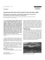
Figure 3 Intraoperative photo following resection of well
differentiated mediastinal liposarcoma. (a) indicates the position
of the innominate vein and (b) indicates the position of the
trachea/larynx.
Gethin-Jones et al. World Journal of Surgical Oncology 2010, 8:13
/>Page 2 of 2