Báo cáo khoa học: "Robot-assisted complete excision of choledochal cyst type I, hepaticojejunostomy and extracorporeal Roux-en-y anastomosis: a case report and review literature" ppsx
Bạn đang xem bản rút gọn của tài liệu. Xem và tải ngay bản đầy đủ của tài liệu tại đây (479.35 KB, 4 trang )
CASE REPO R T Open Access
Robot-assisted complete excision of choledochal
cyst type I, hepaticojejunostomy and
extracorporeal Roux-en-y anastomosis: a case
report and review literature
Thawatchai Akaraviputh
1*
, Atthaphorn Trakarnsanga
1
, Nutnicha Suksamanapun
2
Abstract
For Choledochal cyst type I, complete excision of cyst with Roux-en-Y hepaticojejunostomy anastomosis is the
treatment of choice. It has been performed laparoscopically with the advancement of laparoscopic skill. Recently, a
telemanipulative robotic surgical system was introduced, providing laparoscopic instruments with wrist-arm tech-
nology and 3-dimensional visualization of the operative field. We present a case of robot-assisted total excision of a
choledochal cyst type I and biliary reconstruction in a 14-year-old girl. No intraoperative complications or technical
problems were encountered. An intraabdominal collection occurred and was successfully treated with continuous
percutaneous drainage. At one-year follow-up, she is doing well without evidence of recurrent cholangitis.
Background
Choledochal cyst is a rare congenital anomaly of the
biliary system in the western countries, but has a higher
rate of occurrence in Asia. This disorder is usually diag-
nosed during childhood and is more common in
females. After being described first by Vater in 1723 [1],
choledochal cysts are now classifi ed using the Todani
modification of the Alonzo-Lej classification s ystem [2].
The most common is type I consisting of cystic, fusi-
form dilatation of the extrahepatic common bile duct.
Untreated choledoc hal cysts are associated with compli-
cations such as recurrent cholangitis, acute pancreatitis
and cholangiocarcinoma. The standard procedure is
complete resection of the cyst with a Roux-en-Y hepati-
cojejunostomy anastomosis. Cystoenterostomy is no
longer recommended [3]. Recently, many centers
reported their experience with lapar osco pic resection of
the cyst [ 4]. Although this approach has been shown to
be feasible and safe, most repo rts emphasized the t ech-
nical challenge of the procedure as well as the long
operative times [5]. The use of da Vinci Robotic Surgical
System (Intuitive Surgical, Sunnyvale, California) pro-
vides the advantages of three-dimensional visualization
through a stereoendoscope, tremor reduction, motion
scaling, and wristed instrumentation with additional
degrees of freedom compared to standard laparoscopic
instruments [6,7]. We report the application of da Vinci
Robotic Surgical System in type I chol edochal cyst exci-
sion in a 14-year-old girl.
Case presentation
A 14-year-old, girl presented with recurrent abdominal
dyspepsia and intermittent jaundice. Her b lood labora-
tory examinations were within normal limits. Ser um CA
19-9 was normal. Ultrasonography demonstrated a large
cystic dilatation of common bile duct. An abdominal
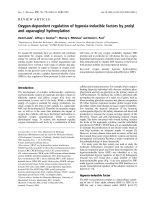
computed tomography (CT) scan revealed a type I cho-
ledochal cyst measuring > 4 cm in diameter (Figure 1).
The patient underwent da Vinci robot-assisted excision
of th e choledochal cyst, hepaticojejunostomy, and extra-
corporeal jejuno-jejunostomy of Roux-en-Y limb.
Surgical technique
The patient was placed in supine position. The pneumo-
peritoneum was created upto 12 mmHg using closed
technique with Veress needle. Three 8-mm robotic
* Correspondence:
1
Minimally Invasive Surgery Center, Division of General Surgery, Department
of Surgery, Faculty of Medicine Siriraj Hospital, Mahidol University, Bangkok,
Thailand
Full list of author information is available at the end of the article
Akaraviputh et al. World Journal of Surgical Oncology 2010, 8:87
/>WORLD JOURNAL OF
SURGICAL ONCOLOGY
© 2010 Akaraviputh et al; licensee BioMed Central Ltd. This is an Open A ccess article distributed under the terms of the Creative
Commons At tribution License ( y/2.0), which permits unrestricted use, distribution, and
reproduction in any medium, provided the original work is properly cited.
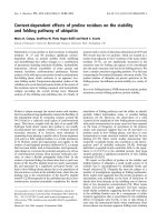
trocars and two 12-mm trocars for camera and acces-
sory device were applied ( Figure 2). After introduction
of the camera and wrist arm instruments, the table was
placed in reverse Trendelenburg position to allow the
intestines to fall caudaully. With the 3
rd
robotic arm
instrument, the liver was retracted more cephalad to
better expose the porta hepatis. The portal dissection
was begun firstly. The cyst was carefully dissected, p re-
serving the hepatic arteries as well as the portal vein
lying posterior to it. It was started on the inferior half of
the cyst. Once the portal vein and hepatic arteries were
separated from the cyst, the dissection was carried infer-
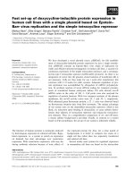
iorly toward the pancreas. The cyst was eventually
found to taper rapidly to a small duct. The common
bile duct was then ligated with plastic clips and trans-
ected (Figure 3). The cy st was then dissected cephalad
until normal calib er common hepa tic duct (CHD) was
identified.
The gallbladder was dissected in top-down fashion. The
cystic artery wa s clipped and divided. The CHD was
transected and then complete cyst excision was done.
The resected specimen was placed in right subdiaphar-
matic s pace. The jejunum was transected at about 20 cm
from duodenojejunal junction by endo GIA staple. An
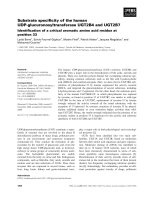
end-to-side hepaticojejunostomy, anticolic route, wa s
created using interrupted 3-0 Vicryl suture (Figure 4).
After completion of the anastomosis, the r obotic system
was undocked and smal l upper midline incision was
made. Side-to-side enteroenterostomy anastomosis was
created outside abdominal cavity. The Roux-en-Y limb
and jejunojejunostomy were re-checked and confirmed
to be in good position witho ut any evidence of torsion,
bleeding, or bile leak. Jackson Pratt drain was placed.
Finally the resected specimen was removed through this
incision. The fascial and skin incisions were closed with
absorbable sutures.
The total procedure time was 180 minutes. The total
robotic setup time (preparation, port placement, dock-
ing) was 30 minutes and the total robotic operative time
was 120 minutes. No intraoperative complications or
technical problems were encountered.
Postoperative course
One week after the operation, the Jackson Pratt drain
was removed. Unfort unately she developed high fever
Figure 1 Computed tomography scan demonstrating the
choledochal cyst type I.
Figure 2 Schematic illustration of the port placement: C, 12-mm
camera port; R1-3, 8-mm robotic instrument ports; A, 12-mm
assisted port.
Figure 3 Intraoperative finding of the narrow pancreatic p art
of common bile duct ligated with a plastic clip.
Akaraviputh et al. World Journal of Surgical Oncology 2010, 8:87
/>Page 2 of 4
and abdominal distension. CT scan revealed small right
subdiaphramatic intraabdominal collection. Percuta-
neous drainage was performed with ultrasound guide
and pigtail 7Fr silicone tube was placed. About 120 ml
of clear yellowish color fluid was aspirated and bile leak-
age was diagnosed. Systemic antibiotic was applied. One
week later, she had no fever and tolerated regular diet
well. Pathological result confirmed c holedochal cyst
without evidence o f malignancy. On postoperative 4th
week, the tube was removed and she was discharged
from the hospital. At one-year follow-up, she is doing
well without any evidence of recurrent cholangitis.
Discussion
Laparoscopic surgery has revolutionized the approach to
abdominal surgery. Technological advanc ements have
resulted i n the application of minimally invasive techni-
ques to increasingly complex procedures. However,
standard laparoscopic approach of hepatobiliary surgery
is still limited due to the technical complexities of thes e
procedures. The rigid nature of the instruments with
limited degrees of freedom, coupled with the fulcr um
effect of laparoscopy and 2-dimensional imaging, cer-
tainly contributes to the limi tations of the laparoscopic
approach. Robotic technology may help overcome these
obstacles.
The robot eliminates surgeon tremor and allows
3-dimensional visualization of the operative environment
[2], which can allow the correct ide ntification of anato-
mical variation. However, the main advantage of the d a
Vinci surgical system is the dexterity afforded by the
Endowrist design, which allows precise control of tech-
nically challenging tasks such as delicate dissection, fine
suturing [4]. It may be that advanced robo tics will be
reserved f or o nly the most complex operations, such as
choledochojejunostomy or pancreaticoduodenectomy.
Robotic surgery can ameliorate the technical difficulties
encountered laparoscopically and may allow surgeons
to perform delicate procedures with shorter operative
time [8-10].
Although robotic-assisted results and o utcomes
abound for many procedures, only limited information
has been published on robotic-assisted choledochal cyst
excision. We found only 4 cases in the literatures (Table 1).
Interestingly, the R oux limb could be created entirely
intracorporeall y by the robot or extracorporeally through
a small incision, which could decrease the robotic time
and total operative times. In our case, we did an extracor-
poreal jejuno-jejunostomy anastomosis, and therefore our
operative time was significantly shorter than the others
report in literature. The minor leakage of hepatico-jeju-
nostomy anastomosis found may be caused by unsecured
suturing technique from the early experiences in robotic
surgery.
Disadvantages include the size of the robotic hardware
in relation to patient body; t he loss of haptic feedback;
and the overall cost of the hardware, drapes, and main-
tenance of the robotic system. The robotic approach in
gastrointestinal tract surgery has also a learning curve
period regard to suturing technique, but we believe that
this might be sho rter than the standard laparoscopic
surgery [11,12].
Finally, the robotic app roach to the complex hepato-
biliary surgery is feasible and safe in selected patients.
Three-dimensional visualization, a rticulating instru-
ments, and fine-motion filtering are the principle advan-
tages.Roboticsurgerymayincreasethevarietyof
Figure 4 The Robot-assisted end-to-side hepaticojejunostomy
(white arrow) was completely performed with Vicryl #3/0
interrupted stitches.
Table 1 The summary of robotic-assisted choledochal cyst excision
No Author Year Age Gender Total OPT
(min.)
No of port Robotic time
(min.)
Roux limb LOH (day) Complication
1 Woo et al.
11
2006 5 F 440 5 390 Extracorporeal 4 no
2 CM Kang et al.
12
2007 63 F 380 5 270 Extracorporeal 15 no
3 JJ Meehan et al.
7
2007 2 N/A 445 5 408 Intracorporeal N/A no
4 JJ Meehan et al.
7
2007 9 N/A 472 5 428 Intracorporeal N/A no
5 The study 2010 14 F 180 5 120 Extracorporeal 20 Collection
Akaraviputh et al. World Journal of Surgical Oncology 2010, 8:87
/>Page 3 of 4
procedures, which can be accomplished with a mini-
mally invasive approach and may also enable more g en-
eral surgeons to perform these complex procedures.
Surgeons need to become familiar with these improve-
ments as the technology continues to progress [13].
Conclusions
In summary, we report the feasibil ity and safety of
robot-assisted laparoscopic resection of a type I chole-
dochal cyst in a child. Compared to total l aparoscopic
surgery, the robot-assist ed technique facilitates the most
difficult part of the procedure, namely the creation of
the hepaticojejunostomy anastomosis. Further experi-
ence is needed to properly e valuate the advantages and
applicability of t his approach, especially in the pediatric
patient.
Consent
Written informed consent was obtained from the patient
for publication of this case report and any accompany-
ing images. A copy of the written c onsent is available
for review by the Editor-in-Chief of this journal.
Author details
1
Minimally Invasive Surgery Center, Division of General Surgery, Department
of Surgery, Faculty of Medicine Siriraj Hospital, Mahidol University, Bangkok,
Thailand.
2
Division of Pediatric Surgery, Department of Surgery, Faculty of
Medicine Siriraj Hospital, Mahidol University, Bangkok, Thailand.
Authors’ contributions
TA was the surgeon who performed the operation. TA and AT draft the
manuscript. AT and NS participated in the operation. All authors read and
approved the final manuscript.
Competing interests
The authors declare that they have no competing interests.
Received: 4 June 2010 Accepted: 12 October 2010
Published: 12 October 2010
References
1. Shimura H, Tanaka M, Shimizu S, Mizumoto K: Laparoscopic treatment of
congenital choledochal cyst. Surg Endosc 1998, 12:1268-71.
2. Tan HL, Shankar KR, Ford WD: Laparoscopic resection of type I
choledochal cyst. Surg Endosc 2003, 17:1495.
3. Akaraviputh T, Boonnuch W, Watanapa P, Lert-Akayamanee N, Lohsiriwat D:
Surgical Management of Adult Choledochal Cysts. J Med Assoc Thai 2005,
88:939-43.
4. Metcalfe MS, Wemyss-Holden SA, Maddern GJ: Management dilemmas
with choledochal cysts. Arch Surg 2003, 138:333-9.
5. Tanaka M, Shimizu S, Mizumoto K, Yokohata K, Chijiiwa K, Yamaguchi K,
Ogawa Y: Laparoscopically assisted resection of choledochal cyst and
Roux-en-Y reconstruction. Surg Endosc 2001, 15:545-52.
6. Ballantyne GH, Moll F: The da Vinci telerobotic surgical system: the virtual
operative field and telepresence surgery. Surg Clin North Am 2003,
83:1293-304.
7. Lanfranco AR, Castellanos AE, Desai JP, Meyers WC: Robotic surgery: a
current perspective. Ann Surg 2004, 239:14-21.
8. Horgan S, Vanuno D: Technical report: robots in laparoscopic surgery. J
Laparoendosc Adv Surg Tech 2001, 11:415-19.
9. Hazey J, Melin WS: Robot-assisted general surgery. Semin Laparosc Surg
2004, 11:107-12.
10. Cadiere GB, Himpens J, Germay O, Izizaw R, Degueldre M, Vandromme J,
Capelluto E, Bruyns J: Feasibility of robotic laparoscopic surgery: 146
cases. World J Surg 2001, 25:1467-77.
11. Woo R, Le D, Albanese CT, Kim SS: Robot-assisted laparoscopic resection
of a type I choledochal cyst in a child. J Laparoendosc Adv Surg Tech A
2006, 16:179-83.
12. Kang CM, Chi HS, Kim JY, Choi GH, Kim KS, Choi JS, Lee WJ, Kim BR: A case
of robot-assisted excision of choledochal cyst, hepaticojejunostomy, and
extracorporeal Roux-en-y anastomosis using the da Vinci surgical
system. Surg Laparosc Endosc Percutan Tech 2007, 17:538-41.
13. Meehan JJ, Elliott S, Sandler A: The robotic approach to complex
hepatobiliary anomalies in children: preliminary report. J Pediatr Surg
2007, 42:2110-2114.
doi:10.1186/1477-7819-8-87
Cite this article as: Akaraviputh et al.: Robot-assisted complete excision
of choledochal cyst type I, hepaticojejunostomy and extracorporeal
Roux-en-y anastomosis: a case report and review literature. World
Journal of Surgical Oncology 2010 8:87.
Submit your next manuscript to BioMed Central
and take full advantage of:
• Convenient online submission
• Thorough peer review
• No space constraints or color figure charges
• Immediate publication on acceptance
• Inclusion in PubMed, CAS, Scopus and Google Scholar
• Research which is freely available for redistribution
Submit your manuscript at
www.biomedcentral.com/submit
Akaraviputh et al. World Journal of Surgical Oncology 2010, 8:87
/>Page 4 of 4