Báo cáo khoa học: "Marjolin''''s ulcers: theories, prognostic factors and their peculiarities in spina bifida patients" ppsx
Bạn đang xem bản rút gọn của tài liệu. Xem và tải ngay bản đầy đủ của tài liệu tại đây (984.66 KB, 5 trang )
RESEARC H Open Access
Marjolin’s ulcers: theories, prognostic factors and
their peculiarities in spina bifida patients
Peter M Nthumba
Abstract
Background: Due to improved care, more and more children born with spina bifida in rural Kenya are surviving
into adulthood. This improved survival has led to significant challenges in their lifestyles, especially the need to
ensure pressure ulcer prevention and treatment. Malignant degeneration of pressure ulcers in spina bifida patients
is very rare. The author describes the clinical presentation of two pressure ulcer carcinomas that are at variance
from classical descriptions.
Materials and methods: An internet/Medline/PubMed search of English literature for theori es on Marjolin’s ulcer
evolution and prognostic features of Marjolin’s ulcers was performed.
A chart review of two young adults with spina bifida who had presented to the author’s hospital between 2004
and August 2010 with chronic pressure ulcers found to be Marjolin’s ulcers on histo-pathological examination was
performed, and the clinical features are reported.
Results: The two ulcers appeared clinically benign: one was a deep ulcer, while the other was shallow; both had
normal, benign-appearing edges, and a foul smelling discharge. The two ulcers were surrounded by induration and
multiple communicating sinuses, with no evidence of chronic osteomyelitis. The internet search revealed a total of
nine theories on Marjolin’s ulcer development, as well as seven clinical and four histological prognostic features.
Discussion: The multifactorial theory, a coalescence of a number of proposed theories, best explains the evolution
of Marjolin’s ulcers. Poor prognostic feature s include pressure ulcer carcinomas, lesions and location in the lower
limbs/trunks, all present in the two patients making their prognosis dim: this is despite the surgical margins being
clear of tumor. Benign appearance, induration and presence of multiple communicating sinuses are features that
have not been previously described as presenting features of pressure ulcers carcinomas.
Conclusion: There is need for spina bifida patients and their guardians/caretakers to receive a close follow-up
throughout life; health education focused on pressure ulcer prevention as well as early treatment of pressure ulcers
when they occur, will avert the development of Marjolin’s ulcers, and save lives.
Background
The population of children with spina bifida surviving
into adulthood in rural Kenya is growing because of
improved health education, care as well as an increas-
ingly supportive environment [1]. Improved survival and
integration into such social structures as schooling,
work, marriage and child-bearing places significant
demands on this populat ion: the need for a lifestyle that
is protective/preventive against the development of such
life-threatening complicatio ns as renal failure and pres-
sure ulcers, amongst others. Prevention requires active
bladder and bowel care, as well as regular shifting of
position to avoid prolonged pressure leading to the
development of pressure ulcers. Failure to adhere to this
‘protective lifestyle’ almost invariably leads to the devel-
opment of pressure ulcers; these ulcers may heal with
appropriate care. Othe rs may suffer either frequent
ulcer relapses or chronic non-healing ulcers that may
degenerate into Marjolin’ s ulcers. A number of hypoth-
eses have been proposed to explain malignant degenera-
tion of chronic wounds and scar tissue (Table 1) [2-16].
Four clinical signs have been proposed as characteristi c
for malignant pressure ulcer d egeneration: the appear-
ance of a mass, new onset of pain, a change in drainage
odor and change in volume, character or appearance of
Correspondence:
Department of Surgery, AIC Kijabe Hospital, Kijabe, Kenya, Africa
Nthumba World Journal of Surgical Oncology 2010, 8:108
/>WORLD JOURNAL OF
SURGICAL ONCOLOGY
© 2010 Nthumba; licensee BioMed Central Ltd. This is an Open Access article distributed under the terms of the Creative Commons
Attribution License ( g/licenses/by/2.0 ), which permits unrestrict ed use, distribution, and reproduction in
any medium, provided the original work is properly cited.
drainage [17]. Unfortunately, most spina bifida patients
lack sensation, a nd they and their caretakers may not
recognize any significant changes in their ulcers. Health
education, with an emphasis on ulcer prevention and
care, should be taught to healthcare workers and parent
(s)/guardian(s); it is ulcers tha t develop i n childhood that
may later degenerate into malignancy [18].
Our understanding of the process of pressure ulcer
development amongst spina bi fida patients, and their
subsequent degeneration into malignant ulcers is lim-
ited. The purpose of this study was to collect and review
the various theories on Marjolin’s ulcers, the different
prognostic factors, with a view to applying these to
spina bifida patients. This understanding would aid the
healthcare worker in developing programs suited to a
growin g population of spina bifida patients, especially in
the low income countries. The author also sought to
describe atypical clinical presentation of Marjolin’ s
ulcers in these patients.
Patients and methods
A chart review of two young adults with spina bifida
who had presented to the author’ s hospital between
2004 and August 2010 with chronic pressure ulcers
found to b e squamous cell carcinomas on histopatholo-
gical examination was performed.
An internet/Medline/PubMed search of English litera-
ture for pressure ulcer theories as well as on the prognos-
tic features of Marjolin’s ulcers was performed. The terms
‘pressure ulcer’, ‘pressure sore’, ‘decubitus ulcer’ indepen-
dently and wit h the term ‘theory’ or ‘theories’ were used,
as were the terms, ‘Marjolin’ s ulcers’, ‘malignant pressure
ulcers’ , ‘ prognosis’ , ‘ prognostic features’,invarious
combinations.
Results
The two patients, both females, w ere aged 20 and
26 years. While one of the patients was ambulant with
bilateral below-knee prostheses [1], the other was wheel-
chair-bound. Both had chro nic pressure ulcers; one had
lasted 16 years, while the secon d patient had had the
ulcer for five years, with a previous history of ulcers
from the same site that had recurred a number of times
in the past, with none having lasted for more than a
year. The ulcer of one patien t was deep, while the other
was a shallow flat ulcer: both had a foul smelling puru-
lent discharge and multiple sinuses that communicated
with the ulcer. The areas with the ulcers and the sinuses
were indurated, and on digital pressure exuded dis-
charge both from the ulcer and sinuses. The margins of
the ulcers were of normal appearance, (not elevated),
and would thus not suggest malignancy to the casual
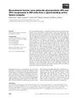
observer (Figure 1 and 2). The excised surgical margins
on both patients were clear of tumor. There was no evi-
dence of underlying chronic osteomyelitis.
The internet/Medline/PubMed search on pressure
ulcer theories revealed a total of nin e different hypoth-
eses (Table 1) [2-16], while a search for prognostic fea-
tures of Marjolin’ s ulcers revealed seven clinical and
four histological features (Table 2) [19-24].
Discussion
A review of theories on Marjolin’s ulcer evolution
reveals that no single theory explains their evolution
Table 1 Theories on Marjolin ’s ulcers [2-16]
Theory Proposed mechanism
Toxin theory Toxins released from damaged tissues later lead to cellular mutations.
Chronic irritation theory Chronic irritation with repeated attempts at re-epithelialization contributes to neoplastic initiation.
Traumatic epithelial elements
implantation theory
Epithelial elements implanted into the dermis, lead to a foreign body response reaction and a disordered
regenerative process.
Co-carcinogen theory Chemical or trauma such as burn injury acts to ‘stir’ pre-existing but dormant neoplastic cells into
proliferation.
Initiation and promotion theory A two-step process that converts normal cells into malignant cells. In the initiation phase, normal cells
become dormant neoplastic cells that may then be subsequently stimulated into neoplastic cells by a co-
carcinogen such as infection, in the promotion phase. This theory overlaps with the co-carcinogen theory.
Immunologic privileged site theory Burn scarring effectively obliterates lymphatics to injured area, preventing normal immunosurveillance and
thus permitting neoplastic growth. These tumors initially grow slowly, but quickly overwhelm the immune
system, metastasize and are rapidly fatal, once they break through the scar barrier.
Heredity theory HLA DR4 is associated with cancer development and p53 gene abnormalities have been demonstrated in
patients with Marjolin’s ulcers. Further, Fas mutations in the apoptosis function region that predispose to
malignant degeneration of scars have been demonstrated in burn scar Marjolin’s ulcers.
Ultraviolet rays theory Ultraviolet rays theory - UV rays cause a reduction in Langerhans cell population leading to a reduction in
cutaneous immuno-surveillance against developing malignancy and also cause p53 tumor suppressor gene
alterations.
Environmental and genetic
interaction theory
Attempts to explain the occurrence of ‘Acute’ Marjolin’s ulcers.
Nthumba World Journal of Surgical Oncology 2010, 8:108
/>Page 2 of 5
fully. These postulates include the toxin, the chronic
irritation, the traumatic epithelial elements implantation,
the co-carcinogen and the initiati on and promotion the-
ory; these theories include trauma as an integral part of
the process of the evolution of Marjolin’sulcers[2-9].
The immunologically privileged site theory, which has a
large number of proponents, attempts to explain the
poor prognosis of Marjolin ’s ulcers [10,11]. The heredi-
tary and ultraviolet rays’ theories were proposed after
genetic changes were found in patients with Marjolin’s
ulcers [12-15]. The environmental and genetic interac-
tion theory seeks to explain the evolution of acute Mar-
jolin’ sulcers[16].Acombinationoftheoriesbetter
expl ains the process: for example, the chronic irritation,
the initiation and promotion, the toxin and the co-carci-
nogen theories when combined together, explain the
evolution of pressure ulcer carcinomas, under which
spina bifida pressure ulcers fall. The current author pro-
poses the multifactorial theory, a combination of any of
the current theories (Table 1) [2-16], as the one that
best explains this process. It is to be noted that some of
these theories may overlap.
Marjolin ’ s ulcers complicating pressure ulcers in spina
bifida patients are rarely reported: there are les s than
ten reported cases in English literature [1]. Marjolin’s
ulcers in general, develop in younger p atients amongst
sub-Saharan patients than those reported from other
regions [18]; therefore, pa tients presenting with pressur e
ulcers should be investigated during the initial evalua-
tion for this possibility. Additionally, at surgery, all the
excised tissue should be submitted for histopathological
investigation. Unfortunately, surgical margins clear of
malignancy do not necessarily improve the prognosis of
pressure ulcer carcinomas [1,18], which have a much
poorer prognosis than Marjolin’ sulcersarisingfrom
other sources [4]. Table 2 highlights prognostic features
of Marjolin’s ulcers in general - it is notable that a pres-
sure ulcer carcinoma is a poor prognostic indicator.
Further, Marjolin’sulcerslocatedonthelowerlimbsor
trunk, those with diameters above two centimeters, and
latency of five years or more, all common features in
the two spina bifida patients presented here, made their
prognosis even poorer, especially in an environment
with limited resources and options [1,3,11,19-24].
Marjolin’s ulcers are characteristically either grossly flat,
indurated, infiltrative shallow ulcers with well-defined, ele-
vated margins, or exophytic proliferative ulcers [ 1]. The
two ulcers in this report had a benign appearance of both
the ulcer edges and the bases, and except for a foul smell,
none of the o ther fo ur hallmark signs of pressure ulcer
carcinoma [17] were found. The ot her common features
in these two ulcers were: induration and multiple sinuses
communicating with the ulcers, two signs that have not
been previously noted in pressure ulcer carcinomas. Pres-
sure ulcer malignancy in spina bifida patients may thus
not present with the classical descriptions, and whereas
the current rarity of Marjolin’ sulcersinspinabifida
patients may be partially explained by the fact that not
many spina bifida patients have survived long enoug h to
develop this complication in the past, these peculiar pre-
sentations of the Marjolin’sulcersismoredifficultto
explain. The extent to which the congenital immobility,
incontinence and lack of sensation, (factors that predis-
pose to pressure ulcer development in both spinal cord
injured patients and those with spina b ifida), differs from
the same factors when these develop secondary to trauma
or tumors, is difficult to determine, but may be another
var iable that could explain the low incidence of pressure
ulcer malignancy in spina bifida patients.
It is conceivable that our environment will see more
such survivors, and lack of preparedness for prevention
of pressure ulcers may lead to increased numbers with
Marjolin’s ulcers. Prevention is better that cure, more so
Figure 1 Marjolin’s ulcer with sinuses included within surgica l
excision margins. Note deep ulcer and benign appearance of ulcer
edges.
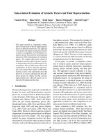
Figure 2 Marjolin’s ulcer with sinuses extending into the thigh
and labia majora. One sinus was found in the anus, and another
in the vagina. Note benign appearance of ulcer margins
surrounding a flat ulcer.
Nthumba World Journal of Surgical Oncology 2010, 8:108
/>Page 3 of 5
when the cure is not possible, especially in an environ-
ment such as rural Kenya. All chronic ulcers should
undergo multiple biopsies, to help define their therapy,
and to avoid missing malignant ulcers [1,18].
Conclusion
The multifactorial theory best explains the malignant
degeneration of pressure ulcers, independent of the
cause. Appropriate Marjolin’s ulcer patient prognost i-
cation should aid in clinical decision making, espe-
cially the utilization of resources in poor income
countries.
There is need for spina bifida patients and their guar-
dians/caretakers to receive a close follow-up throughout
life; health education focused on pressure ulcer preven-
tion as well as early treatment of pressure ulcers when
they occur, will avert the development of Marjolin’ s
ulcers, and save lives.
Consent statement
Publication of these cases without patients consent was
exempted by the AIC Kijabe hospital ethics committee
as the patients consent for publication could not be
obtained.
Competing interests
The author declares he has no competing interests. No grants were given
for this work, and no financial benefits are expected from this work. This
paper has not been presented in any form, in any forum. There is no
association between the author with any commercial firm, and no grants
were granted for this article. There are no competing interests in the
publication of this article.
Received: 4 September 2010 Accepted: 5 December 2010
Published: 5 December 2010
References
1. Nthumba P, Bird G: Marjolin’s ulcer in a spina bifida patient. A case
report. East and Central African Journal of Surgery 2010, 15:127-129.
2. Fleming MD, Hunt JL, Purdue GF, Sandstad J: Marjolin’s ulcer: a review
and reevaluation of a difficult problem. J Burn Care Rehabil 1990,
11:460-469.
3. Treves N, Pack GT: The development of cancer in burn scars. Surg Gynecol
Obstet 1930, 51:749-782.
4. Hill BB, Sloan DA, Lee EY, McGrath PC, Kenady DE: Marjolin’s ulcer of the
foot caused by non-burn trauma. South Med J 1996, 89:707-710.
5. Dupree MT, Boyer JD, Cobb MW: Marjolin’s ulcer arising in a burn scar.
Cutis 1998, 62:49-51.
6. Neuman Z, Ben-Hur N, Shulman J: Trauma and skin cancer: implantation
of epidermal elements and possible cause. Plast Reconstr Surg 1963,
32:649-656.
7. Gadner AW: Trauma and squamous skin cancer. Lancet 1959, 273:760-761.
8. MacKenzie J, Rous P: The experimental disclosure of latent neoplastic
changes in tarred skin. J Exp Med 1941, 73:391-395.
9. Arons MS, Rodin AE, Lynch JB, Lewis SR, Blocker TG Jr: Scar tissue
carcinoma. Part II: an experimental study with special reference to burn
scar carcinoma. Ann Surg 1966, 163:445-460.
10. Bostwick J, Pendergrast WJ, Vasconez LO: Marjolin’s ulcer: an
immunologically priviledged tumor? Plast Reconstr Surg 1975, 57:66-69.
11. Ryan RF, Litwin MS, Krementz ET: A new concept in the management of
Marjolin’s ulcers. Ann Surg 1981, 193:598-604.
12. Czarnecki D, Nicholson I, Tait B, Nash C: HLA DR4 is associated with the
development of multiple basal cell carcinomas and malignant
melanoma. Dermatolgica 1993, 187:16-18.
13. Harland DL, Robinson WA, Franklin WA: Deletion of the p53 gene in a
patient with aggressive burn scar carcinoma. J Trauma 1997, 42:104-107.
14. Lee SH, Shin MS, Kim HS: Somatic mutations of Fas (Apo-1/CD95) gene in
cutaneous cell carcinomas arising from a burn scar. J Invest Dermatol
1999, 114:122-126.
15. Scarlett WL: Ultraviolet Radiation: sun exposure, tanning beds, and
vitamin D levels. What you need to know and how to decrease the risk
of skin cancer. J Am Osteopath Assoc 2003, 103:371-375.
16. Kowal-Vern A, Criswell BK: Burn scar neoplasms: A literature review and
statistical analysis. Burns 2005, 31:403-413.
17. Esther RJ, Lamps L, Schwartz HS: Marjolin ulcers: secondary carcinomas in
chronic wounds. J South Orthop Assoc 1999, 8:181-187.
18. Nthumba PM: Marjolin’s ulcers in sub-Saharan Africa. World J Surg 2010,
34:2272-2277.
19. Fitzgerald RH Jr, Brewer NS, Dahlin DC: Squamous cell carcinoma
complicating chronic osteomyelitis. J Bone Joint Surg 1976, 58:1146.
20. Ozek C, Cankayali R, Bilkay U, G uner U, Gundogan H , Songur E , Akin Y, Cagd as A:
Marjolin’sulcersarisinginburnscars.JBurnCareRehabil2001, 22 :384-389.
21. Templeton AC: Tumours in a tropical country. Berlin: Springer-Verlag 1973,
182-183.
22. Friedman HI, Cooper PH, Wanebo HJ: Prognostic and therapeutic use of
microstaging of cutaneous squamous cell carcinoma of the trunk and
extremities. Cancer 1985, 56:1099-1105.
Table 2 Prognostic factors in Marjolin’s ulcers [19-24]
PROGNOSIS
Variable Better Poorer
Clinical Latency to malignancy Less than 5 years More than 5 years
Tumor location Head, neck, upper extremeties Lower limbs, trunk
Tumor source Post-burn, chronic osteomyelitis Pressure sore carcinomas
Tumor diameter Smaller than 2 cm 2 cm or more
Tumor type Exophytic Infiltrative
Metastases None Present
Tumor recurrence None Present
Histological Degree of differentiation Well differentiated Moderately-well and poorly differentiated
Peritumoral T lymphocyte infiltration Heavy Scarce or absent
Depth of dermal invasion Superficial to reticular dermis Reticular dermis or deeper
Vertical tumor thickness Less than 4 mm thick 4 mm thick or more
Nthumba World Journal of Surgical Oncology 2010, 8:108
/>Page 4 of 5
23. Phillips TJ, Salman SM, Bhawan J, Rogers GS: Burn scar carcinoma.
Diagnosis and management. Dermatol Surg 1998, 24:561-565.
24. Edwards MJ, Hirsch RM: Squamous cell carcinoma arising in previously
burned or irradiated skin. Arch Surg 1989, 124:115-117.
doi:10.1186/1477-7819-8-108
Cite this article as: Nthumba: Marjolin’s ulcers: theories, prognostic
factors and their peculiarities in spina bifida patients. World Journal of
Surgical Oncology 2010 8:108.
Submit your next manuscript to BioMed Central
and take full advantage of:
• Convenient online submission
• Thorough peer review
• No space constraints or color figure charges
• Immediate publication on acceptance
• Inclusion in PubMed, CAS, Scopus and Google Scholar
• Research which is freely available for redistribution
Submit your manuscript at
www.biomedcentral.com/submit
Nthumba World Journal of Surgical Oncology 2010, 8:108
/>Page 5 of 5