Báo cáo y học: "Production of interleukin-1 receptor antagonist by human articular chondrocyte" ppt
Bạn đang xem bản rút gọn của tài liệu. Xem và tải ngay bản đầy đủ của tài liệu tại đây (288.13 KB, 8 trang )
Research article
Production of interleukin-1 receptor antagonist by human
articular chondrocytes
Gaby Palmer
1
, Pierre-Andre Guerne
1
, Francoise Mezin
1
, Michel Maret
1
, Jerome Guicheux
1,3
,
Mary B Goldring
2
and Cem Gabay
1
1
Division of Rheumatology, University Hospital, Geneva, Switzerland
2
New England Baptist Bone and Joint Institute and Rheumatology Division, Beth Israel Deaconess Medical Center, Harvard Institutes of Medicine,
Boston, Massachussetts, USA
3
Present address: INSERM EM 9903, School of Dental Surgery, Nantes, France
Correspondence: Cem Gabay, MD, Division of Rheumatology, University Hospital, 26 avenue de Beau-Sejour, 1211 Geneva 14, Switzerland.
Tel: +41 22 382 3501; fax: +41 22 382 3509; e-mail:
Introduction
Interleukin-1 receptor antagonist (IL-1Ra) is a member of
the IL-1 family that binds to IL-1 receptors but does not
induce any intracellular response. IL-1Ra prevents the
interaction between IL-1 and its cell surface receptors,
and thus competitively inhibits the biological effects of IL-1
[1–3]. The administration of IL-1Ra ameliorates the course
of arthritis in several experimental models [4]. In addition,
therapeutic administration of IL-1Ra has beneficial effects
in patients with rheumatoid arthritis [5].
Abstract
Interleukin-1 receptor antagonist (IL-1Ra) is a natural IL-1 inhibitor possessing anti-inflammatory
properties. IL-1Ra is produced as different isoforms, one secreted (sIL-1Ra) and three intracellular
(icIL-1Ra1, icIL-1Ra2 and icIL-1Ra3), derived from the same gene. We examined the production of
IL-1Ra species by cultured human articular chondrocytes in response to various cytokines. The levels
of IL-1Ra were undetectable in culture supernatants of untreated cells, but were significantly
increased by IL-1β. Cell lysates contained very low levels of IL-1Ra, even in response to IL-1β,
suggesting that chondrocytes produce predominantly sIL-1Ra. IL-6, which had no effect on its own,
enhanced the effect of IL-1β, while dexamethasone prevented the response. We observed by
RT-PCR that IL-1β and IL-6 induced primarily the production of sIL-1Ra mRNA. Furthermore, IL-1β
alone or combined with IL-6 increased the levels of nascent unspliced sIL-1Ra mRNA, suggesting
that sIL-1Ra expression is regulated at the transcriptional level. Reporter gene assays in immortalized
chondrocytes, C-20/A4, consistently showed increased sIL-1Ra promoter activity in response to
IL-1β and IL-6. In conclusion, human articular chondrocytes produce sIL-1Ra in response to IL-1β and
IL-6. The production of sIL-1Ra by chondrocytes may have a protective effect against articular
inflammatory and catabolic responses.
Keywords: cytokines, glucocorticoids, human articular chondrocytes, IL-1 receptor antagonist
Received: 25 October 2001
Revisions requested: 27 November 2001
Revisions received: 20 February 2002
Accepted: 11 March 2002
Published: 8 April 2002
Arthritis Res 2002, 4:226-231
This article may contain supplementary data which can only be found
online at />© 2002 Palmer et al., licensee BioMed Central Ltd
(
Print ISSN 1465-9905; Online ISSN 1465-9913)
bp = base pairs; C/EBP = CCAAT/enhancer binding protein; DMEM = Dubecco’s modified Eagle’s medium; ELISA = enzyme-linked immunosor-
bent assay; F12 = NUT.MIX.F-12 (HAM) Media; FCS = fetal calf serum; icIL-1Ra = intracellular IL-1 receptor antagonist isoform; IL = interleukin;
IL-1Ra = IL-1 receptor antagonist; NF = nuclear factor; PCR = polymerase chain reaction; RT = reverse transcription; sIL-1Ra = secreted IL-1
receptor antagonist isoform; sIL-6R = soluble IL-6 receptor; SEM = standard error of the mean.
Available online />Available online />IL-1Ra exists in four different isoforms derived from the
same gene [6]. One isoform (sIL-1Ra) is secreted, while
the three others (icIL-1Ra1, icIL-1Ra2 and icIL-1Ra3) are
intracellular. The sIL-1Ra and icIL-1Ra1 mRNAs are trans-
cribed from different promoters and contain isoform-
specific 5′ sequences due to alternative splicing [7,8]. The
mRNA for icIL-1Ra2 is transcribed from the icIL-1Ra1
promoter, but contains an additional exon [9]. Finally,
icIL-1Ra3 is produced by alternative translation initiation
from the sIL-1Ra transcript [10].
The expression of these various isoforms is cell-type
specific and stimulus specific (see [6] for a review). In mice,
sIL-1Ra is found predominantly in peripheral blood cells, the
lungs, the spleen and the liver following lipopolysaccharide
injection. icIL-1Ra1 is constitutively expressed in epithelial
cells, and is inducible in monocytes and macrophages. The
mRNA for icIL-1Ra2 has been detected in monocytes,
neutrophils, keratinocytes and activated fibroblasts;
however, the existence of a corresponding protein was
never demonstrated. icIL-1Ra3 is produced in lipopoly-
saccharide-stimulated neutrophils and in monocytes.
The major biological role of extracellular sIL-1Ra is to
modulate the effects of IL-1 at the cell surface. The intra-
cellular isoforms may be released from cells under some
circumstances, but have also been suggested to perform
important regulatory roles within cells.
IL-1 plays an important role in the mechanisms leading to
cartilage breakdown by increasing the production of matrix
metalloproteinases and by inhibiting the production of
type II collagen and aggrecan [11,12]. Exogenous admin-
istration of IL-1Ra exerts chondroprotective effects in
different in vitro and in vivo models [13–15]. However,
little information is available on local IL-1Ra production in
cartilage [16]. In addition, IL-1Ra isoforms expressed in
chondrocytes have not been characterized. The aim of the
present study was to gain further information on the regu-
lation of IL-1Ra isoform production by human articular
chondrocytes in response to various cytokines.
Materials and methods
Materials
Cell culture reagents and DNAse I were purchased from
Life Technologies AG (Basel, Switzerland). Cytokines
were obtained from R&D Systems (Abingdon, Oxon, UK)
and dexamethasone from Sigma (Fluka Chemie AG,
Buchs, Switzerland). FuGENE 6 was purchased from
Roche Molecular Biochemicals (Rotkreuz, Switzerland).
Cell culture
Cartilage was obtained from patients undergoing joint
replacement for osteoarthritis and was cultured as previ-
ously described (see Supplementary material for details)
[17,18]. C-20/A4-immortalized human chondrocytes [19]
were cultured in high-glucose (4.5 g/l) DMEM/F12 (1:1,
v/v), supplemented with 10% FCS.
ELISA for human IL-1Ra
Freshly isolated or subcultured chondrocytes were
seeded in 96-well plates (40,000 cells/well). After incu-
bation with cytokines, culture supernatants were col-
lected and cell lysates were obtained after three cycles of
freezing and thawing. IL-1Ra concentrations in super-
natants and lysates were measured by ELISA [20]. The
sensitivity of this assay is 78 pg/ml. Results shown are
the mean ± SEM of three determinations in a representa-
tive experiment.
RNA isolation and RT-PCR
Total RNA was prepared using the Tripure reagent (Roche
Molecular Biochemicals). RNA (3 µg) was reverse-
transcribed using avian myeloblastosis virus-RT, and PCR
was performed using appropriate primer pairs and condi-
tions (see Supplementary material).
Reporter gene assays
C-20/A4 cells were transfected with 0.4 µg plasmid DNA
using the FuGENE 6 reagent (see Supplementary material
for details). Luciferase activity was determined with the
assay system from Promega (Wallisellen, Switzerland) and
was normalized for protein content measured with the
protein assay reagent from BioRad (Reinach, Switzerland).
Results shown are the mean ± SEM of three determina-
tions in a representative experiment.
Statistical analysis
The significance of differences was calculated by analysis
of variance.
Results
IL-1Ra production in articular chondrocytes
IL-1Ra production was assessed in culture supernatants
and lysates of chondrocytes incubated with various
cytokines. Similar results were obtained with freshly isolated
chondrocytes and with subcultured cells used after one to
seven passages. IL-1Ra was undetectable in supernatants
of untreated cells, but IL-1β stimulated its production
(Fig. 1). IL-1Ra secretion was first detected after 24 hours
of stimulation and increased progressively for at least
72 hours (Fig. 1a). IL-6, which had no effect on its own,
enhanced the stimulatory effect of IL-1β. The response to
IL-1β, alone or combined with IL-6, was dose dependent
(Fig. 1b). In these experiments, IL-6 was used in combina-
tion with its soluble receptor (sIL-6R), which is present in a
high amount in the synovial fluid. Furthermore, as recently
demonstrated, the presence of sIL-6R is required for full
responsiveness of chondrocytes to IL-6 [21].
IL-4, IL-10, and transforming growth factor (TGF)-beta
(10 ng/ml), used either alone or in the presence of IL-1β,
Arthritis Research Vol 4 No 3 Palmer et al.
were devoid of any stimulatory effect on IL-1Ra production
(data not shown).
Cell lysates contained very low levels of IL-1Ra, which
were mostly below the detection limit of our ELISA, even
after stimulation with IL-1β and IL-6/sIL-6R (data not
shown). These results suggest that articular chondrocytes
produce essentially the secreted isoform, sIL-1Ra.
Dexamethasone (10
–7
M) specifically inhibited the stimula-
tory effect of IL-1β and IL-6/sIL-6R on IL-1Ra production
(see Supplementary material).
Expression of IL-1Ra mRNA
We investigated the expression of sIL-1Ra and icIL-1Ra1
transcripts by RT-PCR using isoform-specific primers.
IL-1β induced sIL-1Ra mRNA production, and this effect
was enhanced by IL-6 (Fig. 2a). In contrast, dexametha-
sone inhibited the induction of sIL-1Ra mRNA by IL-1β
and IL-6. In one experiment, these relative levels of
sIL-1Ra mRNA expression were confirmed by quantitative
real-time PCR (Fig. 2b). The presence of icIL-1Ra1 mRNA
could also be detected in response to IL-1β and IL-6, but
at lower levels as compared with sIL-1Ra mRNA. Dexa-
methasone also decreased its expression (Fig. 2a). This
result is consistent with the low levels of IL-1Ra protein in
cell lysates.
To assess whether the observed increase in steady-state
sIL-1Ra mRNA expression was due to increased gene
transcription, we examined the levels of nascent unspliced
sIL-1Ra mRNA (Fig. 3). IL-1β alone or in combination with
IL-6/sIL-6R induced the expression of nascent sIL-1Ra
mRNA, thereby suggesting that sIL-1Ra gene transcription
was increased.
Activity of the human 1680 bp sIL-1Ra promoter in
chondrocytes
We studied sIL-1Ra promoter activity in transiently trans-
fected C-20/A4 chondrocytes using the enh-1680 con-
struct (see Supplementary material for more detailed
information). This construct exhibited constitutive reporter
gene expression in C-20/A4 cells, which was significantly
increased by the combination of IL-1β and IL-6/sIL-6R
(Fig. 4). These findings are consistent with our results
showing increased secretion of IL-1Ra protein and induc-
tion of sIL-1Ra mRNA in response to IL-1β and IL-6.
Discussion
In the present paper, we report that human articular chon-
drocytes produce IL-1Ra in response to IL-1β, thereby
confirming a previously published observation [16]. In
addition, IL-6 in combination with its soluble receptor
sIL-6R enhanced this response, while IL-4, IL-10 and
TGF-beta were devoid of any stimulatory effect. Cell
lysates contained very little IL-1Ra, suggesting that
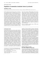
Figure 1
IL-1β and IL-6 stimulate IL-1Ra production. (a) Chondrocytes
(passage 3) were left unstimulated (open squares), or were stimulated
with 1 ng/ml IL-1β (filled diamonds), with 10 ng/ml IL-6 and 100 ng/ml
sIL-6R (filled circles) or with the combination of 1 ng/ml IL-1β,
10 ng/ml IL-6 and 100 ng/ml sIL-6R (filled triangles). IL-1Ra
concentrations in culture supernatants were measured by ELISA.
*P < 0.01 versus control at the same time point,
†
P < 0.01 versus
IL-1β-treated cells at the same time point. (b) Chondrocytes
(passage 1) were stimulated with the indicated doses of IL-1β alone
(white bars) or with IL-1β combined with 10 ng/ml IL-6 and 100 ng/ml
sIL-6R (grey bars) for 72 hours. *P < 0.01 versus control,
†
P < 0.01
versus the same dose of IL-1β alone.
0
250
500
750
1000
020406080
Time (hours)
*†
0
100
200
300
400
500
*†
*†
*
*
*
IL-1Ra (pg/ml)
(a)
(b)
*†
*†
*
IL-1Ra (pg/ml)
Control
IL-1 0.1 ng/mlb
IL-1 1 ng/mlb
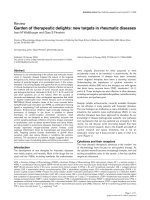
Available online />Figure 2
Effect of IL-1β, IL-6 and dexamethasone on sIL-1Ra and icIL-1Ra1
mRNA expression. (a) Chondrocytes (passage 5) were left
unstimulated (control) or were stimulated for 18 hours with 1 ng/ml
IL-1β (IL-1), with 10 ng/ml IL-6 and 100 ng/ml sIL-6R (IL-6) or with
10
–7
M dexamethasone (dex). Blank, distilled water was used as the
negative control. (b) In one experiment, expression levels of sIL-1Ra
were assessed by quantitative real-time PCR. The graph shows
sIL-1Ra expression corrected for glyceraldehyde-3-phosphate
dehydrogenase (GAPDH) levels.
0
2
4
6
8
(a)
(b)
sIL-1Ra/GAPDH (arbitrary unitsl)
Control
IL-1
dex + IL-1 + IL-6
dex
IL-1 + IL-6
IL-6
sIL-1Ra
icIL-1Ra1
b-actin
521 bp
577 bp
579 bp
Blank
Control
IL-1
IL-6
Il1 + IL-6
dex
dex + IL-1 + IL-6
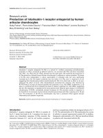
Figure 3
IL-1β and IL-6 increase the expression of nascent sIL-1Ra transcripts.
Chondrocytes (passage 5) were stimulated as described in Figure 2.
Blank, distilled water; w/o RT, PCR performed on nonreverse-
transcribed RNA from IL-1β-stimulated and IL-6-stimulated cells as a
control to exclude genomic DNA contamination; U937-LPS,
lipopolysaccharide-stimulated U937 monocytic cells used as positive
control. MW, molecular weight.
unspliced IL-1Ra
b-actin
303 bp
579 bp
MW marker
Control
IL-1
IL-6
Il1 + IL-6
w/p RT
U937 - LPS
Blank
Figure 4
IL-1β and IL-6 stimulate human sIL-1Ra promoter activity. C-20/A4 cells
were transfected with the enh-1680 reporter gene construct. Negative
controls were performed using empty pGL3-enhancer (pGL3-enh). Cells
were left unstimulated (white bars), or were stimulated with 1 ng/ml IL-1β
(grey bars) or with a combination of 1 ng/ml IL-1β, 10 ng/ml IL-6 and
100 ng/ml sIL-6R (black bars) for 48 hours. *P < 0.001 versus
unstimulated cells transfected with the same plasmid.
0 5000 10000 15000 20000
enh-1680
pGL3-enh
Plasmid
Light units/ g proteinsm
*
Arthritis Research Vol 4 No 3 Palmer et al.
chondrocytes mainly produce the secreted isoform. Using
specific primers, we confirmed that articular chondrocytes
produce predominantly sIL-1Ra mRNA.
A similar stimulation of sIL-1Ra production by IL-1β, which
was also enhanced by IL-6, was observed in hepatocytes,
allowing the classification of IL-1Ra as an acute phase
protein [20]. In both hepatocytes and chondrocytes, IL-1β
and IL-6 stimulated sIL-1Ra expression at the level of gene
transcription. Furthermore, this response was shown to be
mediated by activation of NF-κB and CCAAT/enhancer
binding protein (C/EBP) in hepatocytes. Interactions
between the two transcription factors NF-κB and C/EBP
have been suggested to mediate the synergistic effects of
IL-1 and IL-6 on acute phase protein production [20].
Glucocorticoids have been reported to increase or
decrease IL-1Ra production depending on the cell type
[20,22]. In chondrocytes, we observed a specific inhibi-
tion of IL-1Ra expression by dexamethasone. This finding
suggests that IL-1Ra does not contribute to the anti-
inflammatory effect of glucocorticoids in the cartilage.
The physiologic function of endogenous IL-1Ra has been
clearly demonstrated in several studies using IL-1Ra gene
knockout mice, which have an earlier onset of collagen-
induced arthritis and more severe synovitis, often accom-
panied by tissue damage [23]. Furthermore, when bred
into the BALB/cA background, IL-1Ra knockout mice
develop spontaneous chronic polyarthritis, reproducing
some of the clinical and biological features of rheumatoid
arthritis [24]. These findings suggest that IL-1Ra plays an
important role in the modulation of articular inflammation
and in subsequent cartilage breakdown. Our results show
that IL-1Ra is produced by articular chondrocytes in
response to IL-1 and IL-6, two cytokines present in signifi-
cant amount in inflamed joints. Secreted IL-1Ra in turn
then probably modulates the effects of IL-1 in the cell
microenvironment. Local production of IL-1Ra might thus
be part of a negative feedback mechanism initiated by
these cytokines and may exert a chondroprotective effect
against IL-1-mediated cartilage lesions during physiologic
and pathologic processes, including rheumatoid arthritis
and osteoarthritis.
Conclusion
Human articular chondrocytes produce sIL-1Ra in
response to IL-1β and IL-6. This effect reflects
increased transcription from the sIL-1Ra promoter. The
local production of sIL-1Ra in cartilage may have a pro-
tective effect against articular inflammatory and cata-
bolic responses.
Acknowledgements
Gaby Palmer and Pierre-Andre Guerne contributed equally to this
work. The authors thank Cristiana Juge for her help with quantitative
real-time PCR and Nathalie Pellegrinelli for her expert technical assis-
tance. This work was supported by the Swiss National Science Foun-
dation (grant 3100-064123.00/1 to PAG, and grants 3200-054955.98
and 3231-05454.98 to CG) and the Albert-Boeni Foundation.
References
1. Dripps DJ, Brandhuber BJ, Thompson RC, Eisenberg SP: Inter-
leukin-1 (IL-1) receptor antagonist binds to the 80-kDa IL-1
receptor but does not initiate IL-1 signal transduction. J Biol
Chem 1991, 266:10331-10336.
2. Granowitz EV, Clark BD, Mancilla J, Dinarello CA: Interleukin-1
receptor antagonist competitively inhibits the binding of inter-
leukin-1 to the type II interleukin-1 receptor. J Biol Chem
1991, 266:14147-14150.
3. Greenfeder SA, Nunes P, Kwee L, Labow M, Chizzonite RA, Ju G:
Molecular cloning and characterization of a second subunit of
the interleukin 1 receptor complex. J Biol Chem 1995, 270:
13757-13765.
4. Gabay C: IL-1 inhibitors: novel agents in the treatment of
rheumatoid arthritis. Expert Opin Investig Drugs 2000, 9:113-
127.
5. Bresnihan B, Alvaro-Gracia JM, Cobby M, Doherty M, Domljan Z,
Emery P, Nuki G, Pavelka K, Rau R, Rozman B, Watt I, Williams B,
Aitchison R, McCabe D, Musikic P: Treatment of rheumatoid
arthritis with recombinant human interleukin-1 receptor
antagonist. Arthritis Rheum 1998, 41:2196-2204.
6. Arend WP, Malyak M, Guthridge CJ, Gabay C: Interleukin-1
receptor antagonist: role in biology. Annu Rev Immunol 1998,
16:27-55.
7. Hannum CH, Wilcox CJ, Arend WP, Joslin FG, Dripps DJ,
Heimdal PL, Armes LG, Sommer A, Eisenberg SP, Thompson RC:
Interleukin-1 receptor antagonist activity of a human inter-
leukin-1 inhibitor. Nature 1990, 343:336-340.
8. Haskill S, Martin G, Van Le L, Morris J, Peace A, Bigler CF, Jaffe
GJ, Hammerberg C, Sporn SA, Fong S, Arend WP, Ralph P:
cDNA cloning of an intracellular form of the human interleukin
1 receptor antagonist associated with epithelium. Proc Natl
Acad Sci USA 1991, 88:3681-3685.
9. Muzio M, Polentarutti N, Sironi M, Poli G, De Gioia L, Introna M,
Mantovani A, Colotta F: Cloning and characterization of a new
isoform of the interleukin 1 receptor antagonist. J Exp Med
1995, 182:623-628.
10. Malyak M, Guthridge JM, Hance KR, Dower SK, Freed JH, Arend
WP: Characterization of a low molecular weight isoform of IL-
1 receptor antagonist. J Immunol 1998, 161:1997-2003.
11. Goldring MB, Birkhead J, Sandell LJ, Kimura T, Krane SM: Inter-
leukin 1 suppresses expression of cartilage specific types II
and IX collagens and increases types I and III collagens in
human chondrocytes. J Clin Invest 1988, 82:2026-2037.
12. Tyler JA: Articular cartilage cultured with catabolin (pig inter-
leukin 1) synthetizes a decreased number of normal proteo-
glycan molecules. Biochem J 1985, 227:869-878.
13. Baragi VM, Renkiewicz RR, Jordan H, Bonadio J, Hartman JW,
Roessler BJ: Transplantation of transduced chondrocytes pro-
tects articular cartilage from interleukin 1-induced extracellu-
lar matrix degradation. J Clin Invest 1995, 96:2454-2460.
14. Fernandes J, Tardif G, Martel-Pelletier J, Lascau-Coman V, Dupuis
M, Moldovan F, Sheppard M, Krishnan BR, Pelletier JP: In vivo
transfer of interleukin-1 receptor antagonist gene in
osteoarthritic rabbit knee joints: prevention of osteoarthritis
progression. Am J Pathol 1999, 154:1159-1169.
15. Muller-Ladner U, Roberts CR, Franklin BN, Gay RE, Robbins PD,
Evans CH, Gay S: Human IL-1Ra gene transfer into human
synovial fibroblasts is chondroprotective. J Immunol 1997,
158:3492-3498.
16. Pelletier JP, Mineau F, Ranger P, Tardif G, Martel-Pelletier J: The
increased synthesis of inducible nitric oxide inhibits IL-1ra
synthesis by human articular chondrocytes: possible role in
osteoarthritic cartilage degradation. Osteoarthritis Cartilage
1996, 4:77-84.
17. Lotz M, Clark-Lewis I, Ganu V: HIV-1 transactivator protein Tat
induces proliferation and TGF beta expression in human artic-
ular chondrocytes. J Cell Biol 1994, 124:365-371.
18. Klareskog L, Forsum U, Malmnas Tjernlund UK, Kabelitz D,
Wigren A: Appearance of anti-HLA-DR-reactive cells in normal
and rheumatoid synovial tissue. Scand J Immunol 1981, 14:
183-192.
19. Goldring MB, Birkhead JR, Suen LF, Yamin R, Mizuno S,
Glowacki J, Arbiser JL, Apperley JF: Interleukin-1 beta-modu-
lated gene expression in immortalized human chondrocytes.
J Clin Invest 1994, 94:2307-2316.
20. Gabay C, Smith MF, Eidlen D, Arend WP: Interleukin 1 receptor
antagonist (IL-1Ra) is an acute-phase protein. J Clin Invest
1997, 99:2930-2940.
21. Guerne PA, Desgeorges A, Jaspar JM, Relic B, Peter R,
Hoffmeyer P, Dayer JM: Effects of IL-6 and its soluble receptor
on proteoglycan synthesis and NO release by human articular
chondrocytes: comparison with IL-1. Modulation by dexa-
methasone. Matrix Biol 1999, 18:253-260.
22. Sauer J, Castren M, Hopfner U, Holsboer F, Stalla GK, Arzt E:
Inhibition of lipopolysaccharide-induced monocyte inter-
leukin-1 receptor antagonist synthesis by cortisol: involve-
ment of the mineralocorticoid receptor. J Clin Endocrinol
Metab 1996, 81:73-79.
23. Ma Y, Thornton S, Boivin GP, Hirsh D, Hirsch R, Hirsch E: Altered
susceptibility to collagen-induced arthritis in transgenic mice
with aberrant expression of interleukin-1 receptor antagonist.
Arthritis Rheum 1998, 41:1798-1805.
24. Horai R, Saijo S, Tanioka H, Nakae S, Sudo K, Okahara A, Ikuse T,
Asano M, Iwakura Y: Development of chronic inflammatory
arthropathy resembling rheumatoid arthritis in interleukin 1
receptor antagonist-deficient mice. J Exp Med 2000, 191:313-
320.
Supplementary material
Supplementary materials and methods
Cell culture
Cartilage was obtained from patients undergoing knee
joint or hip joint replacement for osteoarthritis. Chondro-
cytes were isolated by collagenase digestion, as previ-
ously described [17,18], and cultured in DMEM
containing
L-glutamine, streptomycin, penicillin and 10%
FCS. Isolated chondrocytes were either used immediately
or were expanded and subcultured several times. Cells
were stimulated with cytokines 24 hours after seeding.
Proteoglycan synthesis
Proteoglycan synthesis was evaluated by measuring the
aggrecan concentration in culture supernatants by
enzyme-amplified sensitivity immunoassay (Biosource
Europe S.A., Nivelles, Belgium). Results are presented as
the mean ± SEM of three determinations.
Reverse transcription polymerase chain reaction
RNA was digested with DNAse I and reverse-transcribed
using avian myeloblastosis virus-RT and random hexamer
primers (Promega). PCR (40 cycles for IL-1Ra isoforms
and unspliced transcript, 35 cycles for β-actin) was per-
formed using appropriate primer pairs (Supplementary
Figure 1 and Supplementary Table 1) and appropriate
conditions (Supplementary Table 2). The identity of the
amplified products was confirmed by DNA sequencing.
RT-PCR experiments performed on chondrocyte cultures
from four different patients yielded similar results. Quanti-
tative real-time PCR for sIL-1Ra was performed with a
Light Cycler™ (Roche Molecular Biochemicals, Basel,
Switzerland), using primers B and G (Supplementary
Figure S1 and Supplementary Table S1). Expression of
sIL-1Ra mRNA was corrected for glyceraldehyde-3-phos-
phate dehydrogenase levels, which were amplified using
the indicated primers (Supplementary Table 1) and condi-
tions (Supplementary Table 2).
Reporter gene assays
The 1680 bp human sIL-1Ra promoter region was recov-
ered from plasmid psRA1680 [20] by digestion with
HindIII, and it was inserted upstream of the luciferase
reporter gene in pGL3-enhancer (Promega) to yield
plasmid enh-1680. Correct insertion of the promoter frag-
ment was verified by DNA sequencing. C-20/A4 cells
were seeded in 24-well plates (20,000 cells/well) and
transfected 24 hours later in DMEM containing 1% FCS
with 0.4 µg DNA and 1.2 µl FuGENE 6. After 4 hours,
cells were switched to DMEM/F12 containing 10% FCS.
Cytokines were added 24 hours after transfection.
Supplementary results
Specific inhibition of IL-1Ra production by
dexamethasone
Dexamethasone (10
–7
M) inhibited the stimulatory effect
of IL-1β and IL-6/sIL-6R on IL-1Ra production (Supple-
mentary Figure 2a). This inhibitory effect of dexametha-
Available online />Supplementary Figure 1
Schematic representation of the human IL-1RN gene. The IL-1RN gene contains six exons, which are represented by boxes. The icIL-1Ra1 and
icIL-1Ra2 mRNAs are transcribed from a promoter (P ic) located upstream of exon ic1. The icIL-1Ra1 transcript contains exon ic1 spliced to an
internal splice acceptor site within exon 1, followed by exons 2 to 4. The icIL-1Ra2 mRNA contains an additional exon (ic2) inserted downstream of
exon ic1. This differential splicing is symbolized by dotted lines. The sIL-1Ra mRNA is transcribed from a promoter (P s) located immediately
upstream of exon 1 and contains exons 1 to 4. The locations of PCR primers A–D and G (Supplementary Table 1) are indicated by arrows. The
combination of primers A and C allows specific amplification of icIL-1Ra1 and icIL-1Ra2 cDNAs, while primers B and C or B and G specifically
amplify sIL-1Ra mRNA. The combination of primers B and D allows the detection of unspliced primary sIL-1Ra transcripts.
ic1 ic2 1 2 3 4
A B
CD
Pic P s
G
sone was dose dependent and was already present at a
concentration of 10
–9
M (data not shown). In contrast,
dexamethasone significantly enhanced proteoglycan syn-
thesis (Supplementary Figure 2b), indicating that the
inhibitory effect of dexamethasone on IL-1Ra production
was specific and not secondary to cell toxicity.
Activity of the human 1680 bp sIL-1Ra promoter in
chondrocytes
The enh-1680 reporter construct contains 1680 bp
human sIL-1Ra promoter region (Supplementary Figure 1,
region P s). This region was shown previously to contain
DNA cis-acting elements responsible for cell-type-specific
expression of sIL-1Ra [20]. It contains, in particular,
binding sites for the transcription factors NF-κB and
C/EBP, which are involved in the activation of sIL-1Ra
transcription by IL-1 and IL-6 in HepG2 cells [20]. Human
osteoarthritis chondrocytes are difficult to transfect [S1]
and our transfection efficiencies were not sufficient to
yield detectable luciferase activities using the enh-1680
plasmid. As an alternative, we used C-20/A4-immortalized
human chondrocytes for these experiments. These cells
exhibit responses to IL-1 similar to those described in
primary human chondrocytes [19].
Supplementary reference
S1. Madry H, Trippel SB: Efficient lipid-mediated gene transfer to
articular chondrocytes. Gene Ther 2000, 7:286-291.
Arthritis Research Vol 4 No 3 Palmer et al.
Supplementary Table 1
PCR primer location and sequence
Primer Gene GenBank Location (bp)* Sequence 5′–3′
A IL-1RN U65590 16,106–16,131 (s) CAGGTACTGCCCGGGTGCTACTTTAT
B IL-1RN U65590 25,827–25,852 (s) GGCCTCCGCAGTCACCTAATCACTCT
C IL-1RN U65590 30,881–30,856 (as) TACTACTCGTCCTCCTGGAAGTAGAA
D IL-1RN U65590 26,130–26,109 (as) GGTCGCACTATCCACATCTGGG
E β-Actin M10277 1559–1583 (s) CCAAGGCCAACCGCGAGAAGATGAC
F β-Actin M10277 2681–2658 (as) AGGGTACATGGTGGTGCCGCCAGAC
G IL-1RN U65590 29134–29115 (as) TTGACACAGGACAGGCACAT
H GAPDH M32599 140–157 (s) AACGACCCCTTCATTGAC
I GAPDH M32599 330–312 (as) TCCACGACATACTCAGCAC
*Sense (s) or antisense (as) orientation with respect to the corresponding gene is indicated. GAPDH, glyceraldehyde-3-phosphate dehydrogenase.
Supplementary Table 2
PCR conditions
cDNA Forward primer Reverse primer Annealing temperature (°C) Product length (bp)
icIL-1Ra1 A C 55 577
sIL-1Ra B C 55 521
Unspliced IL1-Ra B D 55 303
β-Actin E F 60 579
sIL-1Ra (LC) B G 57 268
GAPDH (LC) H I 57 191
LC, Light Cycler™ (Roche Molecular Biochemicals, Basel, Switzerland). GAPDH, glyceraldehyde-3-phosphate dehydrogenase.
Available online />Supplementary Figure 2
Specific inhibition of IL-1Ra production by dexamethasone. (a) Freshly
isolated chondrocytes were stimulated with 1 ng/ml IL-1β, 10 ng/ml
IL-6 and 100 ng/ml sIL-6R for 72 hours in the absence (black bars) or
presence (white bars) of 10
-7
M dexamethasone. *P < 0.001 versus
control without dexamethasone,
†
P < 0.001 versus IL-1β, IL-6 and
sIL-6R without dexamethasone. (b) Proteoglycan content was
measured in the culture supernatants used for IL-1Ra determination.
*P < 0.01 versus control without dexamethasone.
0
5,000
10,000
15,000
(a)
(b)
*
Proteopglycans (pg/ml)
Control
IL-1 + IL-6 + sIL-6Rb
*
0
500
1000
1500
IL-1Ra (pg/ml)
*
†