Báo cáo khoa học: "A rare coexistence of adrenal cavernous hemangioma with extramedullar hemopoietic tissue: a case report and brief review of the literature" docx
Bạn đang xem bản rút gọn của tài liệu. Xem và tải ngay bản đầy đủ của tài liệu tại đây (926.62 KB, 4 trang )
BioMed Central
Page 1 of 4
(page number not for citation purposes)
World Journal of Surgical Oncology
Open Access
Case report
A rare coexistence of adrenal cavernous hemangioma with
extramedullar hemopoietic tissue: a case report and brief review of
the literature
Nikolaos Arkadopoulos
1
, Maria Kyriazi
1
, Anneza I Yiallourou*
1
,
Vaia K Stafyla
1
, Theodosios Theodosopoulos
1
, Nikolaos Dafnios
1
,
Vassilis Smyrniotis
1
and Agathi Kondi-Pafiti
2
Address:
1
2nd Department of Surgery, Aretaieion Hospital, Athens University School of Medicine, Athens, Greece and
2
Department of Pathology,
Aretaieion Hospital, Athens University School of Medicine, Athens, Greece
Email: Nikolaos Arkadopoulos - ; Maria Kyriazi - ; Anneza I Yiallourou* - ;
Vaia K Stafyla - ; Theodosios Theodosopoulos - ; Nikolaos Dafnios - ;
Vassilis Smyrniotis - ; Agathi Kondi-Pafiti -
* Corresponding author
Abstract
Background: Cavernous hemangiomas of the adrenal gland are rare, benign, non-functioning
neoplastic tumors. To our knowledge, 55 cases have been reported in the literature to date.
Case presentation: We report the first case of a large, non-functioning adrenal cavernous
hemangioma that was incidentally found during the preoperative staging workup of a 75 year old
woman with left breast adenocarcinoma. Imaging with US, CT scan and MRI showed a
heterogeneous 8 cm mass with non-specific radiological features that was located on the left
adrenal gland. The mass was surgically excised and pathology revealed an adrenal hemangioma with
areas of extramedullar hemopoiesis.
Conclusion: Although adrenal hemangiomas are rare and their preoperative diagnosis is difficult,
they should always be included in the differential diagnosis of adrenal neoplasms.
Background
Adrenals are an infrequent location for benign vascular
tumors like cavernous hemangiomas-such tumors are
most commonly situated on the skin or in the liver. Their
clinical presentation is usually vague, with non-specific
abdominal pain being the predominant symptom. Fre-
quently, they are discovered as incidentalomas either dur-
ing imaging or in autopsies. Since 1955, when Johnson
and Jeppesen described the first adrenal cavernous
hemangioma, only 55 cases have been reported in the lit-
erature [1]. We report a case of a large, non-functioning
adrenal hemangioma that was found incidentally during
pre-operative staging of a 75 year old woman with adeno-
carcinoma of the left breast.
Case presentation
A 75 year old female patient with breast cancer was admit-
ted to our hospital for surgical treatment. Her preopera-
tive staging workup with an abdominal ultrasound,
revealed a heterogeneous solid lesion of the left adrenal
gland. Clinical examination and laboratory tests, includ-
ing adrenal hormonal levels (plasma renin 7,40 pg/ml,
Published: 5 February 2009
World Journal of Surgical Oncology 2009, 7:13 doi:10.1186/1477-7819-7-13
Received: 8 November 2008
Accepted: 5 February 2009
This article is available from: />© 2009 Arkadopoulos et al; licensee BioMed Central Ltd.
This is an Open Access article distributed under the terms of the Creative Commons Attribution License ( />),
which permits unrestricted use, distribution, and reproduction in any medium, provided the original work is properly cited.
World Journal of Surgical Oncology 2009, 7:13 />Page 2 of 4
(page number not for citation purposes)
plasma aldosterone 12,7 ng/dl, plasma adrenaline 27 pg/
ml, plasma noradrenaline 243 pg/ml, 24 h urine metane-
phrine excretion 169 μg/24 h), were normal. Abdominal
CT scan showed a well-defined, heterogeneous, retroperi-
toneal mass with speckled calcifications that measured 8
cm and was located on the left adrenal gland. After bolus
IV injection of contrast medium the tumor showed irreg-
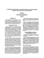
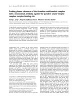
ular enhancement. On subsequent MRI, the tumor dem-
onstrated hyperintensity on both T1- and T2-weighted
images with fat component and irregular peripheral
enhancement (Figure 1, 2). Malignancy could not be
excluded due to the non-specific radiological features,
therefore surgical resection was mandatory.
During the same operation, the patient underwent a left
adrenalectomy through a left subcostal incision followed
by modified radical left mastectomy. Her postoperative
course was uneventful and she was discharged five days
later.
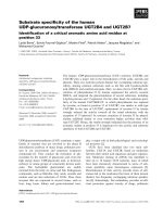
On gross examination, the adrenal tumor appeared as a
red tan mass measuring 8 cm × 6 cm × 4 cm. Focal red-pur-
ple hemorrhagic and cystic areas were present, along with
diffuse calcifications. Normal adrenal gland parenchyma
was noted on the surface of the mass (Figure 3).
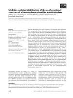
Microscopically, dilated, blood filled vascular spaces were
observed. The spaces were lined by a single layer of thin
endothelial cells with collagenous walls (Figure 4). Inter-
estingly, areas of extramedullar hemopoiesis were also
seen (Figure 5).
The histological diagnosis was that of an adrenal cavern-
ous hemangioma with coexistence of extramedullar
hemopoiesis and no signs of malignancy.
The pathology report on the breast specimen showed a
grade II infiltrating tubular adenocarcinoma, measuring 5
MRI scan of a left adrenal hemangioma demonstrating hyper-intensity on T1-weighted image with a fat componentFigure 1
MRI scan of a left adrenal hemangioma demonstrat-
ing hyperintensity on T1-weighted image with a fat
component.
MRI scan of a left adrenal hemangioma demonstrating hyper-intensity on T2-weighted image and irregular peripheral enhancementFigure 2
MRI scan of a left adrenal hemangioma demonstrat-
ing hyperintensity on T2-weighted image and irregu-
lar peripheral enhancement.
Gross section of adrenal hemangioma showing macrocystic, haemorrhagic surfaceFigure 3
Gross section of adrenal hemangioma showing mac-
rocystic, haemorrhagic surface.
World Journal of Surgical Oncology 2009, 7:13 />Page 3 of 4
(page number not for citation purposes)
cm in greatest diameter. None of the 13 excised lymph
nodes had signs of malignancy.
Discussion
The evolution of radiological imaging in the last 20 years
resulted in increased detection rate of clinically inappar-
ent adrenal masses, also known as adrenal incidentalo-
mas. It is estimated that adrenal masses are an accidental
finding in 1–5% of all abdominal CT scans performed.
Adrenal hemangiomas, however, are extremely rare, and
their differential diagnosis preoperatively is rather chal-
lenging.
Adrenal hemangiomas are most usually cavernous, unilat-
eral lesions of the adrenal glands that appear in the sixth
or seventh decade of life, with a 2:1 female to male ratio
[2-4]. Their size ranges from 2 cm to 25 cm in diameter,
with the majority measuring more than 10 cm [5-7]. They
are most commonly non-functioning tumors, with only
three cases of hormone-secreting adrenal hemangiomas
being reported to date [8-10]. These unusual benign adre-
nal masses are usually detected as incidental radiological
findings in abdominal imaging performed for various
other reasons. They are hardly ever symptomatic, with
abdominal pain due to mechanical mass effects on neigh-
bouring structures being the main symptom. However, in
two cases adrenal haemangiomas presented with sponta-
neous life-threatening retroperitoneal haemorrhage
[3,11]. The adrenal glands are a common site of metasta-
sis for various cancers, therefore adrenal masses must be
excluded in the preoperative staging of several carcinomas
(melanomas, lung, breast, renal and gastrointestinal can-
cers). Three cases of adrenal hemangiomas, coexisting
with malignant tumors of other organs (non-small-cell
lung cancer, common bile duct cancer and gynaecological
cancer) [12-14] have been reported in the literature. This
is the only case of adrenal hemangioma in a patient with
breast cancer reported so far. Histologically, these tumors
are primary mesenchymal vascular neoplasms with
angioblastic cells predominating. Surprisingly, this is the
only case reported with extramedullar hemopoietic tissue
within a hemangioma.
Distinguishing a large adrenal hemangioma from other
lesions of the adrenal glands, and especially from malig-
nant tumors, can be very difficult. In most cases the final
diagnosis is made by histopathology after surgical resec-
tion. However, there are some radiological features that,
although not entirely specific, should raise the suspicion
of adrenal haemangioma. CT scans usually display a char-
acteristic peripheral patchy enhancement with progres-
sion to the centre of the tumor that is a common finding
[15]. Speckled calcifications that appear throughout the
mass are attributed to multiple phleboliths located in
dilated vascular spaces [16,17]. Nonetheless, this is a com-
mon finding in other adrenal lesions, such as pheochro-
mocytoma, carcinoma and adenoma, and cannot,
therefore, be pathognomonic for hemangiomas.
MRI has been proven to be the best diagnostic tool so far.
The most characteristic finding is the peripheral spotty
and centripetal enhancement on dynamic studies. Marked
hyperintensity on T2-weighted images in combination
with focal hyperintensity in T1-weighted images, indicate
areas of calcification and haemorrhage that are associated
with adrenal hemangiomas [2,15,18]. Angiography usu-
ally reveals peripheral pooling of the contrast, persisting
well during the venous phase [16,17].
Histological appearance of the adrenal hemangioma (hema-toxylin-eosin × 25)Figure 4
Histological appearance of the adrenal hemangioma
(hematoxylin-eosin × 25).
Histological section of the adrenal hemangioma showing a focus of extramedullar hemopoiesis (hematoxylin-eosin × 25)Figure 5
Histological section of the adrenal hemangioma
showing a focus of extramedullar hemopoiesis
(hematoxylin-eosin × 25).
Publish with BioMed Central and every
scientist can read your work free of charge
"BioMed Central will be the most significant development for
disseminating the results of biomedical research in our lifetime."
Sir Paul Nurse, Cancer Research UK
Your research papers will be:
available free of charge to the entire biomedical community
peer reviewed and published immediately upon acceptance
cited in PubMed and archived on PubMed Central
yours — you keep the copyright
Submit your manuscript here:
/>BioMedcentral
World Journal of Surgical Oncology 2009, 7:13 />Page 4 of 4
(page number not for citation purposes)
The surgical indication for excision of the tumor is the
size. Adrenal incidentalomas larger than 6 cm in diameter
must be excised because the risk of adrenal cancer is 35%
to 98%. For lesions measuring 4 cm to 6 cm, other imag-
ing features, history of extra-adrenal malignancy, patient's
preference, age and comorbitities should be taken into
consideration. Adrenalectomy and follow-up with imag-
ing are both acceptable in such cases [3]. Most adrenal
hemangiomas reported so far have been treated surgically
due to their size. Other indications for surgery include
mass-effect type symptoms from neighbouring organs
and complications, such as haemorrhage.
Adrenalectomy can be performed laparoscopically for
lesions measuring less than 6 cm [7,19]. Larger tumors,
that are technically challenging and more likely to be
malignant are treated preferably with open technique
through an anterior (subcostal or midline incision), pos-
terior or thoracoabdominal approach.
Conclusion
We presented a rare coexistence of an adrenal cavernous
hemangioma with extramedullar hemopoietic tissue in a
woman treated for breast cancer. Although rare, adrenal
haemangioma should be included in the differential diag-
nosis of adrenal neoplasms. The main indication for sur-
gical removal of an adrenal mass is its size. However, the
risks of haemorrhage, necrosis and thrombosis necessitate
surgical excision in most of the cases, especially for
tumors more than 3 cm.
Consent
Written informed consent was obtained from the patient
for publication of this case report and any accompanying
images. A copy of the written consent is available for
review by the Editor-in-Chief of this journal.
Competing interests
The authors declare that they have no competing interests.
Authors' contributions
NA was responsible for critical revision of scientific con-
tent. MK drafted the manuscript. AIY participated in the
design of the manuscript and helped to draft the manu-
script. VKS contributed substantially to manuscript con-
ception and design. TT assisted in the preparation of the
manuscript.
ND participated in the acquisition of data and prepara-
tion of the manuscript. VS was the surgeon, approved the
final version of the manuscript for publication. AKP per-
formed histopathological and immunohistochemical
analysis and contributed substantially to pathology con-
tent. All authors read and approved the final manuscript.
References
1. Johnson CC, Jeppesen FB: Haemangioma of the adrenal. J Urol
1955, 74:573-577.
2. Heis HA, Bani-Hani KE, Bani-Hani BK: Adrenal cavernous hae-
mangioma. Singapore Med J 2008, 49(9):e236-e237.
3. Forbes TL: Retroperitoneal haemorrhage secondary to rup-
tured cavernous hemangioma. Can J Surg 2005, 48:78-79.
4. Sabanegh E, Harris MJ, Grider D: Cavernous adrenal haemangi-
oma. Urology 1993, 42(3):327-330.
5. Makiyama K, Fukuoka H, Kawamoto K, Suwa Y: Surgical removal
of adrenal haemangioma after five years of follow-up: a case
report. Hinyokika Kiyo 1998, 44:579-581.
6. Hisham AN, Samad SA, Sharifah NA: Huge adrenal haemangi-
oma. Austral Radiol 1998, 42:250-251.
7. Nigri G, Bellagamba R, Giaccaglia V, Felicioni F, Aurello P, D' Angelo
F, Del Gaudio M, Ramacciato G: Minimally invasive adrenalec-
tomy for incidentally discovered cavernous haemangioma.
Minim Invasive Ther Allied Technol 2008, 17(4):255-258.
8. Stumvoll M, Fritsche A, Wehrmann M, Dammann F, Becker HD, Egg-
stein M: A functioning adrenocortical haemangioma. Urol
1996, 155:638.
9. Oh BR, Jeong YY, Ryu SB, Park YI, Kang HK: A case of adrenal cav-
ernous haemangioma. Int J Urol 1997, 4:608-610.
10. Ng AC, Loh HL, Shum CF, Yip SK: A case of adrenal cavernous
haemangioma presenting with progressive enlargement and
apparent hormonal hypersecretion. Endocr Pract 2008,
14(1):104-108.
11. Boraschi P, Campatelli A, Di Vito A, Perri G: Haemorrhage in cav-
ernous haemangioma of the adrenal gland: US, CT and MRI
appearances with pathologic correlation. Eur J Radiol 1995,
21(1):41-43.
12. Alcázar J, Márquez A, Rosales M: An unusual cause of adrenal
mass in a patient with operable non-small-cell pulmonary
carcinoma. Arch Bronconeumol 1998, 34(10):513-514.
13. Päivänsalo M, Siniluoto T, Seppänen U: Cavernous haemangioma
of the adrenal gland. Diagn Imaging Clin Med 1986, 55(3):168-171.
14. Chudácek Z, Kohoutek V: Simultaneous occurrence of a cav-
ernous adrenal gland haemangioma and a bile duct-liver car-
cinoma. Rofo 1980, 132(4):460-462.
15. Yamada T, Ishibashi T, Saito H, Majima K, Tsuda M, Takahashi S,
Moriya T: Two cases of adrenal haemangioma: CT and MRI
findings with pathological correlations. Radiat Med 2002,
20(1):51-56.
16. Rothberg M, Bastidas J, Mattey WE, Bernas E: Adrenal haemangi-
omas: angiographic appearance of a rare tumor. Radiology
1978, 126:341-344.
17. Thiele J, Bodie B: Adrenal haemangioma. Surgery 2001,
1(129):373-374.
18. Marotti M, Sučić Z, Krolo I, Dimanovski J, Klarić R, Ferenčić Ž, Kara-
panda N, Babić N, Pavleković K: Adrenal cavernous haemangi-
oma: MRI, CT and US appearance. Eur Radiol 1997, 7:691-694.
19. Trupka A, Hallfeldt K, Schmidbauer S: Laparoscopic adrenalec-
tomy with lateral approach – a comparison with the conven-
tional dorsal technique. Chirurg 2001, 72:1478-1484.