Báo cáo khoa học: "Mucin pattern reflects the origin of the adenocarcinoma in Barrett''''s esophagus: a retrospective clinical and laboratorial study" ppsx
Bạn đang xem bản rút gọn của tài liệu. Xem và tải ngay bản đầy đủ của tài liệu tại đây (993.81 KB, 8 trang )
BioMed Central
Page 1 of 8
(page number not for citation purposes)
World Journal of Surgical Oncology
Open Access
Research
Mucin pattern reflects the origin of the adenocarcinoma in Barrett's
esophagus: a retrospective clinical and laboratorial study
Sergio Szachnowicz*
1
, Ivan Cecconello
1
, Ulysses Ribeiro
1
, Kiyoshi Iriya
2
,
Roberto El Ibrahim
2
, Flávio Roberto Takeda
1
, Carlos Eduardo
Pereira Corbett
2
and Adriana Vaz Safatle-Ribeiro
1
Address:
1
Department of Gastroenterology, Digestive Surgery Division, University of São Paulo School of Medicine, São Paulo, Brazil and
2
Department of Pathology, University of São Paulo School of Medicine, São Paulo, Brazil
Email: Sergio Szachnowicz* - ; Ivan Cecconello - ;
Ulysses Ribeiro - ; Kiyoshi Iriya - ; Roberto El Ibrahim - ;
Flávio Roberto Takeda - ; Carlos Eduardo Pereira Corbett - ; Adriana Vaz Safatle-
Ribeiro -
* Corresponding author
Abstract
Background: Mucin immunoexpression in adenocarcinoma arising in Barrett's esophagus (BE)
may indicate the carcinogenesis pathway. The aim of this study was to evaluate resected specimens
of adenocarcinoma in BE for the pattern of mucins and to correlate to the histologic classification.
Methods: Specimens were retrospectively collected from thirteen patients who underwent
esophageal resection due to adenocarcinoma in BE. Sections were scored for the grade of intestinal
metaplasia. The tissues were examined by immunohistochemistry for MUC2 and MUC5AC
antibodies.
Results: Eleven patients were men. The mean age was 61 years old (varied from 40 to 75 years
old). The tumor size had a mean of 4.7 ± 2.3 cm, and the extension of BE had a mean of 7.7 ± 1.5
cm. Specialized epithelium with intestinal metaplasia was present in all adjacent mucosas.
Immunohistochemistry for MUC2 showed immunoreactivity in goblet cells, while MUC5AC was
extensively expressed in the columnar gastric cells, localizing to the surface epithelium and
extending to a variable degree into the glandular structures in BE. Tumors were classified according
to the mucins in gastric type in 7/13 (MUC5AC positive) and intestinal type in 4/13 (MUC2
positive). Two tumors did not express MUC2 or MUC5AC proteins. The pattern of mucin
predominantly expressed in the adjacent epithelium was associated to the mucin expression profile
in the tumors, p = 0.047.
Conclusion: Barrett's esophagus adenocarcinoma shows either gastric or intestinal type pattern
of mucin expression. The two types of tumors developed in Barrett's esophagus may reflect the
original cell type involved in the malignant transformation.
Published: 9 March 2009
World Journal of Surgical Oncology 2009, 7:27 doi:10.1186/1477-7819-7-27
Received: 13 November 2008
Accepted: 9 March 2009
This article is available from: />© 2009 Szachnowicz et al; licensee BioMed Central Ltd.
This is an Open Access article distributed under the terms of the Creative Commons Attribution License ( />),
which permits unrestricted use, distribution, and reproduction in any medium, provided the original work is properly cited.
World Journal of Surgical Oncology 2009, 7:27 />Page 2 of 8
(page number not for citation purposes)
Background
Barrett's esophagus (BE) is the eponymous term used to
describe a condition with malignant potential where the
lower esophagus becomes lined with a specialized colum-
nar epithelium as a result of chronic gastroesophageal
reflux. Nowadays, Barrett's esophagus represents the tran-
sition from normal squamous mucosa to columnar epi-
thelium plus the identification of intestinal metaplasia. In
macroscopic form, BE is classified as long, when the
columnar epithelium is longer than 3 cm, and short when
is lower than 3 cm [1,2].
BE is a complex, mosaic of cell, gland, and architectural
types, showing variable degrees of atrophy and matura-
tion toward intestinal and gastric epithelium. Surface
mucous, goblet cells, absorptive, mucous neck, mucous
gland and neuroendocrine cells are randomly distributed
in relation to the gastroesophageal junction [3,4].
Although there are three types of columnar epithelium –
namely, gastric fundic, junctional cardiac and specialized
intestinal epithelium, – it is now accepted that adenocar-
cinoma arises only from the specialized intestinal-type of
metaplasia [3,5-12]. Nonetheless, many of the esophageal
adenocarcinomas in BE (ABE) exhibit a poor differenti-
ated and/or undifferentiated pattern, distinct from the
intestinal type tumors commonly observed in patients
with intestinal metaplasia.
Mucin genes are expressed throughout the human gas-
trointestinal tract in a site specific manner [13]. In special-
ized BE, there is strong expression of MUC2 in the goblet
cells (intestinal mucin pattern) and MUC5AC in the
superficial columnar epithelium (gastric mucin pattern)
[14]. This is the same pattern already described for incom-
plete intestinal metaplasia of the stomach, and is further
evidence that BE and incomplete intestinal metaplasia of
the stomach are the same condition and represent differ-
entiation into a unique epithelial lineage [15,16].
BE is a marker of tissue injury possibly as a consequence
of inflammatory lesions and regeneration. Thus, all cells
of the BE under damage could originate an expansion
clone capable of initiate the carcinogenesis cascade. The
pattern of expression of mucin gene products in adenocar-
cinoma arising in BE has yet to be known.
Thus, we have studied a homogenous group of patients
with adenocarcinoma arising in BE. We sought to deter-
mine whether gastric (MUC5AC) and/or intestinal type
(MUC2) markers, could help improve our understanding
of the carcinogenesis in Barrett's adenocarcinoma.
Patients and methods
This investigation was approved by the Ethical Committee
of the Hospital das Clínicas of São Paulo Medical School.
From January, 1990 to June, 2002, a total of 297 patients
with diagnostic of BE confirmed through endoscopic
biopsies, were treated at the Esophageal Surgery Service of
Digestive Surgery Division of Hospital das Clínicas of the
University of São Paulo School of Medicine. Of those,
Adenocarcinoma was diagnosed in 17 patients, with a
prevalence of 5.7%. We retrospectively review the clinical
charts of these patients regarding the presence of Barrett's
esophagus, clinical characteristics and pathology report.
Gastric tumors with esophageal invasion and esophageal
neoplasias with invasive components to the gastric cardia
were excluded. Carcinomas were deemed to be arising
from the Barrett's esophagus, if, on histological examina-
tion, there was specialized columnar metaplasia proximal
and/or involving the tumor.
Among the 17 patients, three were excluded due to unre-
sectable advanced neoplasia. One underwent argon plas-
matic ablation of the columnar epithelium, including the
tumor, which was not identified in the histopathologic
study of the resected esophagus. The remaining 13
patients underwent esophageal resection, and form the
basis of this study.
Histopathologic study
All the pathological specimens are prepared according to
the Pathology Department guidelines. The resected
esophagus was opened longitudinally, photographed,
stretched in glides of plastic or cardboard surface, BE and
tumor extension were measured. The distances between
tumor's distal margins and gastroesophageal junction
(Dist. Tu-GEJ); and tumor's proximal margins and colum-
nar-squamous transition (Prox. Tu-Tepit) were per-
formed. After this, the specimens were fixed using
formaldehyde solution.
For the histological study, tissue samples were retrieved
from archived paraffin embedded sections of histologi-
cally known Barrett's esophagus. Tumor and adjacent epi-
thelium, were stained by hematoxyline-eosine (HE).
Histology of the adjacent tumor area showed a special-
ized-type mucosa characterized by an epithelial lining
which included columnar epithelium showing a poorly
developed brush border, villous architecture, and goblet
cells. The surface cells were of surface mucous type, with
underlying cardiac/antral glands beneath surfaces covered
by goblet and absorptive cells. Barrett's esophagus could
be classified as specialized epithelium in all studied
patients, with areas with predominance of an intestinal or
gastric type epithelium in each patient.
The tumors were classified according to the grade of differ-
entiation.
World Journal of Surgical Oncology 2009, 7:27 />Page 3 of 8
(page number not for citation purposes)
Immunohistochemical evaluation
Sections of tumors, and corresponding adjacent areas,
developing in patients with Barrett's esophagus were
examined by immunohistochemistry for MUC5AC (NCL
– MUC-5AC, Novocastra, Newcastlle, United Kingdom)
and MUC2 (NCL – MUC-2, Novocastra, Newcastlle,
United Kingdom).
Three to five unstained 4 μm blank histologic sections
were cut from each designated block and used for
MUCAC-5 and MUC-2 immunostaining (using humid
heating). Briefly, immunodetection involved the use of 4
μm thick formalin-fixed paraffin-embedded tissues,
treated with 4% and 2% hydrogen peroxidase (H
2
O
2
) in
methanol for 35 minutes, to eliminate endogenous perox-
idase activity. Sections were rinsed in phosphate-buffered
saline (PBS) and incubated with 10% normal horse serum
to block nonspecific binding. Upon removal of the serum,
the primary monoclonal antibody was applied. Following
further washing with PBS, sections were incubated with
biotinylated anti-mouse immunoglobulin for 30 minutes.
After washing twice with PBS, the sections were treated
with Vectastain Elite horseradish peroxidase complex
(Vector Laboratory, Burlingame, CA) for 30 minutes. Fol-
lowing another rinse with PBS, the sections were incu-
bated with diaminobenzidine 0.05% and 0.04% H
2
O
2
for
20 minutes. After a final wash with distilled water, the sec-
tions were counterstained with Harris Alum Hematoxylin,
dehydrated through graded alcohols to xylene, and cover-
slipped.
All sections were examined by three independent investi-
gators (KY, REI and UR) for the histopathological study
and blindly for immunohistochemical evaluation by the
third one. The mucins were expressed as cytoplasmic
staining. The results were expressed semiquantitatively for
each histological group as the number of sections posi-
tively labeled, the predominant cell type labeled, and the
average score of the positively labeled cells. Positive Con-
trol Sections: control tissues taken from colon and stom-
ach, with previously identified MUC gene expression
patterns were included with each batch of sections for
immunohistochemistry.
Negative Control Sections: the primary antibody was
omitted as a negative control to test the specificity of the
antibodies utilized for each section.
Incubation with Primary Antibody (MUC2 was diluted in
1:100, and the MUC5, 1:400)
Statistical analysis
Results of immunohistochemical alterations were com-
pared to the clinical-pathologic features using chi-square
test for qualitative data, with two tailed p value < 0.05
considered significant.
Results
Eleven patients were men (84.6%) and two women
(15.4%), with proportion of 5.5:1. The age range from 40
to 75 years-old (mean = 61 years ± 9.9).
Histopathological results
Measurements obtained from each resected esophagus are
presented in table 1. Columnar epithelium extension
ranged from 3.5 to 16.0 cm (mean of 7.7 ± 3.3 cm).
Table 1: Lengths of barrett's esophagus epithelium and adenocarcinoma.
Patient Barrett's esophagus length (cm) Adenocarcinoma length (cm) Dist. Tu-GEJ (cm) Prox. Tu-Tepit (cm)
1 16 3.6 14 0.4
210 8 0.51.5
34 3.0 10
4 7 6.5 0 0.5
58 5 03
6 6 7.4 2.2 0
7 3.5 3 0 0.5
8 5 4.5 0.3 0.5
9 10.7 2.2 5.5 2.5
10 8 7 0 1
11 6.5 1.5 1.5 3.5
12 9.5 7 2.5 0
13 6 2.5 0 3.5
Mean
(SD)
7.71
(3.33)
4.67
(2.28)
2.07 1.30
Min 3.5 1.5 0 0
Max 16 7.4 14 3.5
Distances from adenocarcinoma to gastroesophageal junction; distances from adenocarcinoma to squamous-columnar transition.
Dist. Tu-GEJ = Distance from tumors (Adenocarcinoma) distal margin to the gastroesophageal junction.
Prox. Tu-Tepit = Distance from the turmors (Adenocarcinoma) proximal margin to the epithelium (columnar-squamous) transition
World Journal of Surgical Oncology 2009, 7:27 />Page 4 of 8
(page number not for citation purposes)
Tumor extension ranged from 1.5 to 7.4 cm (mean of 4.7
± 2.3 cm). All adenocarcinoma developed in BE longer
than 3.0 cm. The distances between the tumor's distal
margins and gastroesophageal junction (Dist. Tu-GEJ)
ranged from just at the GEJ (5 patients – 38.5%) to tumors
14 cm far from GEJ (mean of 2.1 cm). The distances of
tumor's proximal margins and columnar-squamous tran-
sition (Prox. Tu-Tepit) ranged from tumors that reached
the epithelium transition and tumor 3.5 cm far from Tepit
(mean of 1.30 cm). Eight tumors (61.5%) were located
less than 1.0 cm of the columnar-squamous transition.
Histopathological classifications of adenocarcinomas and
their adjacent columnar epithelium are presented in table
2. Four tumors were well differentiated, two moderated,
five were poorly and two were undifferentiated. The adja-
cent epithelium was specialized columnar type. In five
cases there was predominance of intestinal type areas;
five, with predominance of gastric type areas, and three
with similar distribution.
Immunohistochemical results
Immunohistochemical analysis of mucins is presented in
table 2. Normal esophagus epithelium was usually seen in
the sections, often continuous with the BE epithelium.
The mucins were not expressed in the esophageal normal
stratified epithelium. Intestinal metaplasia with goblet
cells was usually found at the mucosal surface, and in
some cases it was seldom detected. MUC2 was associated
specifically with goblet cells in IM and was usually found
at the mucosal surface (Figure 1). Patches of IM within BE
were characterized by expression of MUC2 within goblet
cells, which is also characteristic for normal intestinal epi-
thelium and for IM in stomach. MUC5AC was extensively
expressed in BE columnar epithelium, localizing to the
surface epithelium and extending to a variable degree into
the glandular structures (Figure 2). No MUC5AC staining
was detected in goblet cells.
According to the pattern of mucin expression, four tumors
were classified as MUC2 positive (Figure 3) indicating an
intestinal type of tumor differentiation, while seven were
MUC5AC positive tumors (Figure 4), indicating a gastric
type of tumor differentiation. Two undifferentiated
tumors had no mucin expression and therefore could not
be classified.
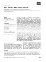
Figure 5, exemplify an exophytic lesion surrounded by an
extensive Barrett's epithelium. Microscopy revealed a well
differentiated type tumor. Immunohistochemistry dem-
onstrated a positive MUC2 expression compatible with an
intestinal type Adenocarcinoma.
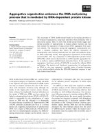
Figure 6, exemplify an ulcerative and depressive lesion
surrounded by an extensive Barrett's epithelium. Micros-
copy revealed an undifferentiated type tumor. Immuno-
histochemistry showed MUC5AC expression denoting a
gastric type Adenocarcinoma.
Table 3 shows the relationship between mucin pattern
predominance in the adjacent epithelium compared to
the mucin tumour expression.
Discussion
The extension of the columnar epithelium in the esopha-
gus is related to the risk of malignant transformation
Table 2: Distribution of 13 ABE patients according to the type of adjacent epithelium and tumor
Characteristics
Patient Cell type (gastric or intestinal) predominance in the specialized
columnar epithelium
Adenocarcinoma
Grade of IHC IHC Type of tumor
differentiation MUC2 MUC5AC according to mucins
1 intestinal well + - Intestinal
2 intestinal moderated - + Gastric
3 similar well - + Gastric
4 intestinal moderated + - Intestinal
5 Gastric poor - + Gastric
6 Gastric undifferentiated - - -
7 Gastric well - + Gastric
8 Intestinal poor + - Intestinal
9 Gastric poor - + Gastric
10 similar well - + Gastric
11 similar undifferentiated - - -
12 Gastric poor - + Gastric
13 Intestinal poor + - Intestinal
IHC = immunohistochemistry
World Journal of Surgical Oncology 2009, 7:27 />Page 5 of 8
(page number not for citation purposes)
[17,18], and there is an increased odds in BE longer than
4 cm [10,19-21]. Some authors describe the adenocarci-
noma in short BE with lower prevalence, since the risk of
malignization area (columnar epithelium) is low [7]. In
this study, adenocarcinoma developed just in long BE
(mean 7.1 cm). This was already observed in our service,
when the mean extension of BE who developed the tumor
was 9.7 cm [21].
The location of ABE was more frequent next to squamous-
columnar transition. Same findings were observed in thir-
teen patients with early adenocarcinoma [20]. This data
suggest that this zone should be specific target during BE
follow up, with multiple endoscopic biopsies.
MUC2 immunoexpression in columnar epithelium adjacent to the AdenocarcinomaFigure 1
MUC2 immunoexpression in columnar epithelium
adjacent to the Adenocarcinoma. Immunohistochemical
staining of MUC2 for columnar epithelium showing goblet
cells as positive control (original magnification × 400)
MUC5AC immunoexpression in columnar epithelium adja-cent to the AdenocarcinomaFigure 2
MUC5AC immunoexpression in columnar epithe-
lium adjacent to the Adenocarcinoma. Immunohisto-
chemical staining of MUC5AC for columnar epithelium
showing glandular structures as positive control (original
magnification × 400)
MUC2 immunoexpression in intestinal type adenocarcinoma arising in Barrett's esophagusFigure 3
MUC2 immunoexpression in intestinal type adeno-
carcinoma arising in Barrett's esophagus. Immunohis-
tochemical staining of MUC2 for adenocarcinoma (original
magnification × 400)
MUC5AC immunoexpression in undifferentiated type adeno-carcinoma (gastric type) arising in Barrett's esophagusFigure 4
MUC5AC immunoexpression in undifferentiated
type adenocarcinoma (gastric type) arising in Bar-
rett's esophagus. Immunohistochemical staining of
MUC5AC for adenocarcinoma (original magnification × 400).
World Journal of Surgical Oncology 2009, 7:27 />Page 6 of 8
(page number not for citation purposes)
Mucins secreted in the esophagus play an important role
in the cytoprotection against reflux of gastric contents
[22]. Barrett's mucosa is characterized by a heterogeneous
mixture of neutral mucins, sialomucins and sulphomu-
cins [23]. Based on this background information, this
study investigated the pattern of expression of MUC2 and
MUC5AC mucin gene protein products using immuno-
histochemistry in patients with adenocarcinoma arising
in BE.
MUC2 and MUC5AC belong to a family of mucin genes
which encode for peptide tandem repeats [22,24]. Mucin
tandem repeats vary in length and sequence, but all char-
acterized to date contain proline, threonine and/or serine
residues which are potential glycosylation sites [25],
which carry the O-linked oligosaccharides characteristic
for these high molecular weight glycoproteins. These
mucins are secreted and form extracelular gels [24].
MUC2 encodes a prototype secretory mucin which is
present in the human intestine, mostly in goblet cells
[26]. The glycopeptide in MUC2 is rich in cysteine resi-
dues with disulphide bonds. This results in polymeriza-
tion and contributes to the intrinsic viscosity and gel-
forming properties required for mucosal surface protec-
tion [27]. MUC2 immunoexpression in Barrett's metapla-
sia was restricted to goblet cells, a pattern specific to
normal rat and human colonic epithelium [28,29], imply-
ing that the mucin in goblet cells of Barrett's metaplasia is
similar if not identical to the native intestinal mucosa.
Several authors have comparable results [22,30]. The pres-
ence of MUC2 in Barrett's metaplasia (goblet cells) is a
feature of cellular differentiation because secretory
mucins are normally produced by highly differentiated
cells [31]. Warson et al, 2002, demonstrated that there is
an association between MUC2 expression and intestinal
metaplasia. Interesting, these authors also found an asso-
ciation between sulphomucin-producing cells and
MUC5AC expression [32].
MUC5AC was extensively immunoexpressed in the
columnar cells, localizing to the surface epithelium and
extending to a variable degree into the glandular struc-
tures in BE, and was more commonly seen than MUC2.
In this investigation BE epithelium showed a mucin pat-
tern similar to human stomach epithelium, in which the
expression of these MUCs has been demonstrated previ-
ously [15,16]. Thus, our finds have been corroborated by
others authors.
A protuding proximal Adenocarcinoma over a long Barrett's EsophagusFigure 5
A protuding proximal Adenocarcinoma over a long
Barrett's Esophagus. Well differentiated adenocarcinoma
arising in a 16 cm lenght Barrett's esophagus. The lesion is
located 14 cm distant from the gastroesophageal junction.
An infiltrative proximal Adenocarcinoma over a long Bar-rett's EsophagusFigure 6
An infiltrative proximal Adenocarcinoma over a long
Barrett's Esophagus. Undifferentiated adenocarcinoma
arising in 10.7 cm lenght Barrett's esophagus, 5.5 cm distant
from gastroesophageal junction.
Table 3: Relationship between Mucin pattern predominance in
the adjacent epithelium compared to the mucin tumor
expression.
Adjacent epithelium
Tumor Intestinal Gastric
Intestinal 40
Gastric 16
P = 0, 01 Fisher Exact Test.
World Journal of Surgical Oncology 2009, 7:27 />Page 7 of 8
(page number not for citation purposes)
The metaplastic epithelium may reflect an adaptative
response to new luminal environment [14]. The esopha-
gus has been shown to increase secretion of mucins from
the submucosal glands in response to stimulation by gas-
tric acid, depending upon the reflux esophagitis [33]. Each
region of the gastrointestinal tract has characteristic func-
tional requirements and the properties of the mucus pro-
duced at each site are adapted to cope with these functions
[34]. Jankowski suggests that incomplete intestinal type
metaplasia may be a response to reflux of gastroduodenal
contents and in particular bile acids [17]. Arul et al. would
support a theory as Barrett's epithelium produces both
MUC5AC and MUC6 associated with protection from gas-
tric acid and MUC2 and MUC3 associated with protection
from bile [14].
Some authors suggested that mucin histochemistry could
be used to establish if a pattern of mucin staining in Bar-
rett's esophagus may be associated with a greater risk of
progression to adenocarcinoma [35]. Three dyes, alciun
blue, high-iron diamine and periodic acid-Schiff reagent
are used to histochemically distinghish the mucins pro-
duced. These dyes are specific for carbohydrates and their
modifications, but do not reveal the underlying molecular
identity of the mucins expressed. Expression of sulpho-
mucin has been associated with an increased malignant
potencial [35,36]. However, Rothery found that 74% of
biopsies of Barrett's esophagus had evidence of sulpho-
mucin and concluded that detection did not help to iden-
tify those at risk of progression to adenocarcinoma [4].
NAKAMURA et al. performed detailed study of gastric
mucosa microcarcinomas, and described the gastric aden-
ocarcinoma histogenesis. They examined stomachs
resected for nonmalignant diseases and identified tumor
less than 2 mm and between 2 and 5 mm. The results con-
firmed that mucocelular adenocarcinoma developed from
own gastric mucosa, and tubular adenocarcinoma, from
atrophic mucosa with IM. After, when he studied tumor
greater than 6 mm, he could observe the same relation of
the tumor with the adjacent columnar epithelium. With
statistical analysis he proved that gastric or undifferenti-
ated adenocarcinoma were related to gastric mucosa (with
pyloric or fundic glands), and the intestinal pattern or dif-
ferentiated adenocarcinoma, with the presence of IM [37].
In this study, the pattern of mucin expression revealed a
specialized type epithelium adjacent to the tumors. There
was an association between the predominance of mucin
expressed in the adjacent epithelium and the pattern of
mucin expression in the tumors, may indicating the route
of carcinogenesis.
This histogenesis description may be utilized in BE, in
order to clarify the presence of gastric mucin type
expressed at seven of the ABE in this investigation. So, an
area with gastric metaplasia within the specialized Bar-
rett's epithelium could originate an expansion clone capa-
ble of initiate the carcinogenesis cascade, developping an
undifferentiated adenocarcinoma, that express MUC5AC.
BE is a columnar epithelium that can be modified as the
gastric mucosa does, and may originate any type of aden-
ocarcinoma.
Conclusion
Currently, histopathologic aspects still remain the best
biologic markers for the BE follow up with the aim of early
ABE diagnosis. The location of the adenocarcinoma next
to the squamous columnar transition point to the most
important zone that should be searched for early adeno-
carcinona during endoscopic examination; and the higher
risk of adenocarcinoma development in long BE, can be
used like a red flag for follow up in this patients. Thus, the
follow up in long (over 3 cm) BE is relevant, and should
be performed in all patients, independently of the type of
columnar epithelium found at the endoscopic biopsy.
Therefore, Barrett's esophagus adenocarcinoma shows
either gastric or intestinal type pattern of mucins expres-
sion. According to the mucins, the two types of tumors
developed in Barrett's esophagus may reflect the original
cell type involved in malignant transformation.
Abbreviations
Dist. Tu-GEJ: Distance from tumors (Adenocarcinoma)
distal margin to the gastroesophageal junction; Prox. Tu-
Tepit: Distance from the turmors (Adenocarcinoma) prox-
imal margin to the epithelium (columnar-squamous)
transition; BE: Barrett's Esophagus; ABE: Adenocarcinoma
developed in Barrett's Esophagus; HE: hematoxyline-
eosine; IM: Intestinal Metaplasia; GEJ: Gastroesophageal
junction.
Competing interests
The authors declare that they have no competing interests.
Authors' contributions
SS participated in the sequence alignment and drafted the
manuscript, design of the study, coordinating data collec-
tion, supervision. IC conceived of the study, and partici-
pated in its design and coordination, department head.
URJ was involved in rewriting, performed the statistical
analysis, carried out the immunoassays. KI, REI and CEPC
were the pathologists and involved in laboratory investi-
gation. AVSR was involved in collecting data, laboratory
investigation, carried out the immunoassays. All authors
read and approved the final manuscript.
References
1. Morales TG, Sampliner RE, Bhattacharyya A: Intestinal metaplasia
of the gastric cardia. Am J Gastroenterol 1997, 92:414-8.
Publish with BioMed Central and every
scientist can read your work free of charge
"BioMed Central will be the most significant development for
disseminating the results of biomedical research in our lifetime."
Sir Paul Nurse, Cancer Research UK
Your research papers will be:
available free of charge to the entire biomedical community
peer reviewed and published immediately upon acceptance
cited in PubMed and archived on PubMed Central
yours — you keep the copyright
Submit your manuscript here:
/>BioMedcentral
World Journal of Surgical Oncology 2009, 7:27 />Page 8 of 8
(page number not for citation purposes)
2. Spechler SJ, Zeroogian JM, Antonioli DA, Wang HH, Goyal RK: Prev-
alence of metaplasia at the gastro-esophageal junction. Lan-
cet 1994, 344:1533-6.
3. Thompson JJ, Zinsser KR, Enterline HT: Barrett's metaplasia and
adenocarcinoma of the esophagus and gastroesophageal
junction. Hum Pathol 1983, 14:42-61.
4. Rothery GA, Patterson JE, Stoddard CJ, Day DW: Histological and
histochemical changes in the columnar lined (Barrett's)
oesophagus. Gut 1986, 27:1062-68.
5. Appelman HD: Is the presence of specialized epithelium nec-
essary for the diagnosis of Barrett's esophagus? In O.E.S.O. –
The esophageal mucosa Edited by: Giuli R, Tytgat GNJ, DeMeester TR,
Galmiche JP. Elsevier Science. Amsterdam; 1994:878-79.
6. Reid BJ: Barrett's esophagus and esophageal adenocarci-
noma. Gastroenterol Clin North Am 1991, 20:817-34.
7. Schnell TG, Sontag SJ, Chejfec G: Adenocarcinomas arising in
tongues or short segments of Barrett's esophagus. Dig Dis Sci
1992, 37:137-43.
8. DeMeester SR, DeMeester TR: Columnar mucosa and intestinal
metaplsia of the esophagus – Fifty years of controversy. Ann
Surg 2000, 231:303-21.
9. Sampliner RE: Practice guidelines on the diagnosis, surveil-
lance, and therapy of Barrett's esophagus. The Practice
Parameters Committee of American College of Gastroen-
terology. Am J Gastroenterol 1998, 93:1028-32.
10. Saubier EC, Gouillat C, Samaniego C, Guillaud M, Moulinier B: Ade-
nocarcinoma in columnar-lined Barrett's esophagus. Analy-
sis of 13 esophagectomies. Am J Surg 1985, 150:365-9.
11. Paraf F, Flejou JF, Potet F, Molas G, Fekete F: Esophageal squa-
mous carcinoma in five patients with Barrett's esophagus.
Am J Gastroenterol 1992, 87:746-50.
12. Paraf F, Fléjou JF, Pignon JP, Fekete F, Potet F: Surgical pathology
of adenocarcinoma arising in Barrett's esophagus. Analysis
of 67 cases.
Am J Surg Pathol 1995, 19:183-91.
13. Audie JP, Janin A, Porchet N, Copin MC, Gosselin B, Aubert JP:
Expression of human mucin genes in respiratory, digestive,
and reproductive tracts ascertained by in situ hybridisation.
J Histochem Cytochem 1993, 41:1479-85.
14. Arul GS, Moorghen M, Myerscough N, Alderson DA, Spicer RD, Cor-
field AP: Mucin gene expression in Barrett's oesophagus: an in
situ hybridisation and immunohistochemical study. Gut 2000,
47:753-61.
15. Reis CA, David L, Correa P, Correa F, de Bolos C, Garcia E, Mandel
U, Clausen H, Sobrinho-Simões M: Intestinal metaplasia of
human stomach displays distinct patterns of mucin (MUC1,
MUC2, MUC5AC, and MUC6) expression. Cancer Research
1999, 59:1003-7.
16. Ho SB, Shekels LL, Toribara NW, Kim YS, Lyftogt C, Cherwitz DL,
Niehans GA: Mucin gene expression in normal, preneopplastic
and neoplastic human gastric epithelium. Cancer Research
1995, 55:2681-90.
17. Jankowski JA, Wright NA, Meltzer SJ, Triadafilopoulos G, Geboes K,
Casson AG, Kerr D, Young LS: Molecular evolution of the meta-
plasia-dysplasia-adenocarcinoma sequence in the esophagus.
Am J Pathol 1999, 154:965-73.
18. Reid BJ, Rubin LE: When is the columnar lined esophagus pre-
malignant? Gastroenterology 1985, 88:A1552.
19. Cameron AJ, Lomboy CT, Pera M, Carpenter HA: Adenocarci-
noma of the esophagogastric junction and Barrett's esopha-
gus. Gastroenterology 1995, 103:1541.
20. Nishimaki T, Holsher AH, Schuler M, Bollschweiler E, Becker K,
Siewet JR: Histopathologic characteristics of early adenocar-
cinoma in Barrett's esophagus. Cancer 1991, 68:1731-6.
21. Szachnowicz S, Cecconello I, Iriya K, Marson AG, Takeda FR, Gama-
Rodrigues JJ: Origin of adenocarcinoma in Barrett's esopha-
gus: p53 and Ki67 expression and histopathologic back-
ground. Clinics 2005, 60(2):103-112.
22. Chinyama CN, Marshall RE, Owen WJ, Mason RC, Kothari D, Wilkin-
son ML, Sanderson JD: Exprerssion of MUC1 and MUC2 mucin
gene products in Barrett's metaplasia, dysplasia and adeno-
carcinoma: an immunopathological study with clinical corre-
lation.
Histopathology 1999, 35:517-24.
23. Lee RG: Mucins in Barrett's esophagus: a histochemical study.
Am J Clin Pathol 1984, 81:500-3.
24. Gum JR: Human mucin glycoproteins: varied structure pre-
dict diverse properties and specific functions. Biochem Soc
Trans 1995, 23:795-9.
25. Gum JR: Mucins genes and the proteins they encode: struc-
ture, diversity, and regulation. Am J Respir Cell Mol Biol 1992,
7:557-64.
26. Gum JR, Byrd JC, Hicks JW, Toribara NW, Lamport DT, Kim YS:
Molecular cloning of humanintestinal mucin cDNAs.
Sequence analysisand evidence for genetic polymorphism. J
Biol Chem 1989, 264:6480-7.
27. Gum JR, Hicks JW, Toribara NW, Siddiki B, Kim YS: Molecular
cloning of human intestinal mucin (MUC2) cDNA. Identifica-
tion of the amino terminus and overall sequence similary to
prepro-von Willebrand factor. J Biol Chem 1994, 269:2440-6.
28. Tytgat KM, Bovelander FJ, Opdam FJ, Einerhand AW, Buller HA,
Dekker J: Biosynthesis of rat MUC2 in colon and its analogy
with human MUC2. Biochem J 1995, 309:221-9.
29. Chang SK, Dohrman AF, Basbaum CB, Ho SB, Tsuda T, Toribara NW,
Gum JR, Kim YS: Localization of mucin (MUC2 and MUC3)
messenger and peptide expression in human normal intes-
tine and colon cancer. Gastroenterology 1994, 107:28-36.
30. Ho SB, Niehans GA, Lyftogt C, Yan PS, Cherwitz DL, Gum ET, Dahiya
R, Kim YS: Heterogeneity of mucin gene expression in normal
and neoplastic tissues. Cancer Research 1993, 53:641-51.
31. Strous GJ, Dekker J: Mucin-type glicoproteins. Crit Rev Biochem
Mol Biol 1992, 27:57-92.
32. Warson C, Bovenkamp JH Van De, Korteland-Van Male AM, Buller
HA, Einerhand AW, Ectors NL, Dekker J: Barrett's esophagus is
characterized by expression of gastric-type mucins
(MUC5AC, MUC6) and TFF peptides (TFF1 and TFF2), but
the risk of carcinoma development may be indicated by the
intestinal-type mucin, MUC2. Hum Pathol 2002, 33:
660-8.
33. Namiot Z, Sarosiek J, Rourk RM, Hetzel DP, McCallum RW: Human
esophageal secretion: mucosal response to luminal acid and
pepsin. Gastroenterology 1994, 106:973-81.
34. Corfield AP, Myerscough N, Gough M, Brockhausen I, Schauer R, Par-
askeva C: Glycosilation patterns of mucins in colonic disease.
Biochem Soc Trans 1995, 23:840-5.
35. Jass JR, Filipe MI: The mucin profiles of normal gastric mucosa,
intestinal metaplasia and its variants and gastric carcinoma.
Histochem J 1981, 13(6):931-9.
36. Duchatelle V, Potet F, Bara J, Ma J, Goldfain D: Mucin immunohis-
tochemistry of the columnar epithelium of the oesophagus
(Barrett's oesophagus). Virchows Archiv A Pathol Anat 1989,
414:359-63.
37. Nakamura K, Sugano H, Takagi K: Carcinoma of the stomach in
the incipient phase: its histogenesis and histological
appearences. Gann 1968, 59(3):251-258.