Báo cáo khoa học: "A pilot study to assess the feasibility of evaluation of markers of response to chemotherapy at one day & 21 days after first cycle of chemotherapy in carcinoma of breast: a prospective non-randomized observational study" potx
Bạn đang xem bản rút gọn của tài liệu. Xem và tải ngay bản đầy đủ của tài liệu tại đây (518.09 KB, 11 trang )
BioMed Central
Page 1 of 11
(page number not for citation purposes)
World Journal of Surgical Oncology
Open Access
Research
A pilot study to assess the feasibility of evaluation of markers of
response to chemotherapy at one day & 21 days after first cycle of
chemotherapy in carcinoma of breast: a prospective
non-randomized observational study
Shekhar Sharma*
1
, KR Hiran
2
, K Pavithran
3
and DK Vijaykumar
1
Address:
1
Department of Surgical Oncology, Amrita Institute of Medical Sciences & Research Center, Amrita Lane, Edapally, Ernakulam – 682026,
Kerala, India,
2
Department of Pathology, Amrita Institute of Medical Sciences & Research Center, Amrita Lane, Edapally, Ernakulam – 682026,
Kerala, India and
3
Department of Medical Oncology, Amrita Institute of Medical Sciences & Research Center, Amrita Lane, Edapally, Ernakulam
– 682026, Kerala, India
Email: Shekhar Sharma* - ; KR Hiran - ; K Pavithran - ;
DK Vijaykumar -
* Corresponding author
Abstract
Background: Interest in translational studies aimed at investigating biologic markers in predicting
response to primary chemotherapy (PCT) in breast cancer has progressively increased. We
conducted a pilot study to evaluate feasibility of evaluating biomarkers of response to PCT at one
& 21 days after first cycle.
Methods: Adult, non-pregnant, non-lactating women with histologically confirmed infiltrating duct
carcinoma underwent serial core biopsies after first cycle of PCT and these were scored for Ki-67,
Bcl-2 and Caspase-3 using immunohistochemistry.
Results: We recruited 30 patients with a mean age of 51 years. We were successful 95.6% times
in performing a core biopsy and of these 84.6% had adequate tissue in the cores harvested. After
a mean of 4 cycles of PCT, 26 patients underwent surgery and good response was noted in 9
patients (30%) using Miller-Payne criteria. There was a trend noted in all markers, which appeared
different in those with good response and poor response. Good responders had significantly higher
Ki-67 and significantly lower Bcl-2 at baseline and a significant decrease in Ki-67 and Caspase-3 at 21
days after the first chemotherapy.
Conclusion: We report a detectable change in biomarkers as early as 24–48 hours after the first
chemotherapy along with a definite trend in change that can possibly be used to predict response
to chemotherapy in an individual patient. The statistical significance and clinical utility of such
changes needs to be evaluated and confirmed in larger trials.
Background
There is increasing interest in the ways and means to pre-
dict the response of an individual patient to primary
chemotherapy (PCT) with an ultimate interest to predict
individual responses to treatment in the minimum time
feasible.
Published: 30 March 2009
World Journal of Surgical Oncology 2009, 7:35 doi:10.1186/1477-7819-7-35
Received: 10 December 2008
Accepted: 30 March 2009
This article is available from: />© 2009 Sharma et al; licensee BioMed Central Ltd.
This is an Open Access article distributed under the terms of the Creative Commons Attribution License ( />),
which permits unrestricted use, distribution, and reproduction in any medium, provided the original work is properly cited.
World Journal of Surgical Oncology 2009, 7:35 />Page 2 of 11
(page number not for citation purposes)
Clinical response has been used as an intermediate, surro-
gate end-point for assessment of the efficacy of PCT in an
individual, although this assessment is far from accurate
[1]. Tools are, therefore, required to better assess the effi-
cacy of chemotherapy regimen.
Ellis et al showed that chemotherapy induced apoptosis in
early breast cancer could be demonstrated soon after the
chemotherapy [2]. In continuation of this, it would
indeed be useful to have a marker of response that can be
evaluated as soon as possible after the first cycle of chem-
otherapy and correlates to the clinical outcome.
We wanted to evaluate if it was feasible to harvest a satis-
factory core biopsy immediately after first cycle and just
prior to second cycle of chemotherapy, when patient is
available in the hospital along with feasibility to evaluate
biomarkers of response to chemotherapy in these biop-
sies. The correlation to response, if proven, would help cli-
nicians to tailor chemotherapy to individual patients and
may provide the opportunity to offer earlier possible alter-
native, non-cross-resistant regimens to those patients not
achieving a response to the initial regimen.
We proposed to explore the change in biomarkers of
response to PCT at one day and 21 days after the first cycle
of PCT in women with breast cancer attending our institu-
tion for care and towards this aim we initiated a pilot
study in our institution, after appropriate scientific and
ethics committee approval, in the patients of breast cancer
undergoing PCT. The primary aims of this pilot study were
to assess the feasibility and reproducibility of performing:
a) Serial core biopsies one day and 21 days after first cycle
of chemotherapy, with emphasis on patient acceptance
and complications of the procedure.
b) Assays of apoptosis (Caspase-3 &Bcl-2) and prolifera-
tion index (Ki-67) in patients of carcinoma of breast on
core biopsy specimens using immunohistochemistry
(IHC).
c) Quantification of extent of change in these biomarkers
of response to chemotherapy one day and 21 days after
first cycle of chemotherapy.
d) Histopathological response grading at final surgical
histopathology using Miller-Payne response assessment
criteria.
Patients and methods
Adult (more than 18 years of age) non-pregnant, non-lac-
tating women with histologically confirmed, previously
untreated infiltrating duct carcinoma (IDC) of breast who
were advised PCT, as per institutional protocol, were eligi-
ble. Patients with inflammatory breast cancer or those
with history of any indigenous form of therapy for breast
cancer were excluded from this study. The study was
approved by the Institute review board.
After an informed written consent, serial core biopsies
were taken before (C0 biopsy), 24–48 hours (C1 biopsy)
and 21 days (C2 biopsy) after first cycle of chemotherapy.
Chemotherapy regimen was at discretion of the treating
medical oncologist. Serial core biopsies were obtained
exclusively for the purpose of this study for determination
of potential predictive surrogate markers of response. A
core biopsy was obtained using Bard Monopty disposable
biopsy instrument (Covington, GA). Three core biopsies
were taken – first before starting chemotherapy (C0), sec-
ond 24–48 h after cycle one (C1), and third 21 days after
cycle one (C2). Biopsy specimens, two cores each time,
were fixed in 10% buffered formalin and embedded in
paraffin and sectioned into 4 μm-thick sections.
Surgery was scheduled after completion of 2–6 cycles of
PCT according to patient's response to chemotherapy and
at discretion of the treating physicians. The study pathol-
ogist carefully evaluated the definitive surgical specimen
for the presence of residual disease and grading of patho-
logical response to chemotherapy was done using Miller-
Payne criteria for assessment of response to chemotherapy
[3]. Miller-Payne response grade 4 & 5 were considered as
good pathological response (GPR) while grades 1 to 3
were considered poor pathological response (PPR). ER,
PR, and HER2/neu were evaluated only on C0 biopsy.
Markers for proliferation (Ki-67), Caspase-3 and Bcl-2 were
evaluated by IHC using appropriate antibodies (Table 1).
Slides were deparaffinized and hydrated. Standard tech-
niques for antigen retrieval, blocking endogenous peroxi-
Table 1: Details of IHC antibodies for Caspase-3, Bcl-2 and Ki-67
Antigen Antibody Manufacturer Scoring
Ki-67 MAb Zymed, San Francisco, CA Nuclear staining; % positive
Bcl-2 MAb DAKO, Carpenteria, CA Cytoplasmic staining, % positive
Caspase-3 Mouse Imgenex, San diego, CA Nuclear and cytoplasm staining, % positive
MAb – Monoclonal antibody
World Journal of Surgical Oncology 2009, 7:35 />Page 3 of 11
(page number not for citation purposes)
dase activity and nonspecific antibody binding were
followed before immuno-staining with commercially
available antibodies (Table 1). Primary antibodies were
pre-diluted except for Caspase-3 for which a dilution of
1:500 was used. Incubation period for all the antibodies
were 1 hour except Ki-67 that was kept for 2 hours at
37°C. Known positive and negative controls were
included for each batch run. Slides were scored for per-
centage of positive cells and relative intensity.
The feasibility of performing serial core biopsies was not
addressed statistically. Non-parametric tests were applied
to assess the other variables. Patient baseline characteris-
tics, the treatment regimen, and molecular markers were
each assessed for an association with pathologic response
using the Mann-Whitney U test. The change in biomarkers
of response from pre-treatment was assessed in the GPR &
PPR groups by paired comparisons, using the Wilcoxon
signed rank test, while within group analysis was per-
formed using Wilcoxon rank sum test.
Results
We recruited 30 patients of breast cancer with a mean age
of 51 years ( ± 8.4) for this study from April 2007 to June
2008.
Patient demographics are mentioned in Table 2. Disease
characteristics are mentioned in Table 3. There were no
significant differences in demographic pattern between
GPR & PPR groups.
ER, PR and Her-2/neu receptors were all positive in four
(13.3%) patients while all three were negative in 10
(33.3%) patients. Of the 10 patients (33.3%) with Stage
IV disease, whole body skeletal scintigraphy detected
metastasis in eight patients (26.7%); chest X-ray in one
patient (3.3%); ultrasound abdomen in four patients
(13.3%) and CT scan chest in two patients (6.7%).
We were successful in harvesting core biopsy tissue with
adequate cellularity in a reasonable proportion of patients
(Table 4). Of the proposed 90 core biopsy procedures
(three each in 30 patients), only four patients (4.44%)
refused the third core biopsy (C2) due to procedure
related pain. We did not observe any other procedure
related complications.
Although paucicellular harvest can be attributed to poor
technique and less number of cores taken, it is interesting
to note that out of the four patients who had a paucicellu-
lar harvest at 21 days after chemotherapy (C2 biopsy),
three had a good response on final histopathology by
Miller-Payne criteria.
Chemotherapy regimens included Adriamycin & Cyclo-
phosphamide followed by Paclitaxel (AC+T) in 12
patients (40%); combination of Docetaxel, Adriamycin
and Cyclophosphamide (TAC) in 10 patients (33.3%); 5-
Flurouracil, Adriamycin (or Epirubicin) and Cyclophos-
phamide (FAC/FEC) in 6 patients (20%) and Docetaxel
alone in 2 patients (6.7%)
Miller-Payne pathological response category could not be
assessed in 4 patients (13.3%) (one expired, one had pro-
gressive disease on chemotherapy, one refused surgery
and one had surgery cancelled due to chemotherapy
induced cardiomyopathy). Details of Miller-Payne patho-
logical response in the remaining 26 patients are shown in
Table 3. Nine patients (30%) had a GPR to chemotherapy
(Table 3).
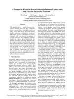
Levels of Ki-67, Bcl-2, and Caspase-3 and their compari-
sons in C0, C1 and C2 biopsy are shown in Figures 1, 2
and 3 respectively.
We observed that GPR group had significantly higher Ki-
67 at baseline (p = 0.042) and both GPR & PPR groups
Table 2: Patient demographics
Characteristic Mean ( ± SD) Range
Age (years) 51 ( ± 8.4) 31–63
Duration of symptoms (months) 10.54 ( ± 10.88) 0.25 – 40
Age at Menarche (years) 14.16 ( ± 1.7) 11–18
Age at Marriage (years) 21.58 ( ± 4.26) 13–33
Age at menopause (years) 47.58 ( ± 3.73) 41–54
Parity (median) 2 0–5
Age at first childbirth 23.81 ( ± 4) 17–32
Duration of breast feeding (months) 43.22 ( ± 22.02) 6–96
Number of PCT cycles (median) 4 3–9
Menstrual status
Premenopausal 19 63.3
Postmenopausal 11 36.7
Laterality
Right 18 60
Left 11 36.7
Bilateral 13.3
World Journal of Surgical Oncology 2009, 7:35 />Page 4 of 11
(page number not for citation purposes)
showed a rise at 24–48 hours after first chemotherapy (in
C1 biopsy). This decreased 21 days after first chemother-
apy to below the baseline values (in GPR group) as well as
C2 values in PPR group, which were static at C1 levels. The
difference between GPR & PPR groups in levels of Ki-67
seen in C2 biopsy was not significant (p = ns), although
the difference in change from C1 to C2 appears striking
with a steep slope in GPR group (Figure 1c).
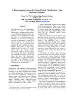
On the converse, Bcl-2 was significantly lower in GPR
group in all the three biopsies (p = 0.015; 0.014; 0.039 for
C0, C1 & C2 respectively). Chemotherapy induced a
steady rise in the entire group, which was steeper in GPR
group from C1 to C2. Bcl-2 peaked at biopsy taken at 24–
48 hours after the first cycle in the PPR group and then
had a plateau to nearly same level at 21 days (Figure 2c).
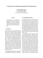
Caspase-3 values peaked at 24–48 hours before falling to
near baseline levels at 21 days after the first chemotherapy
with nearly similar baseline and peak values in both the
groups (p = ns for both C0 & C1 biopsies). The decline in
GPR group for values of Caspase-3 from C1 to C2 biopsy
Table 3: Disease characteristics
Parameter NPercent
Stage of disease at presentation
II B 413.3
III A 723.3
III B 516.7
III C 413.3
IV 10 33.3
Histological type
Infiltrating ductal carcinoma (IDC) 27 90.0
Infiltrating lobular carcinoma (ILC) 26.7
Combined ILC & IDC 13.3
Grade of tumor
Low grade 413.3
Intermediate grade 14 46.7
High grade 12 40.0
Miller-Payne response
#
Poor Grade I (no or <10% response) 5 16.7
pathological Grade II (10–30% response) 4 13.3
response (PPR) Grade III (30–90% response) 8 26.6
Good pathological Grade IV (>90% response) 4 13.3
response (GPR) Grade V (complete response or few isolated tumor cell islands remaining) 5 16.7
#: Miller Payne response could not be assessed in 4 patients who did not undergo surgery
World Journal of Surgical Oncology 2009, 7:35 />Page 5 of 11
(page number not for citation purposes)
was steeper, the difference from PPR group being signifi-
cant (p = 0.024) at this point. In the GPR group, the Cas-
pase-3 values at C2 fell below the baseline values (C0)
(Figure 3c).
In our study, the magnitude of change in Bcl-2 within 24–
48 hours after the first chemotherapy, in the entire group,
was significant (p = 0.04), while that for markers of prolif-
eration (Ki-67) and apoptosis (Caspase-3) was not signifi-
cant (p = ns).
Thus, tumors with a higher Ki-67 at baseline along with a
low Bcl-2 (anti-apoptotic gene) responded better to chem-
otherapy. In other words, high rates of apoptosis and pro-
liferation at baseline were associated with improved
pathological response. Another interesting observation
during this study was that at 21 days, a decrease in Ki-67
and Caspase-3 was predictive of favorable response (p =
0.01 for both).
In this study, ER-positive tumors had a significant associ-
ation with poor response (p = 0.014) and had a higher Bcl-
2 expression at baseline (mean Bcl-2 35.38 in ER-positive
vs. mean Bcl-2 14.35 in ER negative tumors; p = 0.04).
There was no difference in expression of Ki-67 or Caspase-
3 in ER-positive or negative tumors or in expression of
these markers or in response between PR and Her2/neu
positive or negative tumors.
As a word of caution, p values of significance should be
interpreted with caution due to the small sample size. It
was primarily aimed as a pilot study to verify feasibility
and reproducibility of this trial design and to see if
changes in biomarkers could be measured and quantified
at patient-friendly time points, aims that it apparently has
achieved.
We faced problems using Caspase-3 to evaluate the apop-
totic index, as this terminal enzyme of the apoptotic cas-
cade is cytoplasmic in location. This led to a diffuse
staining of slides, which caused difficulty in interpretation
of positive cells and percentage positivity. Additionally,
technical expertise in slide preparation and IHC staining
were other major hurdles in the initial phase of the study.
On the basis of this pilot study, we observe that this trial
design is feasible (in this context, patient acceptable with-
out any specific objective incentive) and quantification of
biomarkers of response to chemotherapy can be per-
formed on these core biopsies. There is a trend towards
change noted in these markers (in this study, Ki-67, Bcl-2,
Caspase-3) both at 24 hours and at 21 days after the first
cycle of chemotherapy, although these results need to be
confirmed in larger studies. In our experience, Miller-
Payne criteria to assess response to chemotherapy, is an
easily reproducible method of grading response objec-
tively.
We hope that we will be able to improve the adequacy of
tissue by increasing the number of cores harvested each
time from two in the present study to three or four in
future studies. A more proactive approach to pain medica-
tion prescription will, hopefully, help us in preventing
dropouts in further trials. However, we would need alter-
Table 4: Feasibility & adequacy of core biopsy procedures
Procedure N%
Core biopsy at baseline (C0 biopsy) 30 100
Core biopsy 24–48 hours after first cycle of chemotherapy (C1 biopsy) 30 100
Core biopsy 21 days after first cycle of chemotherapy (C2 biopsy) 26 86.6
Adequacy
At C0 biopsy 30 100
At C1 biopsy 28 93.3
At C2 biopsy 22 73.3 (84.3% of 26 attempted)
4 (13.3%) refused C2 biopsy; 4 (13.3%) had paucicellular harvest on C2 biopsy
Definitive surgery 26 86.6
4 (13.3%) patients did not undergo surgery due to different reasons.
World Journal of Surgical Oncology 2009, 7:35 />Page 6 of 11
(page number not for citation purposes)
Comparison of Ki-67 levels in (a) C0–C1 biopsy; and (b) C0–C2 biopsy; and (c) Change in mean value over timeFigure 1
Comparison of Ki-67 levels in (a) C0–C1 biopsy; and (b) C0–C2 biopsy; and (c) Change in mean value over
time.
World Journal of Surgical Oncology 2009, 7:35 />Page 7 of 11
(page number not for citation purposes)
Comparison of Bcl-2 levels in (a) C0–C1 biopsy; and (b) C0–C2 biopsy; and (c) Change in mean value over timeFigure 2
Comparison of Bcl-2 levels in (a) C0–C1 biopsy; and (b) C0–C2 biopsy; and (c) Change in mean value over
time.
World Journal of Surgical Oncology 2009, 7:35 />Page 8 of 11
(page number not for citation purposes)
Comparison of Caspase-3 (Csp-3) levels in (a) C0–C1 biopsy; and (b) C0–C2 biopsy; and (c) Change in mean value over timeFigure 3
Comparison of Caspase-3 (Csp-3) levels in (a) C0–C1 biopsy; and (b) C0–C2 biopsy; and (c) Change in mean
value over time.
A
B
C
World Journal of Surgical Oncology 2009, 7:35 />Page 9 of 11
(page number not for citation purposes)
native methods of evaluating apoptotic index (eg TUNEL,
etc) due to problems associated with Caspase-3 in any
future study.
Whether we need to repeat the same design (two biopsies
after baseline – one 24–48 hours and second 21 days after
first chemotherapy) or either one of these biopsies can be
omitted is a matter of debate, although in our opinion a
three point measurement will improve the predictive
power of the larger trial.
Discussion
Pathological complete response to PCT has been corre-
lated with long-term outcome [4,5], although this is seen
in only 3–30% of patients [6]. Bio-molecular predictors of
tumor response to primary CT include S-phase fraction,
ER, PgR, thymidine labeling index, ploidy, p53 and c-
erbB-2 (Her-2/neu) [7-12].
There is preliminary evidence that supports proliferation
& apoptosis-related markers as predictors of long-term
response to PCT [13,14]. These include, among others,
markers for induction of apoptosis, expression of Bcl-2,
and proliferation index (Ki-67 assay) [2,15,16]. However
the exact relationship of the levels of biomarkers in a
tumor in pre and post chemotherapy setting is relatively
under-explored.
Studies have usually evaluated markers for response to
chemotherapy after a significant delay [2]. A time gap of
10 days or more poses a difficult hurdle for investigators
to have the patient come back again for tissue harvesting
alone with most patients being reluctant to do so in
absence of any objective incentive for their extra time,
effort and expenses. This is a more acute issue in the
Indian perspective, where patients often need to travel
great distances to seek medical care.
We chose to evaluate three biomarkers, namely Ki-67
(marker of proliferation), Bcl-2 and Caspase-3 (anti- and
pro-apoptotic markers) as data exist showing a close rela-
tionship between apoptosis and proliferation in untreated
tumours [17,18]. The decision to restrict the number of
biomarkers to three was to keep the study design as simple
as possible in the pilot trial.
Several groups have found that Ki-67 decreases after
chemotherapy over a variable duration [19]. Some studies
have demonstrated a relationship of change in Ki-67 with
response [15,20]. In a similar pilot study where Ki-67 was
measured in 20 patients treated with chemo-endocrine
therapy (mitoxantrone, mitomycin C, methotrexate and
tamoxifen), a decrease at day 10 or 21 after the first course
of treatment correlated with response at 3 months (p =
0.008). Ki-67 changes between the responders and non-
responders were significant for both absolute and percent-
age change in the chemotherapy (p = 0.01 and p = 0.005,
respectively) as well as in chemo-endocrine therapy group
(p = 0.03 and p = 0.06, respectively) [21]. Further follow
up showed that this decrease in Ki-67 after 10–21 days of
therapy had a significant association with good clinical
response on univariate analysis [15]. While significant
associations with response have been revealed in these
studies, none have assessed the predictive power in indi-
vidual patients.
Whilst some studies have shown that a high proliferative
index is a poor prognostic indicator [22,23], others have
debated this with observations that patients with highly
proliferative tumours respond well to chemotherapy [24].
Honkoop et al showed that a high proliferative index in
residual tumours after neoadjuvant chemotherapy and
endocrine therapy was associated with a decreased disease
free survival [25].
In this study, we noted that a higher baseline Ki-67 was
associated with better response to chemotherapy, proba-
bly because a higher fraction of these proliferative tumors
at initiation of chemotherapy were susceptible to chemo-
toxic effects. The low 21-day Ki-67 values, in good
responders, similar to those reported in literature, are
indirect evidence of the efficacy of the chemotherapy in
these patients in eliminating the mitotic fraction. It is
intriguing to note that, as soon as 24 hours after chemo-
therapy, there was a rise in Ki-67 levels, something that, to
our knowledge, has not been reported in literature.
Bcl-2 gene encodes for a 26-kDa protein that mainly
inhibits apoptosis. However, the role of Bcl-2 expression
on clinical outcome following chemotherapy is still under
investigation, since available data are in some instances
contrasting [26]. Also, interpretation of treatment benefit
as a function of biomarkers is difficult in the absence of
randomized, controlled trials.
A number of studies, covering about 5000 patients, with
breast cancer at different stages showed that Bcl-2 over-
expression correlated to a differentiated phenotype and a
favorable prognosis in patients subjected to local-
regional, hormonal or cytotoxic therapies [14,27].
Our data suggests that breast carcinomas with low base-
line apoptosis may respond poorly to chemotherapy. We
observed a significant inverse correlation between expres-
sion of Bcl-2 and response to the chemotherapy. These
results are in general line with the postulated anti-apop-
totic function of Bcl-2 gene, higher levels in poor respond-
ers indicating a possible immunity from chemotherapy
induced apoptosis.
World Journal of Surgical Oncology 2009, 7:35 />Page 10 of 11
(page number not for citation purposes)
Some possible explanations for these paradoxical results
have been mentioned in literature and include a complex
interaction of p53 or its mutant variations with Bcl-2, an
inhibitory effect of Bcl-2 on proliferation along with regu-
lation of Bcl-2 expression by estrogen and presence of
antagonists, which may negate its anti-apoptotic function
[13,28].
The prognostic and predictive value of apoptotic markers
in breast cancer is not yet fully understood. There is some
suggestion that apoptotic index is an independent prog-
nostic factor. Our results are similar to other reports in the
literature that chemotherapy induces early changes in
apoptosis [2].
Data from this study and another similar study [29] sug-
gest that it may be possible, in future, to determine, as
early as 24–48 h after administration of chemotherapy,
whether a woman is likely to respond to a specific agent
or not, information that might help to make an early deci-
sion regarding any change in such treatment. The novel
approach in this study can also answer questions regard-
ing the role of other markers and response to individual
therapies.
This study does have a few limitations like small sample
size (30 patients were recruited as this was planned as a
pilot study only), heterogeneous patient population (no
stratification on the basis of receptor status, chemothera-
peutic regimen received or stage of disease) all of which in
themselves can argue for a different disease biology and
consequently difference in responses to chemotherapy.
However, even with these limitations, results are impres-
sive enough to favor larger, more rigorously controlled tri-
als to confirm these.
Conclusion
In summary, we present a clinical design incorporating
sequential core biopsy after first cycle of PCT in breast can-
cer that can be used as a model in future trials to correlate
surrogate end point biomarkers with response. The model
can also be used to incorporate novel agents with standard
treatments. Changes in biomarkers like apoptosis and
proliferation can then, if validated with larger trials using
standard regimens, be used to determine the efficacy and/
or superiority of the novel combinations compared to
standard treatments.
Whether or not trends observed in this study are signifi-
cant and whether these can be used to tailor chemother-
apy (our ultimate aim) awaits larger trials. Further studies,
including a larger sample size receiving single standard-
ized chemotherapy regimen, are warranted, especially in a
prospective manner with uniform methods of measure-
ment and cut-off points to assess the potential value of
molecular markers in clinical practice. These studies will
need to include multiple assays such as nuclear grade, lev-
els of expression of p53, markers for cell proliferation,
multi-drug resistance, and apoptosis [30].
Competing interests
The authors declare that they have no competing interests.
Authors' contributions
SS was instrumental in design the concept, patient recruit-
ment, data analysis, manuscript preparation and editing.
HKR was instrumental in designing the trial, evaluation of
slides for data generation, manuscript preparation and
editing. PK, and DKV were instrumental in ratifying study
design, patient recruitment, literature search and manu-
script editing & final approval. All authors accept the
responsibility of contents of this manuscript.
Acknowledgements
We wish to acknowledge the funding support for this study from Kerala
State Council for Science, Technology & Environment, Government of Ker-
ala, India. Authors declare that the funding agency was not involved in the
trial at any stage starting from concept to analysis and its role was limited
to providing grant to conduct the research work. Additionally, we also wish
to acknowledge the support of Ms Smitha, Lecturer, Dept of Biostatistics,
AIMS, Cochin in analysis of the data.
References
1. Abu-Farsakh H, Sneige N, Atkinson EN, Hortobagyi G: Pathologic
Predictors of Tumor Response to Pre-operative Chemo-
therapy in Locally Advanced Breast Cancer. Breast 1995,
1:96-101.
2. Ellis PA, Smith IE, McCarthy K, Detre S, Salter J, Dowsett M: Preop-
erative chemotherapy induces apoptosis in early breast can-
cer. Lancet 1997, 349:849.
3. Ogston KN, Miller ID, Payne S, Hutcheon AW, Sarkar TK, Smith I,
Schofield A, Heys SD: A new histological grading system to
assess response of breast cancers to primary chemotherapy:
Prognostic significance and survival. Breast 2003, 12:320-327.
4. Kuerer HM, Newman LA, Smith TL, Ames FC, Hunt KK, Dhingra K,
Theriault RL, Singh G, Binkley SM, Sneige N, Buchholz TA, Ross MI,
Mcneese MD, Buzdar AU, Hortobagyi GN, Singletary SE: Clinical
course of breast cancer patients with complete pathologic
primary tumor and axillary lymph node response to doxoru-
bicin-based neoadjuvant chemotherapy. J Clin Oncol 1999,
17:460-469.
5. Jones RL, Smith ID: Neoadjuvant treatment for early-stage
breast cancer: opportunities to assess tumor response. Lan-
cet Oncology 2006, 7:869-74.
6. Wolmark N, Wang J, Mamounas E, Bryant J, Fisher B: Preoperative
chemotherapy in patients with operable breast cancer: nine-
year results from national surgical adjuvant breast and
bowel project B-18. J Natl Cancer Inst Monogr 2001:96-102.
7. Valero V, Buzdar AU, Hortobagyi GN: Locally advanced Breast
Cancer. Oncologist 1996, 1:8-17.
8. Resnick JM, Sneige N, Kemp BL, Sahin A, Ordonez NG, Frye DK,
Hortobagyi GN: p53 and c-erbB-2 expression and response to
preoperative chemotherapy in locally advanced breast carci-
noma. Breast Disease 1995, 8b:149-158.
9. Wood WC, Budman DR, Korzun AH, Cooper MR, Younger J, Hart
JD, Moore A, Ellerton JA, Norton L, Ferree CR, Ballow AC, Hender-
son IC: Dose and dose intensity of adjuvant chemotherapy for
stage II, node-positive breast carcinoma. N Engl J Med 1994,
330(18):1253-1259.
10. Hortobagyi GN, Buzdar AU: Locally advanced breast cancer: a
review including the M.D. Anderson experience. In High-Risk
Publish with BioMed Central and every
scientist can read your work free of charge
"BioMed Central will be the most significant development for
disseminating the results of biomedical researc h in our lifetime."
Sir Paul Nurse, Cancer Research UK
Your research papers will be:
available free of charge to the entire biomedical community
peer reviewed and published immediately upon acceptance
cited in PubMed and archived on PubMed Central
yours — you keep the copyright
Submit your manuscript here:
/>BioMedcentral
World Journal of Surgical Oncology 2009, 7:35 />Page 11 of 11
(page number not for citation purposes)
Breast Cancer Therapy Edited by: Ragaz J, Ariel IM. Berlin: Springer-
Verlag; 1991:382-415.
11. McCready DR, Hortobagyi GN, Kau SW, Smith TL, Buzdar AU, Balch
CM: The prognostic significance of lymph node metastases
after preoperative chemotherapy for locally advanced
breast cancer. Arch Surg 1989, 124(1):21-25.
12. Valagussa P, Zambetti M, Bonadonna G, Zucali R, Mezzanotte G,
Veronesi U: Prognostic factors in locally advanced non-inflam-
matory breast cancer: long-term results following primary
chemotherapy. Breast Cancer Res Treat 1990, 15(3):137-147.
13. Krajewski S, Blomvqvist C, Franssila K, Krajewska M, Wasenius V-M,
Niskanen E, Reed JC: Reduced expression of pro-apoptotic
gene Bax is associated with poor response rates to combina-
tion chemotherapy and shorter survival in women with met-
astatic breast adenocarcinoma. Cancer Research 1995,
55:4471-4478.
14. Daidone MG, Luisi A, Veneroni S, Benini E, Silvestrini R: Clinical
studies of Bcl-2 and treatment benefit in breast cancer
patients. Endocrine-Related Cancer 1999, 6:61-68.
15. Chang J, Powles TJ, Allred DC, Ashley SE, Clark GM, Makris A, Ass-
ersohn L, Gregory RK, Osborne CK, Dowsett : Biologic markers
as predictors of clinical outcome from systemic therapy for
primary operable breast cancer. J Clin Oncol 1999,
17:3058-3063.
16. Dowsett M, Archer C, Assersohn L, Gregory RK, Ellis PA, Salter J,
Chang J, Mainwaring P, Boeddinghaus I, Johnston SR, Powles TJ, Smith
IE: Clinical studies of apoptosis and proliferation in breast
cancer. Endocr Relat Cancer 1999, 6:25-28.
17. Ellis PA, Smith IE, Detre S, Burton SA, Salter J, A'Hern R, Walsh G,
Johnston SRD, Dowsett M: Reduced apoptosis and proliferation
and increased Bcl-2 in residual breast cancer following pre-
operative chemotherapy. Breast Cancer Res Treat 1998,
48:107-116.
18. Lipponen P, Aaltomaa S, Kosma VM, Syrjanen K: Apoptosis in
breast cancer as related to histopathological characteristics
and prognosis. Eur J Cancer 1994, 30A:2068-2073.
19. Makris A, Powles TJ, Dowsett M, Osborne CK, Trott PA, Fernando
IN, Ashley SE, Ormerod MG, Titley JC, Gregory RK, Allred DC: Pre-
diction of response to neoadjuvant chemoendocrine therapy
in primary breast carcinomas.
Clin Cancer Res 1997, 3:593-600.
20. Makris A, Powles TJ, Allred DC, Ashley S, Ormerod MG, Titley JC,
Dowsett M: Changes in hormone receptors and proliferation
markers in tamoxifen treated breast cancer patients and the
relationship with response. Breast Cancer Res Treat 1998,
48:11-20.
21. Makris A, Powles TJ, Allred DC, Ashley SE, Trott PA, Ormerod MG,
Titley JC, Dowsett M: Quantitative changes in cytological
molecular markers during primary medical treatment of
breast cancer: a pilot study. Breast Cancer Res Treat 1999,
53:51-59.
22. Assersohn L, Salter J, Powles TJ, A'hern R, Makris A, Gregory RK,
Chang J, Dowsett M: Studies of the potential utility of Ki67 as a
predictive molecular marker of clinical response in primary
breast cancer. Breast Cancer Res Treat 2003, 82:113-123.
23. Railo M, Lundin J, Haglund C, von Smitten K, von Boguslawsky K,
Nordling S: Ki67, p53, Er-receptors, ploidy and S-phase as
prognostic factors in T1 node negative breast cancer. Acta
Oncol 1997, 36:369-374.
24. Tewari M, Krishnamuthy M, Shukla HS: Predictive markers of
response to neoadjuvant chemotherapy in breast cancer.
Surg Oncol 2008, 17:301-11.
25. Honkoop AH, Van Diest PJ, De Jong JS, Linn SC, Giaccone G, Hoek-
man K, Wagstaff J, Pinedo HM: Prognostic role of clinical, patho-
logical and biological characteristics in patients with locally
advanced breast cancer. Br J Cancer 1998, 77:621-626.
26. Kariya S, Ogawa Y, Nishioka A, Moriki T, Ohnishi T, Ito S, Murata Y,
Yoshida S: Relationship between hormonal receptors, HER-2,
p53 protein, Bcl-2, and MIB-1 status and the antitumor
effects of neoadjuvant anthracycline-based chemotherapy in
invasive breast cancer patients. Radiation Medicine 2005,
23:189-94.
27. Krajewski S, Krajewska M, Turner BC, Pratt C, Howard B, Zapata JM,
Frenkel V, Robertson S, Ionov Y, Yamamoto H, Perucho M, Takayama
S, Reed JC: Prognostic significance of apoptosis regulators in
breast cancer. Endocrine-Related Cancer 1999, 6:29-40.
28. Reed JC: Balancing cell life and death: Bax, apoptosis, and
breast cancer. J Clinic Inves 1996,
97:2403-2404.
29. Stearns V, Singh B, Tsangaris T, Crawford JG, Novielli A, Ellis MJ,
Isaacs C, Pennanen M, Tibery , Farhad A, Slack R, Hayes DF: A Pro-
spective Randomized Pilot Study to Evaluate Predictors of
Response in Serial Core Biopsies to Single Agent Neoadju-
vant Doxorubicin or Paclitaxel for Patients with Locally
Advanced Breast Cancer. Clinic Cancer Research 2003, 9:124-133.
30. Wolff AC, Davidson NE: Preoperative Therapy in Breast Can-
cer: Lessons from the Treatment of Locally Advanced Dis-
ease. The Oncologist 2002, 7:239-245.