Báo cáo khoa học: "Diffuse anorectal melanoma; review of the current diagnostic and treatment aspects based on a case report" doc
Bạn đang xem bản rút gọn của tài liệu. Xem và tải ngay bản đầy đủ của tài liệu tại đây (544.77 KB, 4 trang )
BioMed Central
Page 1 of 4
(page number not for citation purposes)
World Journal of Surgical Oncology
Open Access
Case report
Diffuse anorectal melanoma; review of the current diagnostic and
treatment aspects based on a case report
Christos N Stoidis
1
, Basileios G Spyropoulos
1
, Evangelos P Misiakos*
1
,
Christos K Fountzilas
2
, Panorea P Paraskeva
3
and Constantine I Fotiadis
1
Address:
1
3rd Department of Surgery, University of Athens Medical School, Attikon University Hospital, Athens, Greece,
2
Department of Internal
Medicine, Athens Navy Hospital, Athens, Greece and
3
2nd Department of Propedeutic Surgery, University of Athens Medical School, Laikon
General Hospital, Athens, Greece
Email: Christos N Stoidis - ; Basileios G Spyropoulos - ;
Evangelos P Misiakos* - ; Christos K Fountzilas - ;
Panorea P Paraskeva - ; Constantine I Fotiadis -
* Corresponding author
Abstract
Primary anorectal melanoma is a rare and aggressive disease. Patients commonly complain for
changes in bowel habits and rectal bleeding, and proctoscopically they mostly appear as non
pigmented or lightly pigmented polypoid lesions. Such a lesion should always raise a high index of
suspicion in any gastroenterologist or surgeon to prompt surgery, since early radical excision is the
only treatment option.
Herein, we report a case of a 57-year-old man with a diffuse anal canal melanoma and give reference
to the current diagnostic and treatment options.
Introduction
Malignant melanoma of the anal canal accounts for 1%–
3% of all anal canal tumors, yet it is an uncommon site of
primary melanoma with almost equal male to female
ratio and with an average age of presentation between the
fifth and the sixth decades of life [1]. It is documented that
it is the third most common site following the skin and
the eye, while the majority of patients in the world litera-
ture are Caucasian [2]. The lesions can be located in the
anal canal, rectum or both with the majority of them aris-
ing from the dentate line of the anal canal. They tend to
spread submucosally, and by the time they cause symp-
toms the extent of invasion is usually beyond surgical
cure.
The etiology of the disease is unknown. A history of sun
exposure is not likely to have had an impact on their
development, while recent epidemiologic data indicate a
bimodal age distribution [3]. Currently, there is little
information whether an infection with the human papil-
loma virus plays a role in the tumorigenesis of anorectal
melanoma.
Prompt surgery seems to be the only treatment option
since current chemotherapy and radiotherapy alone have
been proved ineffective. The development of novel thera-
pies to treat malignant melanoma will hopefully improve
the clinical outcome.
Case report
A 57-year-old male patient was admitted to our depart-
ment with a chief complaint of intermittent rectal bleed-
ing and constipation that had started 6 months ago. All
physical findings were undiagnostic except digital exami-
Published: 11 August 2009
World Journal of Surgical Oncology 2009, 7:64 doi:10.1186/1477-7819-7-64
Received: 20 April 2009
Accepted: 11 August 2009
This article is available from: />© 2009 Stoidis et al; licensee BioMed Central Ltd.
This is an Open Access article distributed under the terms of the Creative Commons Attribution License ( />),
which permits unrestricted use, distribution, and reproduction in any medium, provided the original work is properly cited.
World Journal of Surgical Oncology 2009, 7:64 />Page 2 of 4
(page number not for citation purposes)
nation which revealed a wide-based, fixed, ulcerative mass
2,8 cm above the anal sphincter, just behind the anal
verge, without evidence of invasion in the sphincter,
growth outside the rectum or enlarged lymph nodes. Lab-
oratory tests disclosed red blood cell count: 3,8 × 10⁵/
mm
3
, hemoglobin: 8,3 g/dl, and hematocrit: 27,4%. Proc-
tosigmoidoscopy confirmed digital findings and included
the anorectal melanoma in our differential diagnosis.
Nevertheless, despite the characteristic nature of the
tumor multiple biopsies were taken in order to establish
the diagnosis. The serum level of 5-S-cysteinyldopa (5-S-
CD) was elevated at 60 nmol/l (normal range, 1.5–8.0
nmol/l).
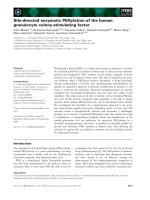
Histopathologically, the tumor consisted of spindle-
shaped cells, resembling fibrosarcoma cells, with melanin
pigment. The tumor cells had invaded the muscularis pro-
pria of the rectum, and lymphatic invasion was noted.
Three perirectal nodes contained histologically evident
metastasis. Thus, the pathologic stage was III Á according
to the AJCC TNM classification (AJCC Cancer Staging
Manual, 6th edition-2002). After surgery, the serum 5-S-
CD level decreased to 5.8 nmol/l.
During the immunohistochemical control the S-100 pro-
tein stain was strongly positive, the HMB-45 stain posi-
tive, the N-Cam stain focally positive in few isolated cells
and negative were the stains for Mart-1 Tyrosynase, Leu7,
Chromogranin, Synaptorysin, CD117, CD34, Actin,
Desmin, EMA, HMWK, LMWK, Ker 8–18, Ker 20 and LCA.
The differential diagnosis included neuroendocrine neo-
plasm, gastrointestinal stromal tumor (GIST), paragangg-
lioma and neoplasm of melanocytic origin. The positive
expression of the HMB-45 and S-100 protein antibodies
set the diagnosis of the malignant neoplasm of melano-
cytic origin (malignant melanoma) as the most prevalent.
CT scans proved no metastatic disease and serum levels of
tumor markers including carcinoembryonic antigen and
Ca 19-9 were within normal ranges. The diagnosis of anal
canal melanoma was based on the proctoscopic biopsies.
An endoluminal ultrasound proved a 10 mm tumor thick-
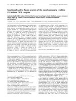
ness. A typical abdominoperineal resection (APR) was
performed with extent resection of the bilateral iguinal,
pelvic sidewall and mesorectal lymph nodes (Figure 1, 2).
The primary treatment was completed with adjuvant
region radiation therapy. The patient recovered unevent-
fully. Six months later during the first follow up examina-
tion, multiple metastatic lesions were recognized. The
relapse site was the liver. The patient submitted to chem-
otherapy and passed away 20 months later from dissemi-
nated disease.
Several questions arose after this aggressive recrudescence
of the disease. Is really anorectal melanoma an incurable
disease and which should be the best surgical treatment
for it? Finally, in what way do the prognostic and clinico-
pathological factors influence the outcome of this disease?
Discussion
As with other anal canal tumors there is often a delay in
diagnosis that results in advanced stage disease at the time
of presentation. Because anorectal melanomas are rare,
staging of the disease has previously been limited to local,
regional, and distant disease [4]. Most patients with such
melanomas complain for bleeding, pain, or an anal mass.
Digital examination provides information concerning
size, fixation and ulceration of the tumor and proctosig-
moidoscopy may be suggestive of anorectal melanoma
The excised specimen of our caseFigure 1
The excised specimen of our case.
Microscopic view of the permanent sectionFigure 2
Microscopic view of the permanent section. Spindle-
shaped cells, resembling fibrosarcoma cells, with
melanin pigment.
World Journal of Surgical Oncology 2009, 7:64 />Page 3 of 4
(page number not for citation purposes)
when pigmentation is obvious. Anal canal melanomas
present no specific clinical manifestations and due to their
polypoid nature they are often misdiagnosed as a throm-
bosed hemorrhoid [5]. Obviously, the inexperienced
endoscopist should always have this rare pathology in
mind in order to avoid misdiagnosing this lesion as a
thrombosed hemorrhoid.
Endoluminal ultrasound is an established mode of evalu-
ation of the tumor thickness and its' nodal status, but the
diagnosis must always be based on the permanent sec-
tions due tendency of amelanotic types to masquerade as
lymphoma, sarcoma or undifferentiated carcinoma [6].
Immunohistochemistry may also be helpful in the diag-
nosis of anorectal melanomas; S 100 protein, HMB-45,
Melan A, and MiTF (microphthalmia-transcription-fac-
tor) are useful immunohistochemical markers.
Metastases occur via lymphatic and hematogenous routes
and it has been reported that 38% of patients have already
metastatic disease at the time of diagnosis [7]. Lymphatic
spread to mesenteric nodes is more common than to
inguinal nodes while lungs, liver and bones are the most
frequent sites of distant metastases.
In the absence of any metastasis surgical therapy is indi-
cated. Most series report no difference in survival in
patients treated by wide local excision or abdominoperi-
neal resection (APR) although the latter has proved more
effective to control the local disease but, without clear
improvement in survival [8,9]. This is caused by the fact
that most recurrences occur systemically regardless of the
initial surgical procedure. However, a recent study sug-
gests that sphincter-sparing local excision and adjuvant
radiation is well tolerated and can effectively control
local-regional disease while avoiding the functional mor-
bidity of APR [10]. The benefits of LE are clear and include
quicker recovery from a less invasive procedure, minimal
impact on bowel function, and no need for a stoma. Pro-
phylactic lymph node resection is of no value whereas
therapeutic lymph node dissection is indicated only in the
presence of positive inguinal nodes.
Introducing sentinel lymph node mapping (SLNM) using
different radioactive tracers and endoscopic ultrasonogra-
phy in recent years has influenced the extent of surgical
resection. Few case reports on the use of SLNM in anal
melanoma have been reported [11]. Although SLNM and
biopsy in anal melanoma has not yet become a standard
of care, it is technically feasible as reported in these case
reports. SLNM seems to be helpful in preventing under-
staging patients who are pathologically node-positive but
clinically node-negative. Long-term follow-up of the
impact of the possible finding of micrometastases is
needed.
In rare cases of anorectal melanomas which tend to block
the anal orifice, palliative surgical treatment is indicated
[12]. In severely ill patients unable to tolerate any surgical
procedure, intramural injections of natural interferon-
beta and systemic administration of dacarbazine has been
proposed with good results [13]. No systemic therapy reg-
imen for metastatic anal melanoma is considered stand-
ard of care. Treatment is based on drugs developed for
advanced cutaneous melanoma and includes cisplatin,
vinblastine, dacarbazine, INF, and interleukin-2,
although given the clinical, biologic, and molecular differ-
ences, mucosal and cutaneous melanomas may be dis-
tinct disease entities [14]. After temozolomide has shown
efficacy comparable to dacarbazine in a randomized trial
of cutaneous melanoma, a combination of temozolo-
mide, cisplatin, and liposomal doxorubicin in one patient
with metastatic anal melanoma was used with encourag-
ing results [15].
The presence of perineural invasion (PNI) is an important
prognostic factor and should be considered in future clin-
ical trials [16]. Notably, tumor thickness seems to be a
strong predictive factor for the risk of local recurrences.
Anorectal melanoma seems to be similar to cutaneous
melanoma, for which tumor thickness is used to plan
therapeutic procedures. Hence, for anorectal melanoma
tumor thickness may also be used as a guideline, i.e. in
early disease with a tumor thickness below 1 mm a local
sphincter-saving excision with a 1-cm safety margin and
in cases of tumor thickness between 1 and 4 mm a local
sphincter-saving excision with a safety margin of 2 cm
seems to be adequate [17]. Tumors with thickness above
4 mm should be treated with APR to avoid local compli-
cations; even so, there will be a stoma and the risk of uri-
nary and sexual dysfunction [18].
Nevertheless, despite sporadic promising reports, regard-
less of surgical approach, anorectal melanoma remains a
highly lethal malignancy with overall 5-years survival rate
less than 20% according to all reported series [19].
Regarding our case, there has been a long debate regarding
the extent of resection, which was necessary to optimally
treat the melanoma. APR procedure, based on the clinico-
pathological features of the tumor was preferred, although
the benefits of LE are clear. The need for regional lym-
phadenectomy has been at the center of the debate. Dur-
ing APR, mesorectal lymph nodes are resected en bloc
with the primary tumor. Although the patient's outcome
was poor, we were able to identify risk factors associated
with survival and prognosis. Clinical symptoms, PNI,
tumor thickness and diameter, spindle cell histology,
mural involvement and necrosis may ultimately impact
outcome. Finally, we hypothesized that systemic dissemi-
nation is an early event in tumorigenesis and by the time
World Journal of Surgical Oncology 2009, 7:64 />Page 4 of 4
(page number not for citation purposes)
the lesion is clinically apparent, micrometastases are well
established.
Conclusion
A standard approach to managing this aggressive tumor
has not been established because of the limited number of
patients and the retrospective design of all anal melanoma
reports. Advanced anorectal melanoma most likely repre-
sents a systemic disease at time of diagnosis. Therefore,
therapy of the primary tumor has no influence on the sys-
temic course of the disease. This is not necessarily true for
early disease.
For anal melanomas, as for any melanoma, the biological
control of the disease is crucial. It is highly desirable that
the new modality of medical, biological or immunologi-
cal therapies will improve the final outcome of these
patients. So far, for this rare tumor, a surgical procedure
which can achieve a complete local excision with the high-
est respect of the functional aspects and quality of life
remains the best therapeutic approach to be applied.
Hence, therapeutic strategies should be adjusted to the
prognosis of the disease. Unfortunately, prognostic
parameters for anorectal melanoma remain to be defined.
Only a few studies addressed this pertinent question.
The aim of this report is to emphasize that early diagnosis
is the key to improved survival rate for patients with these
unusual variants of melanoma.
Consent
Written informed consent was obtained from the patient
for publication of this case report and accompanying
images. A copy of the written consent is available for
review by the Editor-in-Chief of this journal.
Competing interests
The authors declare that they have no competing interests.
Authors' contributions
CIF was the patient's surgeons and has been involved in
drafting the manuscript and revising it critically for impor-
tant intellectual content. EPM, CNS, BGS, PPP and CKF
have made contributions to conception and design. CNS
contributed to the analysis and interpretation of data. All
authors read and approved the final manuscript. All
authors contributed equally to the final draft of the man-
uscript. CIF has given the final approval of the version to
be published.
Acknowledgements
The authors state that there was no extra-institutional funding. Ilias A.
Kouerinis was a major contributor in composing the manuscript.
References
1. Klas JV, Rothenberger DA, Wong WD, Madoff RD: Malignant
tumors of the anal canal: the spectrum of disease, treat-
ment, and outcomes. Cancer 1999, 85:1686-1693.
2. Chang AE, Karnell LH, Menck HR: The National Cancer Data
Base report on cutaneous and noncutaneous melanoma: a
summary of 84,836 cases from the past decade. The Ameri-
can College of Surgeons Commission on Cancer and the
American Cancer Society. Cancer 1998, 83:1664-1678.
3. Helmke BM, Otto HF: Anorectal melanoma. A rare and highly
malignant tumor entity of the anal canal. Pathologe 2004,
25:171-7.
4. Ishizone S, Koide N, Karasawa F, Akita N, Muranaka F, Uhara H, Miy-
agawa S: Surgical treatment for anorectal malignant
melanoma: report of five cases and review of 79 Japanese
cases. Int J Colorectal Dis 2008, 23:1257-1262.
5. Winburn GB: Anal carcinoma or "just hemorrhoids"? Am Surg
2001, 67:1048-58.
6. Hillenbrand A, Barth TFE, Henne-Bruns D, Formentini A: Anorectal
amelanotic melanoma. Colorectal Disease 2007, 10:612-615.
7. Podnos YD, Tsai NC, Smith D, Joshua DI: Factors affecting sur-
vival in patients with anal melanoma. Am Surgeon 2006,
72:917-920.
8. Yeh JJ, Shia J, Hwu WJ, Busam KJ, Paty PB, Guillem JG, Coit DG,
Wong WD, Weiser MR: The role of abdominoperineal resec-
tion as surgical therapy for anorectal melanoma. Ann Surg
2006, 244:1012-7.
9. Yap LB, Neary P: A comparison of wide local excision with
abdominoperineal resection in anorectal melanoma.
Melanoma Res 2004, 14:147-150.
10. Ballo MT, Gershenwald JE, Zagars GK, Lee JE, Mansfield PF, Strom EA,
Bedikian AY, Kim KB, Papadopoulos NE, Prieto VG, Ross MI: Sphinc-
ter-sparing local excision and adjuvant radiation for anal-rec-
tal melanoma. J Clin Oncol 2002, 20:4555-8.
11. Olsha O, Mintz A, Gimon Z, Gold Deutch R, Rabin I, Halevy A, Reiss-
man P: Anal melanoma in the era of sentinel lymph node
mapping: a diagnostic and therapeutic challenge. Tech Colo-
proctol 2005, 9:60-2.
12. Moozar KL, Wong CS, Couture J: Anorectal malignant
melanoma: treatment with surgery or radiation therapy, or
both. Can J Surg 2003, 46:345-9.
13. Ulmer A, Metzger S, Fierlbeck G: Successful palliation of stenos-
ing anorectal melanoma by intratumoral injections with nat-
ural interferon-beta. Melanoma Res 2002, 12:395-8.
14. Yeh JJ, Weiser MR, Shia J, Hwu WJ: Response of stage IV anal
mucosal melanoma to chemotherapy. Lancet Oncol 2005,
6:438-9.
15. Kim KB, Sanguino AM, Hodges C, Papadopoulos NE, Eton O, Cama-
cho LH, Broemeling LD, Johnson MM, Ballo MT, Ross MI, et al.: Bio-
chemotherapy in patients with metastatic anorectal
mucosal melanoma. Cancer 2004, 100:1478-1483.
16. Ueno H, Hase K, Mochizuki H: Criteria for extramural
perineural invasion as a prognostic factor in rectal cancer. Br
J Surg 2001, 88:994-1000.
17. Weyandt GH, Eggert AO, Houf M, Raulf F, Bröcker EB, Becker JC:
Anorectal melanoma: surgical management guidelines
according to tumour thickness. Br J Cancer 2003, 89:2019-2022.
18. Guren MG, Eriksen MT, Wiig JN, Carlsen E, Nesbakken A, Sigurdsson
HK, Wibe A, Tveit KM: Quality of life and functional outcome
following anterior or abdominoperineal resection for rectal
cancer. Eur J Surg Oncol 2005, 31:735-42.
19. Belli F, Gallino G, Tragni G, Andreola S, Leo E: Surgical and path-
ological prognostic factors in anorectal melanoma: The
Experience of the National Cancer Institute of Milano. Pro-
ceedings 12th Congress of the European Society of Surgical Oncology. Buda-
pest 2004:77.