Báo cáo y học: "The molecular mechanism of osteoclastogenesis in rheumatoid arthritis" pptx
Bạn đang xem bản rút gọn của tài liệu. Xem và tải ngay bản đầy đủ của tài liệu tại đây (543.35 KB, 9 trang )
281
CFU-M = colony-forming-unit megakaryocyte; COX-2 = cyclooxygenase; ELISA = enzyme-linked immunosorbent assay; GM-CSF =
granulocyte–macrophage colony-stimulating factor; HuPBL-NOD/SCID = human peripheral blood lymphocyte-nonobese diabetic/severe combined
immunodeficiency; IFN = interferon; IL = interleukin; MAP = mitogen-activated protein; M-CSF = macrophage colony-stimulating factor; NF =
nuclear factor; OA = osteoarthritis; OPG = osteoprotegerin; PBMC = peripheral blood mononuclear cell; PCR = polymerase chain reaction;
PGE
2
= prostaglandin E
2
; RA = rheumatoid arthritis; RANK = receptor for RANKL; RANKL = receptor activator of NF-κB ligand; sIL-6R = soluble
IL-6 receptors; TNF = tumor necrosis factor; TNFR1 = TNF receptor type 1 (p55); TNFR2 = TNF receptor type 2 (p75); TRAF = TNF receptor-
associated factor; TRAP = tartrate-resistant acid phosphatase.
Available online />Introduction
Bone-resorbing osteoclasts originate from hemopoietic cells
probably of the colony-forming-unit megakaryocyte (CFU-M)-
derived monocyte–macrophage family. Osteoclasts are large
multinucleated giant cells that express tartrate-resistant acid
phosphatase (TRAP) activity and calcitonin receptors, and
they have the ability to form resorption pits on bone and
dentine slices. The characteristics of osteoclasts thus differ
from those of macrophage polykaryons.
We have developed a mouse coculture system of hemopoi-
etic cells and primary osteoblasts to investigate osteoclast
formation in vitro [1–3]. In this coculture system, several sys-
temic and local factors induced formation of TRAP-positive
multinucleated cells, which satisfied most of the osteoclast
criteria [4]. Subsequent experiments established that the
target cells of osteotropic factors for inducing osteoclast for-
mation in the coculture were osteoblasts/stromal cells but
not osteoclast precursors. In the coculture system, cell-to-
cell contact between osteoblastic cells and osteoclast prog-
enitors was essential for inducing osteoclastogenesis.
From these experimental findings, we proposed in 1992
that osteoblastic cells induce osteoclast differentiation
factor as a membrane-associated factor in response to
various osteotropic factors [4]. Six years later, we suc-
ceeded in the molecular cloning of osteoclast differentia-
tion factor from a cDNA library of mouse stromal ST2 cells
treated with bone-resorbing factors [5]. Osteoclast differ-
entiation factor is a member of the tumor necrosis factor
(TNF) ligand family, and was found to be identical to
RANKL, TNF-related activation-induced cytokine and
Bone-resorbing osteoclasts are formed from hemopoietic cells of the monocyte–macrophage lineage
under the control of bone-forming osteoblasts. We have cloned an osteoblast-derived factor essential
for osteoclastogenesis, the receptor activator of NF-κB ligand (RANKL). Synovial fibroblasts and
activated T lymphocytes from patients with rheumatoid arthritis also express RANKL, which appears to
trigger bone destruction in rheumatoid arthritis as well. Recent studies have shown that T lymphocytes
produce cytokines other than RANKL such as IL-17, granulocyte–macrophage colony-stimulating
factor and IFN-γ, which have powerful regulatory effects on osteoclastogenesis. The possible roles of
RANKL and other cytokines produced by T lymphocytes in bone destruction are described.
Keywords: granulocyte–macrophage colony-stimulating factor, IFN-γ, IL-17, IL-18, RANKL
Review
The molecular mechanism of osteoclastogenesis in rheumatoid
arthritis
Nobuyuki Udagawa
1
, Shigeru Kotake
2
, Naoyuki Kamatani
2
, Naoyuki Takahashi
3
and Tatsuo Suda
4
1
Department of Biochemistry, Matsumoto Dental University, Nagano, Japan
2
Institute of Rheumatology, Tokyo Women’s Medical University, Tokyo, Japan
3
Institute for Dental Science, Matsumoto Dental University, Nagano, Japan
4
Research Center for Genomic Medicine, Saitama Medical School, Saitama, Japan
Corresponding author: Nobuyuki Udagawa (e-mail: )
Received: 24 January 2002 Revisions received: 14 March 2002 Accepted: 14 March 2002 Published: 12 April 2002
Arthritis Res 2002, 4:281-289
© 2002 BioMed Central Ltd (
Print ISSN 1465-9905; Online ISSN 1465-9913)
Abstract
282
Arthritis Research Vol 4 No 5 Udagawa et al.
osteoprotegerin (OPG) ligand, which were independently
identified by three other research groups [5–9]. The ad
hoc Committee of the American Society of Bone and
Mineral Research has recommended using RANKL as the
standardized name [10].
RANKL induced osteoclast differentiation from mouse
hemopoietic cells and human peripheral blood mononu-
clear cells (PBMCs) in the presence of macrophage
colony-stimulating factor (M-CSF) [5,8,11,12]. RANK, a
receptor for RANKL, is the sole signaling receptor for
RANKL in inducing development and activation of osteo-
clasts [9] (Fig. 1). OPG, which is produced by a variety of
cells including osteoblasts, is a soluble decoy receptor for
RANKL. OPG inhibits osteoclastogenesis to compete
against RANK [9]. The present review article describes
the possible roles of members of the TNF receptor and
ligand superfamily in osteoclastic bone resorption, espe-
cially in rheumatoid arthritis (RA).
Possible roles of TNF receptor and ligand
superfamily members in osteoclastic bone
resorption in RA
RA is a chronic inflammatory disease characterized by the
destruction of articular cartilage and bone in a chronic
phase. Although histologic analyses of periarticular trabec-
ular bone have demonstrated that osteoclastic bone
resorption is greatly stimulated in RA patients, the mecha-
nism of the joint destruction in RA patients remains to be
elucidated.
The levels of monocyte/macrophage-derived cytokines
such as IL-1 and IL-6, together with soluble IL-6 receptors
(sIL-6R), are significantly elevated in the synovial fluids of
patients with RA compared with those patients with
osteoarthritis (OA) [13]. These cytokines may play impor-
tant roles not only in immune responses and in develop-
ment of inflammation, but also in joint destruction.
The role of T cells in the pathogenesis of RA at a chronic
stage, however, has not yet been determined, because
T-cell-derived cytokines such as IL-2 and IFN-γ are un-
detectable in the synovial tissues and fluids [14]. We
recently reported that T cells indirectly regulate osteo-
clastogenesis via IL-17 and IL-18. IL-18 inhibits osteoclast
formation by inducing granulocyte–macrophage colony-
stimulating factor (GM-CSF) in T cells [15,16]. In contrast,
IL-17 first acts on osteoblastic cells, then stimulates
cyclooxgenase (COX)-2-dependent prostaglandin E
2
(PGE
2
) synthesis, and subsequently induces RANKL gene
expression, which in turn induces differentiation of osteo-
clast progenitors into mature osteoclasts [17].
It has been reported that RANKL is expressed in activated
T cells as well as in osteoblastic cells [6,7,18]. These acti-
vated T cells are in fact capable of triggering osteoclasto-
genesis directly through RANKL [18–20]. Kong et al. [8]
found that systemic activation of T cells in vivo leads to a
RANKL-mediated increase in osteoclastogenesis and
bone loss. In a T-cell-dependent model of rat adjuvant
arthritis characterized by severe joint destruction, OPG
treatment prevented bone destruction but not inflamma-
tion [18]. In addition, we demonstrated that the level of the
soluble form of RANKL is elevated, while the level of OPG
is decreased in synovial fluids from RA patients [20]. It is
thus possible to postulate that T cells directly and
indirectly stimulate osteoclastogenesis. Takayanagi et al.
[21] recently reported that T-cell production of IFN-γ
strongly suppresses osteoclastogenesis by disrupting the
RANKL–RANK signaling pathway. They showed that there
is a crosstalk between the TNF and IFN families of
cytokines in bone resorption.
A potential role of IL-17 in joint destruction of
RA patients
We previously reported that IL-6 alone did not induce
osteoclast formation, but sIL-6R together with IL-6
markedly stimulated osteoclast formation in a mouse
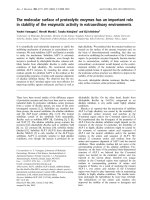
Figure 1
A schematic representation of osteoclast differentiation supported by
osteoblasts/stromal cells. RANKL, which is induced by bone resorbing
factors such as 1-α,25(OH)
2
D
3
, parathyroid hormone (PTH) and IL-11
on the plasma membrane of osteoblasts/stromal cells, binds its
receptor RANK present in osteoclast progenitors and mature
osteoclasts. Osteoprotegerin (OPG), a decoy receptor for RANKL,
strongly and competitively inhibits the RANKL–RANK interaction. The
RANK signaling is transduced via TNF receptor-associated factor 2
(TRAF2) and TNF receptor-associated factor 6 (TRAF6), leading to the
activation of NF-κB and Jun kinase (JNK), which in turn stimulates
differentiation and activation of osteoclasts. M-CSF, macrophage
colony-stimulating factor.
283
coculture system [22,23]. We also examined whether
sIL-6R and IL-6 are involved in joint destruction in RA
patients [13]. The number of osteoclast-like multinucleated
cells found in the synovial tissues derived from the knee
joint was greater in RA patients than in OA patients. These
multinucleated cells from RA synovial tissues expressed
the osteoclast-specific phenotypes such as TRAP, car-
bonic anhydrase II, vacuolar type proton-ATPase and vit-
ronectin receptors at similar levels to those from a human
giant cell tumor of bone. The concentrations of both IL-6
and sIL-6R were significantly higher in the synovial fluids of
patients with RA than in those of patients with OA. The
concentrations of IL-6 and sIL-6R were correlated well with
the roentgenologic grades of joint destruction [13]. These
results suggest that IL-6 in RA synovial fluids is responsi-
ble, at least in part, for joint destruction in the presence of
sIL-6R through osteoclastogenesis (Fig. 2).
IL-17 is a newly discovered cytokine that is secreted by
activated memory CD4
+
T cells and modulates an early
stage of immune responses [24]. Rouvier et al. [25] cloned
cytotoxic T-lymphocyte-associated antigen 8 (rat IL-17)
from a T-cell subtraction library. Mouse IL-17 was subse-
quently cloned from a thymus-derived, activated T-cell
cDNA library [26]. Fossiez et al. [27] reported that IL-17
stimulated epithelial cells, endothelial cells and fibroblastic
stromal cells to secrete several cytokines, including IL-6, IL-
8, granuloctye colony-stimulating factor and PGE
2
. In addi-
tion, IL-17 greatly promoted the proliferation of CD34
+
hemopoietic progenitors in cocultures with synovial fibrob-
lastic cells collected from RA patients [27].
We examined potential roles of IL-17 in osteoclastogenesis
using a mouse coculture system. IL-17 greatly stimulated
osteoclast formation via a cell-to-cell interaction between
osteoclast progenitors and osteoblasts [17]. IL-17
increased PGE
2
synthesis in cultures of osteoblasts. In
addition, IL-17 induced the expression of RANKL mRNA in
osteoblasts. Like OPG, NS398 (a selective inhibitor of
COX-2) completely inhibited IL-17-induced osteoclast dif-
ferentiation in the cocultures.
IL-17 levels were significantly higher in the synovial fluids
of RA patients compared with those of OA patients. Anti-
IL-17 antibody significantly inhibited osteoclast formation
induced by conditioned media of the cultures of RA syn-
ovial tissues in cocultures. Immunostaining of the synovial
tissues from RA patients demonstrated that IL-17-positive
cells were detected in a subset of CD4
+
, CD45RO
+
T cells in the specimens [17]. These findings suggest that
IL-17 first acts on osteoblasts, which stimulates COX-2-
dependent PGE
2
synthesis, and it then induces RANKL
gene expression, which in turn induces differentiation of
osteoclast progenitors into mature osteoclasts. It is proba-
ble that IL-17 is a crucial cytokine for osteoclastic bone
resorption in RA patients (Fig. 2).
Chabaud and co-workers [28,29] examined the contribu-
tion of soluble factors in the interaction between T cells
and synoviocytes in RA patients. IL-17 induced production
of IL-6 and leukemia inhibitory factor in synovial fibroblasts
[28]. IL-17 increased bone resorption and decreased bone
formation in human RA bone explants [29]. Chabaud et al.
also reported that IL-17 was spontaneously produced in
organ cultures of synovial tissues derived from RA patients.
Addition of anti-inflammatory cytokines IL-4 and IL-13
completely inhibited the production of IL-17 in synovial
tissues [30]. Lubberts et al. [31] recently reported the IL-4
gene therapy for collagen-induced arthritis in mice, using a
gene transfer with an IL-4-expressing adenoviral vector.
Local treatment with IL-4 greatly prevented joint damage
and bone erosion, although severe inflammation remained
unchanged. The protective effect of IL-4 was associated
with the decreased formation of osteoclasts and the
downregulation of IL-17 mRNA and RANKL protein
expression [31].
Jovanovic et al. [32] reported that IL-17 induced produc-
tion of matrix metalloproteinase 9 in human monocyte/
macrophages through PGE
2
synthesis. This stimulation
was involved in both phosphorylation of p38 mitogen-
activated protein (MAP) kinase and in NF-κB activation
[32]. They also found that IL-17 stimulated the production
and expression of inflammatory cytokines such as IL-1β,
IL-6, and TNF-β by human macrophages [33]. Ziolkowska
et al. [34] reported that high concentrations of IL-17 were
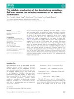
Available online />Figure 2
A possible mechanism of osteoclast formation by activated T cells in
rheumatoid arthritis. Activated T cells present in the synovial tissues
also produce membrane-associated RANKL, some of which are
cleaved enzymatically from the plasma membrane, resulting in soluble
RANKL (sRANKL). Activated T cells also produce IL-17, which induces
RANKL via prostaglandin E
2
synthesis in osteoblasts. IL-6 together
with soluble IL-6 receptors (sIL-6R), IL-1-α and TNF-α derived from
macrophages induce RANKL in osteoblasts. In addition, TNF-α directly
acts on osteoclast progenitors, which then differentiate into
osteoclasts by a mechanism independent of the RANKL–RANK
interaction. IL-1 also induces osteoclast activation directly. OPG,
osteoprotegerin.
284
strongly correlated with those of IL-15 in synovial fluids of
RA patients. IL-15 stimulates IL-17 production by human
PBMCs in primary cultures [34]. These results together
with our recent findings suggest that IL-17 plays an impor-
tant role in the joint destruction of RA patients.
Osteoclastogenesis by activated T cells in RA
Kong et al. [8] reported that RANKL knockout (–/–) mice
showed severe osteopetrosis with total occlusion of the
bone marrow space within endosteal bone. RANKL (–/–)
mice lack osteoclasts but have normal osteoclast progeni-
tors that can differentiate into functionally active osteo-
clasts when cocultured with wild-type osteoblasts. In
addition, RANKL (–/–) mice exhibited defects in early dif-
ferentiation of T cells and B cells, and they lacked all
lymph nodes but had normal splenic structures and
Peyer’s patches [8]. These results suggest that RANKL is
not only a prerequisite for osteoclast development, but
that it also plays an important role in early differentiation of
T cells and B cells.
Several reports have demonstrated that RANKL is
detected in the synovial fibroblasts and activated T lym-
phocytes derived from RA patients [18,20,35–37].
Horwood et al. [19] reported that human PBMC-derived
T cells activated by concanavalin A expressed RANKL,
and that these cells supported osteoclast formation in
cocultures with murine hemopoietic cells. Romas et al.
[38] found that RANKL mRNA was highly expressed by
the synovial cell infiltrate in arthritic joints, as well as by
osteoclasts at the sites of bone erosion in collagen-
induced arthritis. It was recently reported that the degree
of bone erosion in RANKL (–/–) mice was greatly reduced
in a serum transfer model of arthritis, when compared with
the control mice [39].
To elucidate the direct effect of human T cells in inducing
osteoclastogenesis in RA, we conducted coculture experi-
ments of activated human T cells and human adherent
PBMCs [20]. When PBMCs were cultured in the pres-
ence of M-CSF for 3 days and further cocultured for
7 days with activated CD3
+
T cells, vitronectin receptor
(CD51)-positive osteoclasts were formed even in the
absence of exogenous RANKL. Osteoclast formation
induced by activated T cells was completely inhibited by
adding OPG.
Using an ELISA system, we measured the level of a
soluble form of RANKL in the synovial fluids. Concentra-
tions of soluble RANKL in the synovial fluids were signifi-
cantly higher in patients with RA than in patients with
other arthropathies including OA, gout, and trauma. In
contrast, a decreased concentration of OPG was
detected in the synovial fluids from RA patients. The ratio
of the concentration of soluble RANKL to that of OPG
was significantly higher in the synovial fluids of RA
patients than in those of patients with OA or gout [20].
These results suggest that excess production of RANKL
by activated T lymphocytes may contribute to the patho-
genesis of bone destruction in these patients (Fig. 2).
Regulation of RANKL and/or OPG expression in RA
patients will provide a clue for the strategy of the develop-
ment of new treatment for inhibiting of bone destruction in
this disease.
In a T-cell-dependent model of rat adjuvant arthritis char-
acterized by severe joint inflammation and bone and carti-
lage destruction, OPG treatment at the onset of the
disease prevented bone and cartilage destruction but not
inflammation [18]. Teng et al. [40] also reported that
CD4
+
T-cell-mediated immunity is involved in the modula-
tion of periodontal bone destruction in HuPBL-NOD/SCID
mice after oral inoculation of Actinobacillus actino-
mycetemcomitans, a well-known Gram-negative anaerobic
microorganism that causes human periodontitis. OPG
treatment significantly reduced the number of osteoclasts
at the sites of local periodontal infection.
Juji et al. [41] recently reported a simple and effective
method of active immunization against self RANKL as a
potential treatment of bone diseases. Immunization with
RANKL vaccines almost completely prevented the bone
destruction in RA model mice (SKG mice). These results
suggest that a therapeutic vaccine approach targeting
RANKL may be useful for inhibiting bone destruction in a
variety of pathological bone diseases.
Inhibitory cytokines produced by T cells on
osteoclast differentiation
We previously reported that bone-marrow-derived stromal
cell lines, MC3T3-G2/PA6 and ST2, had the capacity to
support osteoclast formation in cocultures with hemo-
poietic cells [2,3]. Chambers and co-workers established
several bone-marrow-derived stromal cell lines from a
transgenic mouse, in which the IFN-inducible major mouse
histocompatibility complex H-2Kb promoter drives the
temperature-sensitive immortalizing gene of simian virus
40 [42,43]. These cell lines differed in their osteoclast-
inductive activity in cocultures with hematopoietic cells.
To identify genes in osteoblasts/stromal cells that are
involved in the process of osteoclastogenesis, we used
differential display of PCR to compare mRNA populations
between osteoclast-inductive and osteoclast-noninductive
cell lines [15]. Using this approach, we identified IL-18
(IFN-γ-inducing factor) as a product of osteoblastic
stromal cells. IL-18 has been reported to induce produc-
tion of IFN-γ and GM-CSF in T cells, both of which exhibit
a potent inhibitory activity of osteoclastogenesis, at least
in vitro [44]. IL-18 strongly inhibited osteoclast formation
induced by bone-resorbing factors in cocultures. IL-18
Arthritis Research Vol 4 No 5 Udagawa et al.
285
was found to inhibit osteoclast formation in cocultures
with osteoblasts and spleen cells from IFN-γ receptor type
II-deficient mice, similarly to those with wild-type
osteoblasts and spleen cells In contrast, IL-18 was unable
to inhibit osteoclast formation in cocultures of osteoblasts
and spleen cells from GM-CSF-deficient mice (Fig. 3).
Since T cells comprise a large proportion of the spleen
cell population, the role of T cells in osteoclastogenesis
was examined. T cells were removed from spleen cell
preparations using a monoclonal antibody against Thy 1.2
membrane antigen, which was predominantly expressed
on T lymphocytes. The complete absence of T cells abol-
ished the action of IL-18 on osteoclast formation in cocul-
tures of osteoblasts and spleen cells from wild-type mice
(Fig. 3). Addition of wild-type T cells but not GM-CSF-
deficient T cells to the coculture restored the inhibition by
IL-18 of osteoclastogenesis (Fig. 3). These results
suggest that IL-18 inhibits osteoclast formation by making
T cells promote the release of GM-CSF, which then acts
on osteoclast precursors to limit osteoclast differentiation
[15,16] (Fig. 4).
Horwood et al. [45] found that, like IL-18, IL-12 strongly
inhibited osteoclast formation in cocultures, as well as in
spleen cell cultures treated with M-CSF and RANKL. An
unknown inhibitory molecule was found to be secreted
from T cells in response to IL-12 and IL-18. Transwell
experiments in which T cells were separated from hemo-
poietic cells suggested that the inhibitory molecule was a
secreted factor, but not a membrane-associated factor.
Although a number of cytokines (IL-4, IL-10, IL-13, GM-
CSF and IFN-γ) expressed by T cells have the capacity to
inhibit osteoclast formation, the present inhibitory factor
has not been identified. IL-12 and IL-18 are detected in
the RA synovial membrane [46]. It was also reported that
IL-18 stimulated expression of OPG mRNA in osteoblasts
and bone marrow stromal cells [47]. IL-12 and IL-18 may
therefore protect the joint destruction via osteoclast-
mediated erosion. IL-18 is effective in inhibiting bone
destruction in murine models of breast cancer metastasis
in bone [48]. These results suggest that IL-12 and/or IL-18
therapy may be useful for reducing pathological bone loss.
Takayanagi et al. [21] demonstrated that activated T cells
are capable of inhibiting osteoclastogenesis through IFN-γ
production, which interferes with the RANKL–RANK sig-
naling pathway. In that study, osteoclast formation was
strongly inhibited in the coculture of activated T cells and
bone marrow cells in the presence of RANKL and M-CSF
[21]. When activated T cells were cocultured with bone
marrow cells derived from IFN-γ receptor knockout mice in
the presence of RANKL and M-CSF, the inhibitory effect
of activated T cells was completely canceled.
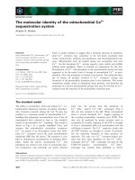
Available online />Figure 3
Effects of IL-18 on osteoclast formation. Mouse spleen cells and
osteoblasts from wild-type mice (WT), IFN-γ receptor type II-knockout
mice (IFNγR KO) or granulocyte–macrophage colony-stimulating
factor-knockout mice (GM-CSF KO) were cocultured with
1-α,25(OH)
2
D
3
and prostaglandin E
2
(PGE
2
) in the presence or
absence of IL-18. In some cocultures, T-cell-depleted spleen cells and
osteoblasts were cocultured in the presence and absence of WT
T cells or GM-CSF KO T cells. In each coculture, numbers of tartrate-
resistant acid phosphatase-positive osteoclasts formed were scored.
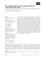
Figure 4
A proposed mechanism of the inhibitory action of IL-18 on osteoclast
differentiation. IL-18 secreted from osteoblasts acts on T lymphocytes,
which generate granulocyte–macrophage colony-stimulating factor
(GM-CSF) and IFN-γ. Both GM-CSF and IFN-γ are potent inhibitors of
osteoclast formation, at least in vitro. When GM-CSF binds its
receptor, GM-CSFR (present in osteoclast progenitors), osteoclast
formation is completely inhibited. In contrast, the target molecule of
IFN-γ is TNF receptor-associated factor 6 (TRAF6). The degradation of
TRAF6 by IFN-γ leads to the inhibition of osteoclastogenesis. The
inhibitory action of IL-18 on osteoclast differentiation occurs via GM-
CSF, but not via IFN-γ.
286
The expression of TNF receptor-associated factor (TRAF)6
was markedly inhibited by IFN-γ in osteoclast progenitors
stimulated by RANKL and M-CSF, indicating that TRAF6
is a target molecule of IFN-γ. IFN-γ appears to inhibit
osteoclastogenesis by decomposing TRAF6. In fact,
TRAF6-deficient mice exhibited severe osteopetrosis
[49,50]. It was also reported that IFN-γ receptor knockout
mice developed collagen-induced arthritis more readily
than wild-type mice [51]. These results suggest that
TRAF6 is the critical target molecule in the IFN-γ-mediated
suppression of osteoclast formation, and that the balance
between RANKL and IFN-γ action may regulate osteo-
clastogenesis (Fig. 4).
Possible roles of TNF-
αα
in osteoclast
differentiation
We have reported that TNF-α induced osteoclast formation
via a mechanism independent of the RANKL–RANK signal-
ing pathway [52] (Fig. 5). When mouse bone marrow cells
were cultured with M-CSF for 3 days and nonadherent
cells removed, adherent cells of uniform size and shape
remained on the culture dish. The M-CSF-dependent bone
marrow macrophage preparation contained no appreciable
number of alkaline phosphatase-positive osteoblastic cells.
When M-CSF-dependent bone marrow macrophages
were further cultured for 3 days with several bone-resorb-
ing cytokines, mouse TNF-α as well as RANKL induced
osteoclast formation in the presence of M-CSF.
IL-1-α failed to induce osteoclast formation in macrophage
cultures even in the presence of M-CSF. These osteoclast
progenitors expressed not only RANK and c-Fms (M-CSF
receptor), but also TNF receptor type 1 (TNFR1, p55) and
TNF receptor type 2 (TNFR2, p75). Osteoclast formation
induced by RANKL was completely inhibited by adding
OPG, but osteoclastogenesis induced by TNF-α was not.
Adding antibodies against TNFR1 and TNFR2 blocked
osteoclast formation induced by TNF-α but not by RANKL.
Bone marrow macrophages prepared from TNFR1 knock-
out mice differentiated into osteoclasts in response to
RANKL, but they failed to differentiate into osteoclasts in
response to TNF-α. Similarly, TNFR2 knockout mouse-
derived bone marrow macrophages differentiated into
osteoclasts in response to RANKL, but osteoclast differenti-
ation induced by TNF-α was markedly decreased in TNFR2
knockout mouse-derived macrophage cultures [52].
These results suggest that TNF-α stimulates osteoclast
formation via a mechanism independent of the RANKL
pathway (Fig. 5). It was also shown that TNF-α as well as
RANKL stimulated differentiation of RAW 264.7 cells into
osteoclasts [53,54]. RANK-mediated signals for osteo-
clastogenesis are transduced via either TRAF6 or TRAF2,
whereas TNFR1-mediated and TNFR2-mediated signals
are transduced via TRAF2. TRAF-2-mediated signals may
play important roles in osteoclast differentiation induced
by TNF-α. Using TRAF6-deficient mice, Kaji et al. [55]
recently found that TRAF6 is also involved in TNF-α-
induced osteoclastogenesis (Fig. 5). Further studies are
necessary to determine the relationship between TRAF2
and TRAF6 in TNF-α-induced osteoclastogenesis.
To examine, whether TNF-α induces not only osteoclast
differentiation, but also osteoclast activation, macro-
phages were cultured on dentine slices in the presence of
TNF-α, M-CSF, and OPG [52]. Some cultures were also
treated with IL-1-α. After culture for 6 days, similar
numbers of osteoclasts were formed on dentine slices
irrespective of the presence or absence of IL-1-α.
However, no resorption pits were detected in macrophage
cultures treated with TNF-α and M-CSF. Resorption pits
on dentine slices were observed only in the presence of
TNF-α and M-CSF together with IL-1-α.
These results suggest that TNF-α stimulates differentia-
tion, but not activation, of osteoclasts. In contrast, IL-1-α
does not induce differentiation of osteoclasts in
macrophage cultures that do not contain osteoblasts/
stromal cells, but it does stimulate pit-forming activity of
the osteoclasts formed [52,56] (Fig. 5). Since IL-1R binds
TRAF6 but not TRAF2, these results indicate that TRAF6
is a prerequisite for osteoclast activation.
Arthritis Research Vol 4 No 5 Udagawa et al.
Figure 5
Signal transduction of TNF-α, RANKL and IL-1 in osteoclast
differentiation and activation. TNF-α binds TNF receptor type 1
(TNFR1) and TNF receptor type 2 (TNFR2), RANKL binds RANK, and
IL-1 binds IL-1 receptor (IL-1R). Both TNFR1 and TNFR2 bind TNF
receptor-associated factor 2 (TRAF2), whereas IL-1R binds TNF
receptor-associated factor 6 (TRAF6). RANK binds not only TRAF6,
but also TRAF2 and other TNF receptor-associated factors (TRAFs).
M-CSF, macrophage colony-stimulating factor.
287
Pacifici and co-workers [57,58] recently demonstrated that
estrogen deficiency induces bone loss by enhancing
TNF-α production by T cells. Ovariectomy failed to induce
bone loss in T-cell-deficient athymic nude (nu/nu) mice as
well as in TNFR1 knockout mice. They also found that
ovariectomy increased the number of TNF-producing
T cells in the bone marrow of normal mice without altering
the TNF production per T cell [58]. These results suggest
that T-cell-produced TNF and its interaction with TNFR1
play a key role in bone loss induced by estrogen deficiency.
Conclusion
Under physiological conditions, osteoclast formation
requires cell-to-cell contact between hemopoietic cells
(osteoclast progenitors) and osteoblastic cells, in which
osteoblastic cells generate RANKL as a membrane-bound
factor in response to several bone resorbing factors
(Fig. 6). In contrast, like in RA, T cells appear to secrete a
soluble form of RANKL in pathological bone resorption
that acts directly on osteoclast progenitors without cell-to-
cell contact. Furthermore, TNF-α directly stimulates osteo-
clast differentiation via a mechanism independent of the
RANKL–RANK interaction. IL-1-α induces osteoclast acti-
vation via its own receptors (Fig. 6).
Takeuchi et al. [59] recently established a coculture
system with nurse-like cells obtained from synovial tissues
of patients with RA. These cells promote survival of B cells
and maintain the growth of myeloid cells. In addition, the
nurse-like cells supported the generation of TRAP-positive
osteoclasts from PBMCs [60]. These results suggest that,
like bone-marrow-derived stromal cells, the nurse-like cells
from RA synovial tissues also possess a novel ability to
support osteoclast differentiation.
In conclusion, control of the production of RANKL, of OPG
and of other T-cell-derived cytokines in RA patients will
provide a clue for strategies of the development of new
treatment for inhibiting bone destruction in this disease.
References
1. Takahashi N, Akatsu T, Udagawa N, Sasaki T, Yamaguchi A,
Moseley JM, Martin TJ, Suda T: Osteoblastic cells are involved
in osteoclast formation. Endocrinology 1988, 123:2600-2602.
2. Udagawa N, Takahashi N, Akatsu T, Sasaki T, Yamaguchi A,
Kodama H, Martin TJ, Suda T: The bone marrow-derived
stromal cell lines MC3T3-G2/PA6 and ST2 support osteo-
clast-like cell differentiation in cocultures with mouse spleen
cells. Endocrinology 1989, 125:1805-1813.
3. Udagawa N, Takahashi N, Akatsu T, Tanaka H, Sasaki T, Nishihara
T, Koga T, Martin TJ, Suda T: Origin of osteoclasts: mature
monocytes and macrophages are capable of differentiating
into osteoclasts under a suitable microenvironment prepared
by bone marrow-derived stromal cells. Proc Natl Acad Sci
USA 1990, 87:7260-7264.
4. Suda T, Takahashi N, Martin TJ: Modulation of osteoclast differ-
entiation. Endocr Rev 1992, 13:66-80.
5. Yasuda H, Shima N, Nakagawa N, Yamaguchi K, Kinosaki M,
Mochizuki S, Tomoyasu A, Yano K, Goto M, Murakami A, Tsuda E,
Morinaga T, Higashio K, Udagawa N, Takahashi N, Suda T: Osteo-
clast differentiation factor is a ligand for osteoprotegerin/osteo-
clastogenesis-inhibitory factor and is identical to TRANCE/
RANKL. Proc Natl Acad Sci USA 1998, 95:3597-3602.
6. Wong BR, Rho J, Arron J, Robinson E, Orlinick J, Chao M,
Kalachikov S, Cayani E, Bartlett FS III, Frankel WN, Lee SY, Choi
Y: TRANCE is a novel ligand of the tumor necrosis factor
receptor family that activates c-Jun N-terminal kinase in T
cells. J Biol Chem 1997, 272:25190-25194.
7. Anderson DM, Maraskovsky E, Billingsley WL, Dougall WC,
Tometsko ME, Roux ER, Teepe MC, DuBose RF, Cosman D,
Galibert L: A homologue of the TNF receptor and its ligand
enhance T-cell growth and dendritic-cell function. Nature
1997, 390:175-179.
8. Kong YY, Yoshida H, Sarosi I, Tan HL, Timms E, Capparelli C,
Morony S, Oliveira-dos-Santos AJ, Van G, Itie A, Khoo W,
Wakeham A, Dunstan CR, Lacey DL, Mak TW, Boyle WJ, Pen-
ninger JM: OPGL is a key regulator of osteoclastogenesis,
lymphocyte development and lymph-node organogenesis.
Nature 1999, 397:315-323.
9. Suda T, Takahashi N, Udagawa N, Jimi E, Gillespie MT, Martin TJ:
Modulation of osteoclast differentiation and function by the
new members of the tumor necrosis factor receptor and
ligand families. Endocr Rev 1999, 20:345-357.
10. The American Society for Bone and Mineral Research President’s
Committee on Nomenclature: Proposed standard nomenclature
for new tumor necrosis factor family members involved in the
regulation of bone resorption. J Bone Miner Res 2000, 15:
2293-2296.
11. Matsuzaki K, Udagawa N, Takahashi N, Yamaguchi K, Yasuda H,
Shima N, Morinaga T, Toyama Y, Yabe Y, Higashio K, Suda T:
Osteoclast differentiation factor (ODF) induces osteoclast-like
cell formation in human peripheral blood mononuclear cell
cultures. Biochem Biophys Res Commun 1998, 246:199-204.
12. Quinn JM, Elliott J, Gillespie MT, Martin TJ: A combination of
osteoclast differentiation factor and macrophage-colony stim-
ulating factor is sufficient for both human and mouse osteo-
clast formation in vitro. Endocrinology 1998, 139:4424-4427.
13. Kotake S, Sato K, Kim KJ, Takahashi N, Udagawa N, Nakamura I,
Yamaguchi A, Kishimoto T, Suda T, Kashiwazaki S: Interleukin-6
and soluble interleukin-6 receptors in the synovial fluids from
Available online />Figure 6
Involvement of TNF ligand family members in physiological and
pathological bone resorption. RANKL appears to play a major role in
physiological bone resorption. In contrast, both RANKL-dependent and
RANKL-independent pathways appear to be involved in pathological
bone resorption. At present, the contribution ratio of the RANKL-
dependent and RANKL-independent pathways to the pathological
bone resorption is not known. M-CSF, macrophage colony-stimulating
factor.
288
rheumatoid arthritis patients are responsible for osteoclast-
like cell formation. J Bone Miner Res 1996, 11:88-95.
14. Fox DA: The role of T cells in the immunopathogenesis of
rheumatoid arthritis: new perspectives. Arthritis Rheum 1997,
40:598-609.
15. Udagawa N, Horwood NJ, Elliott J, Mackay A, Owens J, Okamura
H, Kurimoto M, Chambers TJ, Martin TJ, Gillespie MT: Inter-
leukin-18 (interferon-
γγ
-inducing factor) is produced by
osteoblasts and acts via granulocyte/macrophage colony-
stimulating factor and not via interferon-gamma to inhibit
osteoclast formation. J Exp Med 1997, 185:1005-1012.
16. Horwood NJ, Udagawa N, Elliott J, Grail D, Okamura H, Kurimoto
M, Dunn AR, Martin T, Gillespie MT: Interleukin 18 inhibits
osteoclast formation via T cell production of granulocyte
macrophage colony-stimulating factor. J Clin Invest 1998, 101:
595-603.
17. Kotake S, Udagawa N, Takahashi N, Matsuzaki K, Itoh K, Ishiyama
S, Saito S, Inoue K, Kamatani N, Gillespie MT, Martin TJ, Suda T:
IL-17 in synovial fluids from patients with rheumatoid arthritis
is a potent stimulator of osteoclastogenesis. J Clin Invest
1999, 103:1345-1352.
18. Kong YY, Feige U, Sarosi I, Bolon B, Tafuri A, Morony S, Cappar-
elli C, Li J, Elliott R, McCabe S, Wong T, Campagnuolo G, Moran
E, Bogoch ER, Van G, Nguyen LT, Ohashi PS, Lacey DL, Fish E,
Boyle WJ, Penninger JM: Activated T cells regulate bone loss
and joint destruction in adjuvant arthritis through osteoprote-
gerin ligand. Nature 1999, 402:304-309.
19. Horwood NJ, Kartsogiannis V, Quinn JM, Romas E, Martin TJ, Gille-
spie MT: Activated T lymphocytes support osteoclast formation
in vitro. Biochem Biophys Res Commun 1999, 265:144-150
20. Kotake S, Udagawa N, Hakoda M, Mogi M, Yano K, Tsuda E, Taka-
hashi K, Furuya T, Ishiyama S, Kim KJ, Saito S, Nishikawa T, Takahashi
N, Togari A, Tomatsu T, Suda T, Kamatani N: Activated human T
cells directly induce osteoclastogenesis from human monocytes:
possible role of T cells in bone destruction in rheumatoid arthritis
patients. Arthritis Rheum 2001, 44:1003-1012.
21. Takayanagi H, Ogasawara K, Hida S, Chiba T, Murata S, Sato K,
Takaoka A, Yokochi T, Oda H, Tanaka K, Nakamura K, Taniguchi
T: T-cell-mediated regulation of osteoclastogenesis by sig-
nalling cross-talk between RANKL and IFN-
γγ
. Nature 2000,
408:600-605.
22. Tamura T, Udagawa N, Takahashi N, Miyaura C, Tanaka S,
Yamada Y, Koishihara Y, Ohsugi Y, Kumaki K, Taga T, Kishimoto
T, Suda T: Soluble interleukin-6 receptor triggers osteoclast
formation by interleukin 6. Proc Natl Acad Sci USA 1993, 90:
11924-11928.
23. Udagawa N, Takahashi N, Katagiri T, Tamura T, Wada S, Findlay
DM, Martin TJ, Hirota H, Taga T, Kishimoto T, Suda T: Interleukin
(IL)-6 induction of osteoclast differentiation depends on IL-6
receptors expressed on osteoblastic cells but not on osteo-
clast progenitors. J Exp Med 1995, 182:1461-1468.
24. Broxmeyer HE: Is interleukin 17, an inducible cytokine that
stimulates production of other cytokines, merely a redundant
player in a sea of other biomolecules? J Exp Med 1996, 183:
2411-2415.
25. Rouvier E, Luciani MF, Mattei MG, Denizot F, Golstein P: CTLA-8,
cloned from an activated T cell, bearing AU-rich messenger
RNA instability sequences, and homologous to a herpesvirus
saimiri gene. J Immunol 1993, 150:5445-5456.
26. Kennedy J, Rossi DL, Zurawski SM, Vega F Jr, Kastelein RA,
Wagner JL, Hannum CH, Zlotnik A: Mouse IL-17: a cytokine
preferentially expressed by
ααββ
TCR + CD4-CD8-T cells. J Inter-
feron Cytokine Res 1996, 16:611-617.
27. Fossiez F, Djossou O, Chomarat P, Flores-Romo L, Ait-Yahia S,
Maat C, Pin JJ, Garrone P, Garcia E, Saeland S, Blanchard D,
Gaillard C, Das Mahapatra B, Rouvier E, Golstein P, Banchereau
J, Lebecque S: T cell interleukin-17 induces stromal cells to
produce proinflammatory and hematopoietic cytokines. J Exp
Med 1996, 183:2593-2603.
28. Chabaud M, Fossiez F, Taupin JL, Miossec P: Enhancing effect
of IL-17 on IL-1-induced IL-6 and leukemia inhibitory factor
production by rheumatoid arthritis synoviocytes and its regu-
lation by Th2 cytokines. J Immunol 1998, 161:409-414.
29. Chabaud M, Lubberts E, Joosten L, van Den Berg W, Miossec P:
IL-17 derived from juxta-articular bone and synovium con-
tributes to joint degradation in rheumatoid arthritis. Arthritis
Res 2001, 3:168-177.
30. Chabaud M, Durand JM, Buchs N, Fossiez F, Page G, Frappart L,
Miossec P: Human interleukin-17: A T cell-derived proinflam-
matory cytokine produced by the rheumatoid synovium.
Arthritis Rheum 1999, 42:963-970.
31. Lubberts E, Joosten LA, Chabaud M, van Den Bersselaar L,
Oppers B, Coenen-De Roo CJ, Richards CD, Miossec P, van Den
Berg WB: IL-4 gene therapy for collagen arthritis suppresses
synovial IL-17 and osteoprotegerin ligand and prevents bone
erosion. J Clin Invest 2000, 105:1697-1710.
32. Jovanovic DV, Martel-Pelletier J, Di Battista JA, Mineau F, Jolicoeur
FC, Benderdour M, Pelletier JP: Stimulation of 92-kd gelatinase
(matrix metalloproteinase 9) production by interleukin-17 in
human monocyte/macrophages: a possible role in rheuma-
toid arthritis. Arthritis Rheum 2000, 43:1134-1144.
33. Jovanovic DV, Di Battista JA, Martel-Pelletier J, Jolicoeur FC, He Y,
Zhang M, Mineau F, Pelletier JP: IL-17 stimulates the production
and expression of proinflammatory cytokines, IL-
ββ
and TNF-
αα
,
by human macrophages. J Immunol 1998, 160:3513-3521.
34. Ziolkowska M, Koc A, Luszczykiewicz G, Ksiezopolska-Pietrzak K,
Klimczak E, Chwalinska-Sadowska H, Maslinski W: High levels of
IL-17 in rheumatoid arthritis patients: IL-15 triggers in vitro
IL-17 production via cyclosporin A-sensitive mechanism.
J Immunol 2000, 164:2832-2838.
35. Gravallese EM, Manning C, Tsay A, Naito A, Pan C, Amento E,
Goldring SR: Synovial tissue in rheumatoid arthritis is a
source of osteoclast differentiation factor. Arthritis Rheum
2000, 43:250-258.
36. Takayanagi H, Iizuka H, Juji T, Nakagawa T, Yamamoto A, Miyazaki
T, Koshihara Y, Oda H, Nakamura K, Tanaka S: Involvement of
receptor activator of nuclear factor
κκ
B ligand/osteoclast dif-
ferentiation factor in osteoclastogenesis from synoviocytes in
rheumatoid arthritis. Arthritis Rheum 2000, 43:259-269.
37. Shigeyama Y, Pap T, Kunzler P, Simmen BR, Gay RE, Gay S:
Expression of osteoclast differentiation factor in rheumatoid
arthritis. Arthritis Rheum 2000, 43:2523-2530.
38. Romas E, Bakharevski O, Hards DK, Kartsogiannis V, Quinn JM,
Ryan PF, Martin TJ, Gillespie MT: Expression of osteoclast dif-
ferentiation factor at sites of bone erosion in collagen-
induced arthritis. Arthritis Rheum 2000, 43:821-826.
39. Pettit AR, Ji H, von Stechow D, Muller R, Goldring SR, Choi Y,
Benoist C, Gravallese EM: TRANCE/RANKL knockout mice are
protected from bone erosion in a serum transfer model of
arthritis. Am J Pathol 2001, 159:1689-1699.
40. Teng YT, Nguyen H, Gao X, Kong YY, Gorczynski RM, Singh B,
Ellen RP, Penninger JM: Functional human T-cell immunity and
osteoprotegerin ligand control alveolar bone destruction in
periodontal infection. J Clin Invest 2000, 106:R59-R67.
41. Juji T, Aoki K, Horie D, Ohya K, Herz M, Gautam A, Mouritsen S, Oda
H, Nakamura K, Tanaka S: A novel therapeutic vaccine that pre-
vents pathological bone destruction in models of osteoporosis
and RA [abstract]. J Bone Miner Res 2001, 16(suppl 1):S150.
42. Chambers TJ, Owens JM, Hattersley G, Jat PS, Noble MD: Gen-
eration of osteoclast-inductive and osteoclastogenic cell lines
from the H-2KbtsA58 transgenic mouse. Proc Natl Acad Sci
USA 1993, 90:5578-5582.
43. Owens JM, Gallagher AC, Chambers TJ: Bone cells required for
osteoclastic resorption but not for osteoclastic differentiation.
Biochem Biophys Res Commun 1996, 222:225-229.
44. Nakanishi K, Yoshimoto T, Tsutsui H, Okamura H: Interleukin-18
is a unique cytokine that stimulates both Th1 and Th2
responses depending on its cytokine milieu. Cytokine Growth
Factor Rev 2001, 12:53-72.
45. Horwood NJ, Elliott J, Martin TJ, Gillespie MT: IL-12 alone and in
synergy with IL-18 inhibits osteoclast formation in vitro.
J Immunol 2001, 166:4915-4921.
46. Gracie JA, Forsey RJ, Chan WL, Gilmour A, Leung BP, Greer MR,
Kennedy K, Carter R, Wei XQ, Xu D, Field M, Foulis A, Liew FY,
McInnes IB: A proinflammatory role for IL-18 in rheumatoid
arthritis. J Clin Invest 1999, 104:1393-1401.
47. Makiishi-Shimobayashi C, Tsujimura T, Iwasaki T, Yamada N,
Sugihara A, Okamura H, Hayashi S, Terada N: Interleukin-18 up-
regulates osteoprotegerin expression in stromal/osteoblastic
cells. Biochem Biophys Res Commun 2001, 281:361-366.
48. Nakata A, Tsujimura T, Sugihara A, Okamura H, Iwasaki T, Shinkai
K, Iwata N, Kakishita E, Akedo H, Terada N: Inhibition by inter-
leukin 18 of osteolytic bone metastasis by human breast
cancer cells. Anticancer Res 1999, 19:4131-4138.
Arthritis Research Vol 4 No 5 Udagawa et al.
289
49. Naito A, Azuma S, Tanaka S, Miyazaki T, Takaki S, Takatsu K,
Nakao K, Nakamura K, Katsuki M, Yamamoto T, Inoue J: Severe
osteopetrosis, defective interleukin-1 signalling and lymph
node organogenesis in TRAF6-deficient mice. Genes Cells
1999, 4:353-362.
50. Lomaga MA, Yeh WC, Sarosi I, Duncan GS, Furlonger C, Ho A,
Morony S, Capparelli C, Van G, Kaufman S, van der Heiden A, Itie
A, Wakeham A, Khoo W, Sasaki T, Cao Z, Penninger JM, Paige
CJ, Lacey DL, Dunstan CR, Boyle WJ, Goeddel DV, Mak TW:
TRAF6 deficiency results in osteopetrosis and defective inter-
leukin-1, CD40, and LPS signaling. Genes Dev 1999, 13:1015-
1024.
51. Vermeire K, Heremans H, Vandeputte M, Huang S, Billiau A,
Matthys P: Accelerated collagen-induced arthritis in IFN-
γγ
receptor-deficient mice. J Immunol 1997, 158:5507-5513.
52. Kobayashi K, Takahashi N, Jimi E, Udagawa N, Takami M, Kotake
S, Nakagawa N, Kinosaki M, Yamaguchi K, Shima N, Yasuda H,
Morinaga T, Higashio K, Martin TJ, Suda T: Tumor necrosis
factor-
αα
stimulates osteoclast differentiation by a mechanism
independent of the ODF/RANKL–RANK interaction. J Exp Med
2000, 191:275-286.
53. Matsumoto M, Sudo T, Maruyama M, Osada H, Tsujimoto M: Acti-
vation of p38 mitogen-activated protein kinase is crucial in
osteoclastogenesis induced by tumor necrosis factor. FEBS
Lett 200, 486:23-28.
54. Quinn JM, Itoh K, Udagawa N, Hausler K, Yasuda H, Shima N,
Mizuno A, Higashio K, Takahashi N, Suda T, Martin TJ, Gillespie
MT: Transforming growth factor
ββ
affects osteoclast differenti-
ation via direct and indirect actions. J Bone Miner Res 2001,
16:1787-1794.
55. Kaji K, Katogi R, Azuma Y, Naito A, Inoue JI, Kudo A: Tumor
necrosis factor-
αα
-induced osteoclastogenesis requires tumor
necrosis factor receptor-associated factor 6. J Bone Miner Res
2001, 16:1593-1599.
56. Jimi E, Nakamura I, Duong LT, Ikebe T, Takahashi N, Rodan GA,
Suda T: Interleukin 1 induces multinucleation and bone-
resorbing activity of osteoclasts in the absence of
osteoblasts/stromal cells. Exp Cell Res 1999, 247:84-93.
57. Cenci S, Weitzmann MN, Roggia C, Namba N, Novack D,
Woodring J, Pacifici R: Estrogen deficiency induces bone loss
by enhancing T-cell production of TNF-
αα
. J Clin Invest 2000,
106:1229-1237.
58. Roggia C, Gao Y, Cenci S, Weitzmann MN, Toraldo G, Isaia G,
Pacifici R: Up-regulation of TNF-producing T cells in the bone
marrow: A key mechanism by which estrogen deficiency
induces bone loss in vivo. Proc Natl Acad Sci USA 2001, 98:
13960-13965.
59. Takeuchi E, Tomita T, Toyosaki-Maeda T, Kaneko M, Takano H,
Hashimoto H, Sugamoto K, Suzuki R, Ochi T: Establishment and
characterization of nurse cell-like stromal cell lines from syn-
ovial tissues of patients with rheumatoid arthritis. Arthritis
Rheum 1999, 42:221-228.
60. Toyosaki-Maeda T, Takano H, Tomita T, Tsuruta Y, Maeda-
Tanimura M, Shimaoka Y, Takahashi T, Itoh T, Suzuki R, Ochi T:
Differentiation of monocytes into multinucleated giant bone-
resorbing cells: two-step differentiation induced by nurse-like
cells and cytokines. Arthritis Res 2001, 3:306-310.
Correspondence
Nobuyuki Udagawa, PhD, DDS, Department of Biochemistry, Mat-
sumoto Dental University, 1780 Hiro-oka Gobara, Shiojiri, Nagano
399-0781, Japan. Tel: +81 263 51 2072; Fax: + 81 263 51 2199;
e-mail:
Available online />Find what you’re looking for by using our website’s
‘search’ or ‘detailed search’ functions. Limit it to
Arthritis Research (including supplements), BioMed
Central, PubMed or PubMed Central. Then store your
search so you can run it every time you return.
Arthritis-Research.com is more than an online version of the
printed journal. Not only are research articles free to access
online but you can also download references, search
PubMed for articles by our authors, e-mail articles to
colleagues, find relevant conferences worldwide, link to
online resources, and search across BioMed Central’s
products – including our scientific supplements of abstracts
and articles.
To learn more register at
Searching for something specific?