Báo cáo y học: "p53 tumor suppressor gene mutations in fibroblast-like synoviocytes from erosion synovium and non-erosion synovium in rheumatoid arthritis" ppsx
Bạn đang xem bản rút gọn của tài liệu. Xem và tải ngay bản đầy đủ của tài liệu tại đây (187.08 KB, 7 trang )
Open Access
Available online />R12
Vol 7 No 1
Research article
p53 tumor suppressor gene mutations in fibroblast-like
synoviocytes from erosion synovium and non-erosion synovium in
rheumatoid arthritis
Yuji Yamanishi
1,2
, David L Boyle
2
, Douglas R Green
3
, Edward C Keystone
4
, Alison Connor
4
,
Susan Zollman
4
and Gary S Firestein
2
1
Department of Rheumatology, Hiroshima City Hospital, Hiroshima, Japan
2
Division of Rheumatology, Allergy, and Immunology, School of Medicine, University of California at San Diego, La Jolla, California, USA
3
La Jolla Institute of Allergy and Immunology, La Jolla, California, USA
4
Department of Medicine, University of Toronto, Canada
Corresponding author: Gary S Firestein,
Received: 9 May 2004 Revisions requested: 8 Jun 2004 Revisions received: 9 Aug 2004 Accepted: 8 Sep 2004 Published: 29 Oct 2004
Arthritis Res Ther 2005, 7:R12-R18 (DOI 10.1186/ar1448)
http://arthrit is-research.com /content/7/1/R12
© 2004 Yamanishi et al., licensee BioMed Central Ltd.
This is an Open Access article distributed under the terms of the Creative Commons Attribution License ( />2.0), which permits unrestricted use, distribution, and reproduction in any medium, provided the original work is cited.
Abstract
Abnormalities in the p53 tumor suppressor gene have been
detected in rheumatoid arthritis (RA) and could contribute to the
pathogenesis of chronic disease. To determine whether
synoviocytes from invasive synovium in RA have an increased
number of mutations compared with non-erosion synoviocytes,
p53 cDNA subclones from fibroblast-like synoviocytes (FLS)
derived from erosion and non-erosion sites of the same
synovium were examined in patients requiring total joint
replacement. Ten erosion FLS lines and nine non-erosion FLS
lines were established from nine patients with RA. Exons 5–10
from 209 p53 subclones were sequenced (114 from erosion
FLS, 95 from non-erosion FLS). Sixty percent of RA FLS cell
lines and 8.6% of the p53 subclones isolated from FLS
contained p53 mutations. No significant differences were
observed between the erosion and non-erosion FLS with regard
to the frequency or type of p53 mutation. The majority of the
mutations were missense transition mutations, which are
characteristic of oxidative damage. In addition, paired intact RA
synovium and cultured FLS from the same joints were evaluated
for p53 mutations. Matched synovium and cultured synoviocytes
contained p53 mutations, although there was no overlap in the
specific mutations identified in the paired samples. Clusters of
p53 mutations in subclones were detected in some FLS,
including one in codon 249, which is a well-recognized 'hot
spot' associated with cancer. Our data are consistent with the
hypothesis that p53 mutations are randomly induced by
genotoxic exposure in small numbers of RA synoviocytes
localized to erosion and non-erosion regions of RA synovium.
The determining factor for invasiveness might be proximity to
bone or cartilage rather than the presence of a p53 mutation.
Keywords: erosion, fibroblast-like synoviocytes, invasiveness, p53 mutation, rheumatoid arthritis
Introduction
Rheumatoid arthritis (RA) is a chronic inflammatory disease
characterized by synovial tissue proliferation with progres-
sive joint destruction. The etiology of RA remains unknown,
but many factors, including autoimmunity, cytokines and
genetic factors, participate in its pathogenesis [1,2].
Although inflammation and joint destruction can be inti-
mately related, the two processes might also be independ-
ent in some circumstances [3,4]. This observation might be
explained, at least in part, by autonomous behavior by
fibroblast-like synoviocytes (FLS) [5]. These cells exhibit
some features of transformation in RA, including loss of
contact inhibition, anchorage-independent growth, onco-
gene activation, autonomous invasion into cartilage and
somatic gene mutations [4,6-9]. One potential gene that
might contribute to this phenotype is the p53 tumor sup-
pressor gene, which plays a critical role in cell-cycle regu-
lation, DNA repair, senescence, genomic stability and
apoptosis [10]. p53 is expressed in many inflammatory and
autoimmune diseases [11-13], and it may serve a protec-
tive function by suppressing cytokine production and matrix
destruction [14,15]. For instance, mice lacking the p53
DMEM = Dulbecco's modified Eagle's medium; ELISA = enzyme-linked immunosorbent assay; FLS = fibroblast-like synoviocytes; IL = interleukin;
MMP-1 = matrix metalloproteinase-1; PCR = polymerase chain reaction; RA = rheumatoid arthritis
Arthritis Research & Therapy Vol 7 No 1 Yamanishi et al.
R13
gene have significantly greater joint destruction compared
with wild-type controls in the collagen-induced arthritis
model [16].
The function of p53 can be altered through somatic muta-
tions in both neoplasia and non-malignant conditions such
as ulcerative colitis, sun-exposed skin and chronic RA
[8,17,18]. The mutations in RA synovium reside primarily in
the intimal lining and have also been identified in cultured
FLS, which are thought to originate from this region [8,9].
p53 sequencing studies in RA have until now focused on
synoviocytes derived from non-erosion regions of the syn-
ovium that are readily obtained at the time of joint replace-
ment surgery. Because some reports suggest that
monoclonal expansion and oligoclonal expansion of synovi-
ocytes occur at sites of invasion [19], we evaluated the rel-
ative frequency of p53 mutations in FLS in paired erosion
and non-erosion synoviocytes from the same patients. Our
data suggest that mutations are present in both regions
and that the tendency to invade may be related to proximity
to the extracellar matrix.
Materials and methods
Synovial tissues and preparation of FLS
Synovial tissue samples were collected with informed con-
sent at the time of joint replacement from patients with RA.
The diagnosis of RA conformed to the 1987 revised Amer-
ican College of Rheumatology criteria [20]. Separate sam-
ples were obtained, at the University of Toronto, from within
erosions at the periphery of the articular surface and from
non-erosion sites collected from the intracapsular non-
articular surface. Erosion FLS lines and non-erosion FLS
lines were then prepared from each patient.
FLS cell lines were established as previously described
[21]. Briefly, tissues were minced by sterilized scissors,
and were incubated with 1 mg/ml collagenase in serum-
free DMEM for 2 hours at 37°C, were filtered through a
nylon mesh, were extensively washed, and were cultured in
DMEM containing 10% fetal calf serum, 2 mmol/l
glutamine, 50 µg/ml gentamicin, 100 U/ml penicillin, and
100 µg/ml streptomycin, in a humidified 5% CO
2
atmos-
phere. After overnight culture, cells were trypsinized, split in
a 1:3 ratio, and were recultured in medium. FLS from pas-
sages 5–8 were used in these experiments, during which
time they represented a homogeneous population of FLS
(< 1% CD 11b, < 1% phagocytic, and < 1% Fc-gamma RII
receptor-positive). A second set of FLS samples obtained
from the University of California at San Diego were
obtained with a matched sample of synovium, which was
embedded in TissueTek OCT compound (Miles Diagnos-
tics, Elkhart, IN, USA), snap frozen, and stored at -80°C
until use. Frozen tissues with approximate area of 10 mm
2
were cut into 10 µl sections at the time of PCR analysis for
p53 mutations.
Production of immunoreactive MMP-1
FLS were cultured to near confluence in six-well tissue cul-
ture plates (Falcon, Bedford, MA, USA). IL-1β (3 ng/ml) or
medium was added to the wells and was incubated for 72
hours at 37°C in a humidified 5% CO
2
atmosphere. Super-
natants were collected and MMP-1 concentrations were
determined by ELISA according to the manufacturer's
instructions (Total MMP1Biotrak; Amersham Biosciences,
Piscataway, NJ, USA) [22].
Statistical analysis
Comparisons between two groups were analyzed by the
Wilcoxon signed rank test. P < 0.05 was considered
significant.
Results
p53 mutations in erosion FLS and non-erosion FLS
To determine whether invasive synovium in RA has an
increased number of mutations, p53 cDNA subclones from
FLS derived from erosion sites and non-erosion sites were
examined. Ten erosion FLS lines and nine non-erosion FLS
lines were established from nine patients with RA (two ero-
sion lines and one non-erosion line were examined in one
patient). A total of 209 p53 subclones were subjected to
sequence analysis (114 from erosion FLS, 95 from non-
erosion FLS), and p53 exons 5–10 were examined. As
shown in Table 1, p53 mutations were identified in 11 out
of 19 FLS lines (four of 10 erosion FLS lines, and seven of
nine non-erosion FLS lines). Eighteen subclones out of the
total 209 (8.6%) contained mutations. There were no sig-
nificant differences in the frequency of p53 mutations
between erosion FLS and non-erosion FLS (7.9% and
9.5%, respectively). Nested PCR was required for one of
the FLS lines (RA4 non-erosion FLS line). The rate of muta-
tion with this line was similar to the other erosion and non-
erosion FLS.
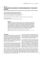
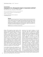
As in previous reports [8,9,23], most p53 mutations (eight
of nine in erosion FLS and eight of nine in non-erosion FLS)
were transition mutations (i.e. G>A or C>T), which are
characteristic of mutations caused by oxidative damage
[24,25], and no transversion mutations (i.e. G>T, A>T,
C>A, C>G) were seen (see Fig. 1 for the pooled data).
One single base deletion and one multinucleotide insertion
were detected. The majority of p53 mutations (78%) were
missense (see Fig. 1 for pooled data). Most of the muta-
tions were identified in a single subclone, although multiple
copies of one mutation in codon 321 AAA to GAA were
observed in an erosion FLS line. These data suggest that
the frequency and types of mutations are similar in FLS iso-
lated from either sites of erosion or from regions that are
not invading into bone or cartilage.
Available online />R14
MMP-1 expression in erosion and non-erosion FLS
Two matched erosion and non-erosion FLS lines were also
evaluated for medium-stimulated and IL-1β-stimulated col-
lagenase gene expression (MMP-1). There were no differ-
ences between the erosion or non-erosion samples with
regard to either basal or cytokine-stimulated MMP-1 pro-
tein concentrations in the culture supernatants (basal, 5.5
± 1.2 ng/ml; IL-1 stimulated, 24.4 ± 3.2 ng/ml).
p53 mutations in matched RA FLS and synovial tissue
samples
After evaluating the matched FLS lines, we then examined
the mutations in whole RA synovium and FLS isolated from
the same joint. The matched erosion and non-erosion syn-
ovia from the preceding analysis were not available, so sub-
clones from four additional paired RA FLS and synovial
tissues from non-erosion regions were sequenced for p53
mutations (see Table 2). Mutations were detected in 12 of
the 43 p53 subclones from RA FLS (28%), and in five of
46 subclones from the paired RA synovial tissues (11%).
The relatively higher percentage of mutations in this limited
number of lines compared with those presented in Table 1
is within the range observed for RA FLS in other studies. A
few of the subclones contained two mutations (two of the
RA FLS subclones, and one of the RA tissue subclones).
Distinct patterns were found in the RA FLS compared with
the paired tissue p53 subclones (see Table 2), although
the frequency of mutations was somewhat higher in these
samples compared with those presented in Table 1 and
previous reports [8,9,23].
Mutation analysis of FLS revealed eight transitions, three
transversions, and three deletions among 14 mutations in
RA FLS. Of the base changes, 11 were missense and three
were silent (Table 2). The RA tissue samples had five tran-
sition mutations (four A>G, one C>T) and one transversion
mutation (G>T), with four mutations identified as missense
and two as silent. Nested PCR was required for two FLS
lines, RA11 and RA12 FLS, and these results were similar
to the cell lines that did not require nested PCR. RA13 syn-
ovial tissue had no mutations identified despite the use of
nested PCR, indicating the fidelity of this process.
Interestingly, FLS from one patient had multiple subclones
containing a mutation at codon 249 (AGG>GGG
[Arg>Gly]) (see Table 2). Codon 249 missense mutations
have been detected frequently in malignant tissues [26,27].
In another patient, a silent codon 213 base change
Table 1
p53 mutations in matched erosion fibroblast-like synoviocytes (FLS) and non-erosion FLS from rheumatoid arthritis(RA) patients
Patient Erosion FLS Non-erosion FLS
Mutation Frequency Mutation Frequency
RA1 (two lines examined)
Line 1 Codon 318 CCA>TCA (Pro>Ser) 1/12 None 0/11
Line 2 None 0/11
RA2 Codon 194 CTT>TTT (Leu>Phe) 1/13 Codon 274 GTT>ATT (Val>Ile) 1/9
Codon 226 GGC>GAC (Gly>Asp) 1/13
RA3 None 0/13 Codon 186 GAT>AAT (Asp>Asn) 1/12
RA4 Codon 155 ACC>GCC (Thr>Ala) 1/12 Codon 266 GGA>AGA (Gly>Arg) 1/12
Codon 321 AAA>GAA (Lys>Glu) 2/12
RA5 None 0/10 None 0/11
RA6 None 0/9 Codon 276 GCC>GTC (Ala>Val) 1/7
RA7 None 0/11 Codon 167 CAG>CAA (Gln>Gln) 1/10
Codon 312 ATC>ATT (Ile>Ile) 1/10
RA8 Codon 212 TTT>TTC (Phe>Phe) 1/12 Codon 176 TGC>TAC (Cys>Tyr) 1/12
Codon 309 CCC>CCT (Pro>Pro) 1/12 Codon 239 AAC>AGC (Asn>Ser) 1/12
Codon 317 CAG>AG (deletion) 1/12
RA9 None 0/11 Codon 261-C262 AGT (Ser) insertion 1/11
Total 9/114 9/95
Data presented as number of mutant subclones/total number of subclones analyzed. Nested PCR used for the RA4 non-erosion FLS.
Arthritis Research & Therapy Vol 7 No 1 Yamanishi et al.
R15
(CGA>CGG [Arg>Arg]) was detected in 50% of p53 sub-
clones from both FLS and synovial tissues. This same base
change was also present in peripheral blood mononuclear
cells of the patient (data not shown) and probably repre-
sents a known p53 germline polymorphism [28].
Discussion
The aggressive nature of RA synovium and the ability of cul-
tured synoviocytes to invade autonomously in cartilage
suggest that these cells might be permanently altered or
imprinted by their sojourn through the rheumatoid joint
[4,5]. Additional data evaluating expression of X-linked
genes indicate that oligoclonal or monoclonal expansion of
synoviocytes can occur in chronically inflamed rheumatoid
synovial tissue, especially at sites of erosions [19]. More
recently, we showed that islands of cells expressing mutant
p53 genes are present in the rheumatoid synovial intimal
lining and that these regions produce significantly higher
amounts of IL-6 [9]. However, the relationship between
mutations and the synovial invasion has not been systemat-
ically examined.
In the present study, we first examined paired samples of
synoviocytes isolated from the erosive front of synovium
and from regions not directly adherent to bone or cartilage
for p53 mutations. Sixty percent of the cell lines examined
had mutations, and 8.6% of the subclones isolated from
either site contained mutations. No significant differences
were observed between erosion and non-erosion FLS with
regard to the frequency or type of mutation. Previous
reports describe p53 base changes in 15–50% of RA FLS
lines, with a frequency of mutations within the p53 cDNA
pool varying from 0% to 30% [8,23,29]. The broad range
might relate to the methods used to identify mutations,
some of which are less sensitive (e.g. single-strand confor-
mation polymorphism), or may be due to the evaluation of
different numbers of exons. Other investigators failed to
find mutations, perhaps because the experiments focused
on sequencing the unfractionated p53 cDNA pool rather
than subclones, on evaluation of less severe disease, or on
sequencing a limited number of subclones [30,31].
The mutations observed in this study are mainly transition
missense base changes, as previously described [8,9,23].
These are characteristic of oxidative deamination by nitric
oxide or oxygen radicals [24,25] and are consistent with
the hypothesis that the p53 mutations are caused by oxida-
tive stress in the inflammatory environment, although this
still has not been proven [32]. Relaxation of the DNA
mismatch repair mechanisms in synoviocytes also probably
contributes to susceptibility to DNA damage. For instance,
suppression of hMSH6 expression in RA synovium has
been associated with synovial microsatellite instability [33].
The majority of mutations in the present study were only
identified in individual subclones. However, multiple copies
were detected in the sequenced subclones from three
Table 2
p53 mutations in paired rheumatoid arthritis (RA) fibroblast-like synoviocytes and synovial tissue samples
Patient Fibroblast-like synoviocytes Synovial tissue
Mutation Frequency Mutation Frequency
RA10 Codon 119 GCC>ACC (Ala>Thr) 1/10 Codon 196 CGA>TGA (Arg>stop) 1/12
Codon 178 CAC>AC (deletion) 1/10
Codon 223 CCT>CAT (Pro>His) 1/10
Codon 360 GGG>AGG (Gly>Arg) 1/10
Long deletion codon 143 – codon 220 1/10
Codon 213 CGA>CGG (Arg>Arg) 5/10
a
Codon 213 CGA>CGG (Arg>Arg) 6/12
RA11 Codon 307 GCA>GCT (Ala>Ala) 2/9 Codon 147 GTT>ATT (Val>IIe) 1/12
Deletion codon 304 – codon 337 1/9 Codon 225 GTT>GTG (Val>Val) 1/12
RA12 Codon 355 GCT>GCC (Ala>Ala) 1/12 Codon 213 CGA>CAA (Arg>Gln) 1/11
Codon 213 CGA>CGG (Arg>Arg) 1/11
Codon 269 AGC>AAC (Ser>Asn) 1/11
RA13 Codon 134 TTT>CTT (Phe>Leu) 1/12 None 0/11
Codon 216 GTG>ATG (Val>Met) 1/12
Codon 249 AGG>GGG (Arg>Gly) 3/12
Data presented as number of mutant subclones/total number of subclones analyzed. Nested PCR required for RA12 and RA13 FLS and RA10 –
RA13 synovial tissues.
a
Recognized p53 polymorphism also detected in normal peripheral blood mononuclear cells (most probably germline).
Available online />R16
patients. One of these at codon 249 is a well-recognized
'hot spot' associated with lung cancer and hepatocellular
carcinoma [26,27]. Inazuka and colleagues previously iden-
tified another 'hot spot' codon 245 transition mutation in
RA FLS lines from two patients [23]. Table 3 summarizes
p53 mutation clusters (i.e. detected in more than one sub-
clone) in the present study and in the literature in cultured
RA FLS or synovial tissue. More than 90% of the repeat
p53 mutations are missense and have been frequently
detected in malignant tissues.
Nishioka and colleagues demonstrated oligoclonal expan-
sion or monoclonal expansion of synoviocytes at the sites
of erosion in RA [19]. Furthermore, p53 expression tends
to be greatest at sites of invasion in the severe combined
immunodeficiency mouse model where cultured FLS erode
into cartilage explants [34]. We expected to see an
increased number of mutations at these sites as well. How-
ever, there were no differences between matched erosion
and non-erosion FLS with regard to the number or types of
p53 mutations. The mutations in very late stage of disease
are therefore equally abundant in all regions of the rheuma-
toid synovium. Although they are clearly present at the inva-
sive front, which might contribute to local tissue
destruction, they are not over-represented compared with
other sites that have been exposed to the genotoxic envi-
ronment for the same period of time. The lack of association
between invasion into bone and p53 mutations is consist-
ent with recent data suggesting that osteoclasts, rather
than synoviocytes, are the primary mediators of bone ero-
sions [35]. FLS might play a more important role in cartilage
damage through the elaboration of proteolytic enzymes and
cytokines, which are increased in cells lacking functional
p53 protein. Mutations in erosion had unique sequences
when compared with the paired non-erosion mutations
from the same patient, which may not be surprising given
the results of microdissection studies demonstrating multi-
ple independent islands of mutant cells rather than diffuse
monoclonal expansion [9].
In addition to studying paired erosion and non-erosion FLS,
we analyzed a second set of samples where we had the
opportunity to examine paired whole non-erosion synovium
with FLS isolated from the same joint. Mutations were iden-
tified in the matched samples, with similar ratios of transi-
tions and missense changes between cell lines and
tissues. There was no overlap in the specific base changes.
Because cells in the lining form islands with oligoclonal
expansion of individual mutations, cell lines derived from a
different fragment of synovium would be expected to have
different mutations compared with another region. The per-
centage of cDNA subclones with mutations was higher in
the FLS than in matched synovium. This is most probably
because the cells bearing mutations in the intact tissue
(synoviocytes in the intimal lining) represent only about
20% of the total compared with 100% of the cells in the
homogeneous cell cultures.
Our data are consistent with the proposed hypothesis that
p53 mutations are randomly induced by genotoxic expo-
sure in small numbers of RA synovial lining cells in both
erosion and non-erosion regions. Based on the association
of these same mutations with neoplasia and our previous
studies showing that these mutations can be dominant
Table 3
p53 mutation clusters identified in greater than one subclone isolated from rheumatoid arthritis (RA) fibroblast-like synoviocytes
(FLS) or from synovial tissue
p53 mutation Origin Reference
Codon 138 GCC>GTC (Ala>Val) Microdissected synovial tissue [9]
Deletion codon 143 – codon 220 Microdissected synovial tissue [9]
Codon 144 CAG>TAG (Gly>stop) Microdissected synovial tissue [9]
Codon 176 TGC>CGC (Cys>Arg) Microdissected synovial tissue [9]
Codon 213 CGA>TGA (Arg>stop) Microdissected synovial tissue [9]
Codon 245 GGC>GAC (Gly>Asp) Cultured FLS [23]
Codon 249 AGG>GGG (Arg>Gly) Cultured FLS Present study
Codon 300 CCC>TCC(Pro>Ser) Microdissected synovial tissue [9]
Codon 307 GCA>GCT (Ala>Ala) Cultured FLS Present study
Codon 321 AAA>GAA (Lys>Glu) Cultured FLS Present study
Codon 337 CGC>AGC (Arg>Ser) Microdissected synovial tissue [9]
Arthritis Research & Therapy Vol 7 No 1 Yamanishi et al.
R17
negative [36], it is reasonable to suggest that some of the
p53 mutant cells in RA have selective growth advantage
and thus form clusters in RA synovial tissue. Subsequently,
the islands can influence cells in the environment through
the elaboration of cytokines and factors that are normally
suppressed by wild-type p53 (such as IL-6). A careful eval-
uation of erosion and non-erosion sites suggests that cells
in both regions are equally likely to contain mutant cells,
and that the expression of proteins related to matrix inva-
sion, like MMP-1, was similar in the two cell populations.
The determining factor for invasiveness might be proximity
to bone or cartilage rather than the presence of a p53 muta-
tion. Hence, cells directly adjacent to the matrix can poten-
tially adhere and invade, whereas those cells in non-erosion
regions would only have an indirect influence on
destruction.
Conclusions
Mutations of the p53 tumor suppressor gene were present
in synoviocytes isolated from both erosion and non-erosion
sites in longstanding RA. Clusters of mutations can occur
in RA synovium, but the abnormal clones are not unique to
sites of joint destruction. The determining factor for inva-
siveness might be proximity to bone or cartilage rather than
the presence of a p53 mutation. Hence, cells directly adja-
cent to the matrix can potentially adhere and invade
whereas those cells in non-erosion regions would only have
an indirect influence on destruction.
Competing interests
The author(s) declare that they have no competing
interests.
Authors' contributions
Yuji Yamanishi designed and executed the study and pre-
pared the manuscript. Edward Keystone, Alison Connor,
and Susan Zollman developed erosion and non-erosion
FLS. David Boyle assisted with the design of experiments
and obtained UCSD clinical samples. Douglas Green eval-
uated and interpreted data and assisted with preparation of
the manuscript. Gary Firestein supervised the project, eval-
uated and interpreted data, and prepared the manuscript.
References
1. Firestein GS: Evolving concepts of rheumatoid arthritis. Nature
2003, 423:356-361.
2. Feldmann M, Maini RN, Bondeson J, Taylor P, Foxwell BM, Bren-
nan FM: Cytokine blockade in rheumatoid arthritis. Adv Exp
Med Biol 2001, 490:119-127.
3. Joosten LA, Helsen MM, Saxne T, van De Loo FA, Heinegard D,
van Den Berg WB: IL-1 alpha beta blockade prevents cartilage
and bone destruction in murine type II collagen-induced arthri-
tis, whereas TNF-alpha blockade only ameliorates joint
inflammation. J Immunol 1999, 163:5049-5055.
4. Muller-Ladner U, Kriegsmann J, Franklin BN, Matsumoto S, Geiler
T, Gay RE, Gay S: Synovial fibroblasts of patients with rheuma-
toid arthritis attach to and invade normal human cartilage
when engrafted into SCID mice. Am J Pathol 1996,
149:1607-1615.
5. Firestein GS: Invasive fibroblast-like synoviocytes in rheuma-
toid arthritis. Passive responders or transformed aggressors?
Arthritis Rheum 1996, 39:1781-1790.
6. Lafyatis R, Remmers EF, Roberts AB, Yocum DE, Sporn MB,
Wilder RL: Anchorage-independent growth of synoviocytes
from arthritic and normal joints. Stimulation by exogenous
platelet-derived growth factor and inhibition by transforming
growth factor-beta and retinoids. J Clin Invest 1989,
83:1267-1276.
7. Muller-Ladner U, Kriegsmann J, Gay RE, Gay S: Oncogenes in
rheumatoid arthritis. Rheum Dis Clin North Am 1995,
21:675-690.
8. Firestein GS, Echeverri F, Yeo M, Zvaifler NJ, Green DR: Somatic
mutations in the p53 tumor suppressor gene in rheumatoid
arthritis synovium. Proc Natl Acad Sci U S A 1997,
94:10895-10900.
9. Yamanishi Y, Boyle DL, Rosengren S, Green DR, Zvaifler NJ,
Firestein GS: Regional analysis of p53 mutations in rheumatoid
arthritis synovium. Proc Natl Acad Sci U S A 2002,
99:10025-10030.
10. Levine AJ: p53, the cellular gatekeeper for growth and division.
Cell 1997, 88:323-331.
11. Maacke H, Kessler A, Schmiegel W, Roeder C, Vogel I, Deppert
W, Kalthoff H: Overexpression of p53 protein during
pancreatitis. Br J Cancer 1997, 75:1501-1504.
12. Firestein GS, Nguyen K, Aupperle KR, Yeo M, Boyle DL, Zvaifler
NJ: Apoptosis in rheumatoid arthritis: p53 overexpression in
rheumatoid arthritis synovium. Am J Pathol 1996,
149:2143-2151.
13. Tak PP, Smeets TJ, Boyle DL, Kraan MC, Shi Y, Zhuang S, Zvaifler
NJ, Breedveld FC, Firestein GS: p53 overexpression in synovial
Figure 1
Types of p53 mutations in rheumatoid arthritis fibroblast-like synovio-cytes (FLS)Types of p53 mutations in rheumatoid arthritis fibroblast-like synovio-
cytes (FLS). Because no differences were observed between erosion
FLS and non-erosion FLS (see Table 1), results were pooled.
Available online />R18
tissue from patients with early and longstanding rheumatoid
arthritis compared with patients with reactive arthritis and
osteoarthritis. Arthritis Rheum 1999, 42:948-953.
14. Santhanam U, Ray A, Sehgal PB: Repression of the interleukin
6 gene promoter by p53 and the retinoblastoma susceptibility
gene product. Proc Natl Acad Sci U S A 1991, 88:7605-7609.
15. Sun Y, Sun Y, Wenger L, Rutter JL, Brinckerhoff CE, Cheung HS:
p53 down-regulates human matrix metalloproteinase-1 (colla-
genase-1) gene expression. J Biol Chem 1999,
274:11535-11540.
16. Yamanishi Y, Boyle DL, Pinkoski MJ, Mahboubi A, Lin T, Han Z,
Zvaifler NJ, Green DR, Firestein GS: Regulation of joint destruc-
tion and inflammation by p53 in collagen-induced arthritis. Am
J Pathol 2002, 160:123-130.
17. Krishna M, Woda B, Savas L, Baker S, Banner B: Expression of
p53 antigen in inflamed and regenerated mucosa in ulcerative
colitis and Crohn's disease. Mod Pathol 1995, 8:654-657.
18. Ling G, Persson A, Berne B, Uhlen M, Lundeberg J, Ponten F: Per-
sistent p53 mutations in single cells from normal human skin.
Am J Pathol 2001, 159:1247-1253.
19. Imamura F, Aono H, Hasunuma T, Sumida T, Tateishi H, Maruo S,
Nishioka K: Monoclonal expansion of synoviocytes in rheuma-
toid arthritis. Arthritis Rheum 1998, 41:1979-1986.
20. Arnett FC, Edworthy SM, Bloch DA, McShane DJ, Fries JF, Cooper
NS, Healey LA, Kaplan SR, Liang MH, Luthra HS, et al.: The Amer-
ican Rheumatism Association 1987 revised criteria for the
classification of rheumatoid arthritis. Arthritis Rheum 1988,
31:315-324.
21. Alvaro-Gracia JM, Zvaifler NJ, Firestein GS: Cytokines in chronic
inflammatory arthritis. V. Mutual antagonism between inter-
feron-gamma and tumor necrosis factor-alpha on HLA-DR
expression, proliferation, collagenase production, and granu-
locyte macrophage colony-stimulating factor production by
rheumatoid arthritis synoviocytes. J Clin Invest 1990,
86:1790-1798.
22. Rosengren S, Firestein GS, Boyle DL: Measurement of inflam-
matory biomarkers in synovial tissue extracts by enzyme-
linked immunosorbent assay. Clin Diagn Lab Immunol 2003,
10:1002-1010.
23. Inazuka M, Tahira T, Horiuchi T, Harashima S, Sawabe T, Kondo M,
Miyahara H, Hayashi K: Analysis of p53 tumour suppressor
gene somatic mutations in rheumatoid arthritis synovium.
Rheumatology 2000, 39:262-266.
24. Wink DA, Kasprzak KS, Maragos CM, Elespuru RK, Misra M,
Dunams TM, Cebula TA, Koch WH, Andrews AW, Allen JS, et al.:
DNA deaminating ability and genotoxicity of nitric oxide and its
progenitors. Science 1991, 254:1001-1003.
25. Nguyen T, Brunson D, Crespi CL, Penman BW, Wishnok JS, Tan-
nenbaum SR: DNA damage and mutation in human cells
exposed to nitric oxide in vitro. Proc Natl Acad Sci U S A 1992,
89:3030-3034.
26. Bressac B, Kew M, Wands J, Ozturk M: Selective G to T muta-
tions of p53 gene in hepatocellular carcinoma from southern
Africa. Nature 1991, 350:429-431.
27. Puisieux A, Lim S, Groopman J, Ozturk M: Selective targeting of
p53 gene mutational hotspots in human cancers by etiologi-
cally defined carcinogens. Cancer Res 1991, 51:6185-6189.
28. Carbone D, Chiba I, Mitsudomi T: Polymorphism at codon 213
within the p53 gene. Oncogene 1991, 6:1691-1692.
29. Reme T, Travaglio A, Gueydon E, Adla L, Jorgensen C, Sany J:
Mutations of the p53 tumour suppressor gene in erosive rheu-
matoid synovial tissue. Clin Exp Immunol 1998, 111:353-358.
30. Kullmann F, Judex M, Neudecker I, Lechner S, Justen HP, Green
DR, Wessinghage D, Firestein GS, Gay S, Scholmerich J, Muller-
Ladner U: Analysis of the p53 tumor suppressor gene in rheu-
matoid arthritis synovial fibroblasts. Arthritis Rheum 1999,
42:1594-1600.
31. Kitasato H, Okamoto R, Kawai S: Absence of p53 mutation in
Japanese patients with rheumatoid arthritis: comment on the
article by Han et al. Arthritis Rheum 2000, 43:469-470.
32. Tak PP, Zvaifler NJ, Green DR, Firestein GS: Rheumatoid arthri-
tis and p53: how oxidative stress might alter the course of
inflammatory diseases. Immunol Today 2000, 21:78-82.
33. Lee SH, Chang DK, Goel A, Boland CR, Bugbee W, Boyle DL,
Firestein GS: Microsatellite instability and suppressed DNA
repair enzyme expression in rheumatoid arthritis. J Immunol
2003, 170:2214-2220.
34. Seemayer CA, Kuchen S, Neidhart M, Kuenzler P, Rihoskova V,
Neumann E, Pruschy M, Aicher WK, Muller LU, Gay RE, et al.: p53
in rheumatoid arthritis synovial fibroblasts at sites of invasion.
Ann Rheum Dis 2003, 62:1139-1144.
35. Walsh NC, Gravallese EM: Bone loss in inflammatory arthritis:
mechanisms and treatment strategies. Curr Opin Rheumatol
2004, 16:419-427.
36. Han Z, Boyle DL, Shi Y, Green DR, Firestein GS: Dominant-neg-
ative p53 mutations in rheumatoid arthritis. Arthritis Rheum
1999, 42:1088-1092.