Báo cáo y học: "Circulating tumour necrosis factor-α bioactivity in rheumatoid arthritis patients treated with infliximab: link to clinical respone" pot
Bạn đang xem bản rút gọn của tài liệu. Xem và tải ngay bản đầy đủ của tài liệu tại đây (348.23 KB, 7 trang )
Open Access
Available online />R149
Vol 7 No 1
Research article
Circulating tumour necrosis factor-α bioactivity in rheumatoid
arthritis patients treated with infliximab: link to clinical response
Hubert Marotte
1
, Wlodzimierz Maslinski
2
and Pierre Miossec
1
1
Departments of Immunology and Rheumatology and Unité Mixte Hospices Civils de Lyon-BioMérieux, Hôpital Edouard Herriot, Lyon, France
2
Institute of Rheumatology, University of Warsaw, Warsaw, Poland
Corresponding author: Pierre Miossec,
Received: 14 Apr 2004 Revisions requested: 7 May 2004 Revisions received: 18 Sep 2004 Accepted: 25 Oct 2004 Published: 1 Dec 2004
Arthritis Res Ther 2005, 7:R149-R155 (DOI 10.1186/ar1465)
http://arthr itis-research.com/conte nt/7/1/R149
© 2004 Marotte et al., licensee BioMed Central Ltd.
This is an Open Access article distributed under the terms of the Creative Commons Attribution License ( />2.0), which permits unrestricted use, distribution, and reproduction in any medium, provided the original work is cited.
Abstract
Our objective was to clarify the heterogeneity in response to
infliximab treatment in rheumatoid arthritis (RA); to this end, a
bioassay was designed to explore the contribution of circulating
tumour necrosis factor (TNF)-α bioactivity and its possible link
to response. The bioassay is based on the induction of IL-6 and
osteoprotegerin (OPG) production by synoviocytes in response
to TNF-α. RA synoviocytes were cultured with TNF-α (5 ng/ml)
and 42 RA plasma samples collected just before starting
therapy. Levels of IL-6 and OPG were measured in
supernatants. In 20 of the patients, plasma samples collected
before and 4 hours after the first and the ninth infusions were
tested in the same way. Plasma concentrations of TNF-α and
p55 and p75 soluble receptors were measured using ELISA.
TNF-α induced IL-6 and OPG production by synoviocytes,
which was further increased with patient plasma dilutions and
inhibited by infliximab. With plasma samples obtained before the
first infusion, the IL-6-induced production was greater in
patients with a good clinical response than in the poor
responders (44.4 ± 23.3 ng/ml versus 27.4 ± 20.9 ng/ml; P =
0.05). This high circulating TNF-α bioactivity was strongly
inhibited with the first infliximab infusion. The difference between
IL-6 levels induced with plasma samples obtained before and 4
hours after the first infusion was greater in patients with a good
clinical response (40.0 ± 23.7 ng/ml versus 3.4 ± 10.0 ng/ml;
P = 0.001). Similar findings were obtained for OPG production
(7.0 ± 6.2 ng/ml versus 0.0 ± 3.0 ng/ml; P < 0.05). Levels of
circulating TNF-α bioactivity were predictive of clinical response
to TNF-α inhibition, confirming a key role for TNF-α in these RA
patients.
Keywords: TNF, Infliximab, Bioactivity, Response, Treatment
Introduction
Rheumatoid arthritis (RA) is a chronic disease character-
ized by synovial inflammation that leads to progressive joint
damage. Knowledge concerning the role played by
cytokines in mediating cell–cell interactions in rheumatoid
synovium has led to the rational development of treatment
with anticytokine agents. Among these proinflammatory
cytokines, tumour necrosis factor (TNF)-α has emerged as
a major therapeutic target, based on clinical studies with
biological inhibitors such as monoclonal antibodies and
soluble receptors. In large proportions of patients, TNF-α
inhibitors strongly reduced symptoms of synovitis, biologi-
cal markers of inflammation and bone destruction [1-4].
However, the improvement varied between patients.
In an attempt to explain these differences between patients,
we explored whether heterogeneity exists in the contribu-
tion of circulating TNF-α bioactivity, with the hypothesis
that patients with higher levels of bioactive TNF-α would be
more sensitive to the systemic administration of a specific
inhibitor. Such circulating TNF-α activity would reflect local
joint production. The goal of the present study was to eval-
uate circulating TNF-α bioactivity in RA patients before inf-
liximab treatment and to assess its acute modulation by
infliximab. Indeed, the remaining TNF-α activity would rep-
resent the difference between total TNF-α and its fraction
bound to specific and nonspecific inhibitors. Therefore, a
bioassay was developed using the properties of synovio-
cytes to produce IL-6 and osteoprotegerin (OPG) in
response to TNF-α [5,6]. Finally, we looked for a possible
ACR = American College of Rheumatology; ELISA = enzyme-linked immunosorbent assay; IL = interleukin; OPG = osteoprotegerin; RA = rheumatoid
arthritis; TNF = tumour necrosis factor.
Arthritis Research & Therapy Vol 7 No 1 Marotte et al.
R150
link between changes in OPG and IL-6 levels and the rate
of clinical improvement during infliximab treatment.
Methods
Patients
Forty-two patients with RA (35 women and 7 men, median
age 46.8 years [range 20–67 years], disease duration 9.0
years [range 1–31 years]), diagnosed according to the
revised criteria of the American College of Rheumatology
(ACR) [7], were enrolled. Rheumatoid factor was present in
31 of the patients. All received infliximab according to the
ATTRACT (Anti-TNF Trial in RA with Concomitant Therapy)
protocol at 3 mg/kg every 8 weeks, combined with meth-
otrexate [8]. The following indices were measured: tender
joint count, swollen joint count, patient's assessment of
pain, patient's global assessment of disease activity, physi-
cian's global assessment of disease activity, the Disability
Index of the Health Assessment Questionnaire, serum lev-
els of C-reactive protein and erythrocyte sedimentation
rate. ACR response was recorded at 54 weeks [9]. RA
patients were divided into two groups: good responders,
with an ACR response equal to or greater than 50 (n = 24);
and poor responders, with an ACR response equal to or
less than 20 (n = 18). EDTA-treated venous blood was col-
lected before infliximab therapy in all patients (n = 42). In
20 patients, blood samples were collected during infliximab
treatment before and 4 hours after the first and ninth infu-
sions. Plasma samples obtained by centrifugation were
stored at -20°C and thawed before use. The main charac-
teristics of the patients are summarized in Table 1.
Bioassay for circulating TNF-α bioactivity
A functional assay for TNF-α activity was designed using
the ability of RA synoviocytes to produce IL-6 and OPG in
response to TNF-α [5]. To isolate synoviocytes, RA synovial
tissues were finely minced and digested with 4 mg/ml col-
lagenase (Worthington, Freehold, NJ, USA) in phosphate-
buffered saline (Life Technologies, Grand Island, NY, USA).
These synovium pieces were obtained from RA patients
undergoing joint replacement.
Synoviocytes were used at passages four to eight. RA syn-
oviocytes (10
4
cells/well) were cultured in 96-well plates
(Falcon, Lincoln Park, NJ, USA) in a final volume of 200 µl
in minimum essential medium (Life Technologies) supple-
mented with 10% heat-inactivated foetal calf serum (Life
Technologies), 25 000 UI penicillin, 25 000 µg streptomy-
cin and 250 µg fungizone.
TNF-α (0–10 ng/ml) was added to RA synoviocytes with or
without infliximab (10 µg/ml). Then, TNF-α (5 ng/ml) was
combined with 20 µl plasma per well in order to increase
the sensitivity of the synoviocyte response. Plasma samples
were collected just before the first infusion. In addition, RA
synoviocytes were stimulated with plasma samples col-
lected before and 4 hours after the first and the ninth infu-
sions from 20 RA patients. TNF-α (5 ng/ml), with or without
infliximab (10 µg/ml), was preincubated with these four
plasma samples for 1 hour before being added to the
culture.
IL-6 and OPG production were measured by ELISA in 48-
hour supernatants, as previously described [5,6]. At base-
line, in the 20 patients with a 1-year follow up, plasma con-
centrations of TNF-α p55 and p75 soluble receptors were
measured using commercial ELISA kits (Biosource,
Camarillo, CA, USA), in accordance with the manufac-
turer's instructions.
Statistical analysis
Statistical analysis was performed using the Statview soft-
ware (Abacus Concept Inc., Cary, NC, USA). Means were
Table 1
Patient characteristics
Characteristic All RA patients (n = 42) RA patients with a 1-year follow up (n = 20) P
Age (years) 46.81 ± 10.78 48.05 ± 9.47 0.51
Sex (% female) 83 ± 11.27 70 ± 20.08 0.38
Disease duration (years) 8.98 ± 8.39 8.80 ± 8.01 0.82
Previous DMARD treatment (n) 2.78 ± 1.41 2.70 ± 2.70 0.47
Swollen joint count (0–28) 4.57 ± 3.16 4.55 ± 2.70 0.43
Tender joint count (0–28) 9.21 ± 5.78 8.35 ± 6.07 0.80
DAS28 score 5.33 ± 1.12 5.31 ± 1.19 0.76
ESR (mm/hour) 37.59 ± 23.18 37.45 ± 23.71 0.91
CRP (mg/l) 26.93 ± 24.87 31.30 ± 27.24 0.65
Values are expressed as mean ± standard deviation. CRP, C-reactive protein; DAS28, Disease Activity Score 28; DMARD, disease-modifying
antirheumatic drug; ESR, erythrocyte sedimentation rate; RA, rheumatoid arthritis.
Available online />R151
compared using a nonparametric test. Spearman's correla-
tion was used to determine a relationship between the
changes in IL-6 and OPG levels and those of TNF-α or TNF
soluble receptors detected by ELISA. χ
2
test was per-
formed to detect differences between different subsets.
Results
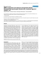
Principle of the bioassay
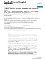
The principle of the bioassay, as shown in Fig. 1, is based
on the ability of TNF-α to induce IL-6 and OPG production
by RA synoviocytes. With TNF-α concentrations ranging
from 0.1 to 100 ng/ml, IL-6 production by synoviocytes
increased in a dose-dependent manner. Addition of inflixi-
mab at 10 µg/ml completely inhibited the effect of TNF-α at
1 ng/ml, and reduced that of TNF-α at 10 ng/ml by 74%
(32.9 ng/ml without versus 8.5 ng/ml with infliximab; Fig.
1a). Similar studies were performed for OPG production.
As for IL-6, OPG production by synoviocytes increased in
a dose-dependent manner in response to TNF-α (Fig. 1b).
Maximal concentrations of IL-6 and OPG were in the same
range up to 35 ng/ml. With regard to IL-6, addition of inflix-
imab inhibited OPG production induced by TNF-α at 10
ng/ml.
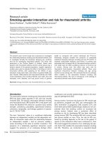
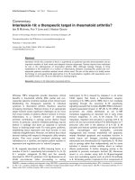
Addition of plasma further increased the effect of exoge-
nous TNF-α. With RA plasma concentrations ranging from
0% to 20% used alone (n = 4), IL-6 production by synovi-
ocytes increased in a dose-dependent manner (Fig. 2a).
This effect was further increased when exogenous TNF-α
at 5 ng/ml was added. The greatest effect was observed
with 10% plasma. An inhibitory effect was often observed
with concentrations of plasma at 20%. In further experi-
ments, 5 ng/ml TNF-α and 10% plasma concentration were
used. With RA plasma samples collected before infliximab
therapy used alone, IL-6 production was 6.5 ± 4.8 ng/ml (n
= 20).
The contribution of TNF bioactivity is shown in Fig. 2b.
Combination of 10% concentration of RA plasma with
TNF-α at 5 ng/ml increased IL-6 production to 43.5 ng/ml.
This effect was inhibited by infliximab (6.5 ng/ml), demon-
strating the specificity for TNF-α.
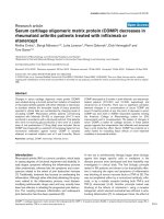
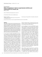
Effect of patient plasma samples collected just before
the first infliximab infusion
Samples were obtained from 42 patients before the first inf-
liximab infusion. The levels of IL-6 produced by stimulation
with 10% plasma and TNF-α (5 ng/ml) are shown in Fig. 3,
stratified by ACR response observed at week 54. Levels
were higher for the good responders (ACR response ≥ 50)
than for poor responders (ACR ≤ 20; 44.4 ± 23.3 ng/ml
versus 27.4 ± 20.9 ng/ml; P = 0.05).
Circulating TNF-α bioactivity modulation by the first
infliximab infusion
For 20 of the patients, samples were collected before and
after the first and the ninth infusions. There was no signifi-
cant clinical difference between the 20 patients and the
rest of the 42 patient cohort (Table 1).
The high IL-6-inducing activity found in samples before
treatment was strongly reduced in samples obtained 4
hours after the first infusion, following TNF-α /anti-TNF-α
complex formation induced by the infliximab infusion (Fig.
4a,4b). This reduction demonstrates the contribution of
TNF-α to the activity following in vivo administration of the
TNF-α inhibitor. This pattern of reduction was associated
with a very good clinical response at 54 weeks for all
patients (ACR 50; n = 11) except one (ACR 20). Con-
versely, no modulation in circulating bioactivity was
observed in patients with a low level of TNF-α bioactivity
Figure 1
Principle of the bioassayPrinciple of the bioassay. Rheumatoid arthritis synoviocytes (10
4
cells/
well) were cultured in 96-well plates and stimulated with increasing
doses of tumour necrosis factor (TNF)-α (0–100 ng/ml). Levels of (a)
IL-6 and (b) osteoprotegerin (OPG) were measured in 48-hour super-
natants. Infliximab at 10 µg/ml was preincubated for 1 hour with TNF-α
before its addition to the culture.
Arthritis Research & Therapy Vol 7 No 1 Marotte et al.
R152
(Fig. 4c). This pattern was associated with a poor clinical
response (ACR <20) except in one patient (ACR 50).
The difference between the IL-6 levels induced with TNF-α
at 5 ng/ml and plasma obtained before and 4 hours after
the first infusion correlated with good clinical response
(40.0 ± 23.7 ng/ml versus 3.4 ± 10.0 ng/ml; P = 0.001;
Fig. 5a). Similarly, OPG levels were measured in the same
supernatants. With regard to IL-6, the difference between
the levels of OPG with plasma samples obtained before
and 4 hours after the first infliximab infusion correlated with
good clinical response (7.0 ± 6.2 ng/ml versus 0.0 ± 3.0
ng/ml; P < 0.05; Fig. 5b). A positive correlation was
observed between changes in IL-6 and OPG production (n
= 20; r = 0.843; P < 0.001).
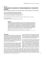
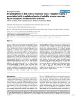
Circulating TNF-α bioactivity modulation by the ninth
infusion
Patients with high circulating bioactivity at the first infusion
could be separated into two subsets. For the first group,
before the ninth infusion, no circulating bioactivity was
detected (Fig. 4a). This pattern was associated with very
good clinical response at 54 weeks in all patients (ACR 50;
n = 6). In the second group, circulating bioactivity was still
present before the ninth infusion but remained sensitive to
further infliximab administration (Fig. 4b). With this pattern,
five out of six patients had an ACR 50 response but one
had a poor response (ACR ≤ 20). No modulation in circu-
lating bioactivity was observed in patients with low circulat-
ing TNF-α bioactivity before infliximab therapy (n = 8; Fig.
4c). A link was observed between these patterns and the
clinical response (χ
2
= 16.6; P < 0.001).
Various clinical and biological parameters were analyzed
according to these patterns. Before treatment, no differ-
ence was observed between joint counts, erythrocyte sed-
imentation rate, C-reactive protein, rheumatoid factor
positivity, or Disease Activity Score 28 (5.4 ± 0.9 in good
responders versus 5.2 ± 1.4 in poor responders; P = 0.6).
Absence of correlation between circulating TNF-α
bioactivity and ELISA levels of TNF-α or soluble
receptors
Levels of TNF-α and of soluble receptors (p55 and p75)
were measured in plasma samples obtained at baseline
from 20 RA patients. The mean TNF-α level was 144.87 ±
130.36 pg/ml in good responders and 153.75 ± 132.93
Figure 2
Pattern of plasma tumour necrosis factor (TNF) bioactivity in rheuma-toid arthritis (RA) patients and healthy individualsPattern of plasma tumour necrosis factor (TNF) bioactivity in rheuma-
toid arthritis (RA) patients and healthy individuals. Using the ability of
TNF-α to stimulate RA synoviocytes, TNF-α (5 ng/ml) was combined
with increasing concentrations of plasma from RA patients (0–20%).
(a) Four RA plasma samples were obtained before infliximab therapy.
(b) RA plasma samples (n = 2) were used at 10% dilution. IL-6 produc-
tion was measured by ELISA in 48-hour supernatants.
Figure 3
Link between circulating tumour necrosis factor (TNF)-α bioactivity and clinical responseLink between circulating tumour necrosis factor (TNF)-α bioactivity and
clinical response. Using the ability of TNF-α to stimulate rheumatoid
arthritis synoviocytes, TNF-α (5 ng/ml) was combined with 20 µl
plasma per well in order to increase the sensitivity of synoviocyte
response. Levels of circulating TNF-α bioactivity were estimated with
plasma samples obtained before infliximab therapy and separated
according to American College of Rheumatology (ACR) clinical
response (good or poor) at 54 weeks (n = 42). A good clinical
response was defined as an ACR 50 response or better (n = 24).
Available online />R153
pg/ml in poor responders (P = 0.97). Similar results were
observed with p55 (2534 ± 1074 pg/ml versus 2436 ±
953 pg/ml; P = 0.57) and p75 (3054.8 ± 673.8 pg/ml ver-
sus 2332.2 ± 921.3 pg/ml; P = 0.84) soluble receptors in
good responders versus poor responders. No correlation
was observed between circulating TNF-α bioactivity and
TNF-α or soluble receptors, or the difference between
TNF-α and p55 and p75 levels. Similarly, no negative cor-
relation was observed between levels of circulating TNF-α
bioactivity and of p55 or p75 soluble receptors.
Discussion
Prediction of the response of RA to treatment remains a hot
topic. There is no evidence that simple determination of
plasma TNF-α levels by ELISA allows such prediction for
treatment with TNF-α inhibitors. However, it still makes
sense that patients producing high levels of TNF-α will be
more sensitive to TNF-α inhibition.
Figure 4
Patterns of plasma tumour necrosis factor (TNF)-α bioactivity in rheu-matoid arthritis (RA) patients treated with infliximabPatterns of plasma tumour necrosis factor (TNF)-α bioactivity in rheu-
matoid arthritis (RA) patients treated with infliximab. Using this bioassay
three patterns were observed and linked to the clinical response in the
20 patients with a 1-year follow up. (a) The first pattern showed good
ability of the plasma to induce IL-6 production before infliximab therapy,
followed by complete inhibition 4 hours after the first infusion. Plasma
samples before the ninth infusion had low IL-6-inducing activity. This
pattern was associated with an American Colege of Rheumatology
(ACR) 50 response at 54 weeks (n = 6). (b) In the second pattern the
effect of the first infusion was similar to that in the first pattern. How-
ever, high IL-6 inducing activity was still present before the ninth infu-
sion but remained sensitive to treatment. In this pattern, five patients
had an ACR 50 response but one had no response. (c) In the last pat-
tern the first plasma samples had a moderate or no effect on IL-6 pro-
duction. This activity was not sensitive to infliximab infusion. All patients
(n = 8) had a poor clinical response (ACR <20). A link was observed
between these patterns and clinical response (χ
2
= 16.6; P < 0.001).
Figure 5
Link between changes in IL-6 and osteoprotegerin (OPG) production by rheumatoid arthritis (RA) synoviocytes and clinical responseLink between changes in IL-6 and osteoprotegerin (OPG) production
by rheumatoid arthritis (RA) synoviocytes and clinical response.
Changes, expressed as ∆IL-6 and ∆OPG production by RA synovio-
cytes before and 4 hours after the first infliximab infusion, are separated
according to the American College of Rheumatology (ACR) clinical
response (good or poor) at 54 weeks (n = 20). A good clinical
response was defined as an ACR 50 response or better (n = 12).
Arthritis Research & Therapy Vol 7 No 1 Marotte et al.
R154
A system was established to evaluate the circulating TNF-
α-related bioactivity in plasma. Exogenous TNF-α alone
stimulates IL-6 production and this effect can be abrogated
by first incubating TNF-α with infliximab before exposure to
the synovial cells. When plasma is combined, cells respond
to free TNF-α and not to inactive TNF-α bound to specific
soluble receptors and to other less specific binding sites
on proteins. This bioassay was based on the IL-6 produc-
tion induced by the combination of TNF-α and plasma.
Addition of exogenous TNF-α was used to detect the pres-
ence of circulating inhibitors. Such inhibitors in plasma are
probably involved in the lower effect of 20% plasma con-
centration as compared with the effect seen with a 10%
concentration (Fig. 2a).
When the system was applied to explain part of the heter-
ogeneity in treatment response, a third of the patients
showed strong and prolonged inhibition in circulating TNF-
α bioactivity, suggesting the critical contribution of sys-
temic TNF-α in these patients. In another third of the
patients circulating TNF-α bioactivity was inhibited by inflix-
imab infusion for a short time, because bioactive TNF-α
reappeared but was again inhibited by the next infliximab
infusion. This profile suggested partial inhibition, although
the clinical benefit was still very significant. In two thirds of
the samples, the bioassay measured a strong inhibition of
circulating TNF-α-related activity during the first infusion.
The link between strong anti-TNF-α activity induced by the
first infusion and the good clinical response confirms the
key role played by TNF-α in approximately two thirds of the
RA patients. This result is in accord with results from the
ATTRACT study [8]. In contrast, in the last third of the
patients the assay suggested no contribution or a reduced
contribution of TNF-α bioactivity either before or after the
first infusion of infliximab. All plasma samples from these
patients inhibited the IL-6 production usually induced by
TNF-α. This profile suggests that these patients may have
a high level of innate neutralizing TNF-α activity and/or no
circulating active TNF-α. Patients with this pattern
exhibited a poor clinical response, suggesting that their RA
was not much driven by TNF-α activity alone but probably
by other cytokines or mechanisms.
The heterogeneity in these patterns may be explained by
higher disease activity for responders than nonresponders,
but no difference was observed in clinical and biological
parameters before treatment. One way to view the
difference among patients with high circulating TNF-α bio-
activity initially is the link observed between higher trough
concentrations of infliximab in RA patient serum and good
response and a reduced progression of radiographic joint
damage [10]. This latter study suggested that RA patients
with a poor clinical response tend to eliminate infliximab
more rapidly from their circulation.
RA synoviocytes produce higher levels of OPG than do
peripheral blood mononuclear cells or synovial fluid mono-
nuclear cells, but they do not produce soluble receptor acti-
vator of nuclear factor-κB ligand [5]. Accordingly, we
focused on OPG, which is produced in response to TNF-
α. Similar results were observed for IL-6 and OPG produc-
tion, although changes in IL-6 levels appeared more sensi-
tive. This is related to the very low levels of IL-6 produced
by resting synoviocytes. Accordingly, the predictive value
of changes in IL-6 was better than that of OPG. Our find-
ings extended previous studies indicating a correction in
high OPG serum levels in RA patients treated with inflixi-
mab [5].
No correlation was observed between circulating TNF-α
bioactivity and its protein concentration in plasma meas-
ured by ELISA. Circulating TNF-α bioactivity levels could
not be calculated as free protein TNF-α taking into account
the levels of TNF-α and soluble receptors (p55 and p75)
measured by ELISA. This discrepancy further indicates the
usefulness of a bioassay when function is the key. Such
complexity has been observed when trying to explain loss
of infliximab response by the induction of anti-mouse
antibodies [11]. Once again, only the demonstration of
inhibitory activity in vivo would allow such a conclusion to
be drawn.
Conclusion
In conclusion, this bioassay was able to predict correctly
the clinical response in 69% of cases (29/42). Taking into
account the effect of the first infusion increased the value
to 90%. However, a simplified assay would need to be
designed for routine application.
Competing interests
The author(s) declare that they have no competing
interests.
Authors' contributions
HM conducted the experiments and wrote the paper. WM
took OPG measurements. PM directed the research.
References
1. Elliott MJ, Maini RN, Feldmann M, Kalden JR, Antoni C, Smolen JS,
Leeb B, Breedveld FC, Macfarlane JD, Bijl H: Randomised dou-
ble-blind comparison of chimeric monoclonal antibody to
tumour necrosis factor alpha (ca2) versus placebo in rheuma-
toid arthritis. Lancet 1994, 344:1105-1110.
2. Lipsky PE, van der Heijde DM, St Clair EW, Furst DE, Breedveld
FC, Kalden JR, Smolen JS, Weisman M, Emery P, Feldmann M, et
al.: Infliximab and methotrexate in the treatment of rheumatoid
arthritis. Anti-tumor necrosis factor trial in rheumatoid arthritis
with concomitant therapy study group. N Engl J Med 2000,
343:1594-1602.
3. Weinblatt ME, Keystone EC, Furst DE, Moreland LW, Weisman
MH, Birbara CA, Teoh LA, Fischkoff SA, Chartash EK: Adalimu-
mab, a fully human anti-tumor necrosis factor alpha mono-
clonal antibody, for the treatment of rheumatoid arthritis in
patients taking concomitant methotrexate: the ARMADA trial.
Arthritis Rheum 2003, 48:35-45.
Available online />R155
4. Bathon JM, Martin RW, Fleischmann RM, Tesser JR, Schiff MH,
Keystone EC, Genovese MC, Wasko MC, Moreland LW, Weaver
AL, et al.: A comparison of etanercept and methotrexate in
patients with early rheumatoid arthritis. N Engl J Med 2000,
343:1586-1593.
5. Ziolkowska M, Kurowska M, Radzikowska A, Luszczykiewicz G,
Wiland P, Dziewczopolski W, Filipowicz-Sosnowska A, Pazdur J,
Szechinski J, Kowalczewski J, et al.: High levels of osteoprote-
gerin and soluble receptor activator of nuclear factor kappa B
ligand in serum of rheumatoid arthritis patients and their nor-
malization after anti-tumor necrosis factor alpha treatment.
Arthritis Rheum 2002, 46:1744-1753.
6. Chabaud M, Miossec P: The combination of tumor necrosis fac-
tor alpha blockade with interleukin-1 and interleukin-17 block-
ade is more effective for controlling synovial inflammation and
bone resorption in an ex vivo model. Arthritis Rheum 2001,
44:1293-1303.
7. Arnett FC, Edworthy SM, Bloch DA, McShane DJ, Fries JF, Cooper
NS, Healey LA, Kaplan SR, Liang MH, Luthra HS: The american
rheumatism association 1987 revised criteria for the classifi-
cation of rheumatoid arthritis. Arthritis Rheum 1988,
31:315-324.
8. Maini R, St Clair EW, Breedveld F, Furst D, Kalden J, Weisman M,
Smolen J, Emery P, Harriman G, Feldmann M, Lipsky P: Infliximab
(chimeric anti-tumour necrosis factor alpha monoclonal anti-
body) versus placebo in rheumatoid arthritis patients receiv-
ing concomitant methotrexate: a randomised phase III trial.
ATTRACT study group. Lancet 1999, 354:1932-1939.
9. Felson DT, Anderson JJ, Boers M, Bombardier C, Furst D, Gold-
smith C, Katz LM, Lightfoot R Jr, Paulus H, Strand V: American
College of Rheumatology. Preliminary definition of improve-
ment in rheumatoid arthritis. Arthritis Rheum 1995, 38:727-735.
10. St Clair EW, Wagner CL, Fasanmade AA, Wang B, Schaible T,
Kavanaugh A, Keystone EC: The relationship of serum inflixi-
mab concentrations to clinical improvement in rheumatoid
arthritis: results from ATTRACT, a multicenter, randomized,
double-blind, placebo-controlled trial. Arthritis Rheum 2002,
46:1451-1459.
11. Baert F, Noman M, Vermeire S, Van Assche G, D'Haens G, Car-
bonez A, Rutgeerts P: Influence of immunogenicity on the long-
term efficacy of infliximab in Crohn's disease. N Engl J Med
2003, 348:601-608.