Báo cáo y học: "A2B adenosine receptor activity is reduced in neutrophils from patients with systemic sclerosis" ppt
Bạn đang xem bản rút gọn của tài liệu. Xem và tải ngay bản đầy đủ của tài liệu tại đây (305.46 KB, 7 trang )
Open Access
Available online />R189
Vol 7 No 2
Research article
A
2B
adenosine receptor activity is reduced in neutrophils from
patients with systemic sclerosis
Laura Bazzichi
1
, Letizia Trincavelli
2
, Alessandra Rossi
2
, Francesca De Feo
2
, Antonio Lucacchini
2
,
Stefano Bombardieri
1
and Claudia Martini
2
1
Department of Internal Medicine, Division of Rheumatology, University of Pisa, Pisa, Italy
2
Departments of Psychiatry, Neurobiology, Pharmacology and Biotechnology, University of Pisa, Pisa, Italy
Corresponding author: Laura Bazzichi,
Received: 20 Apr 2004 Revisions requested: 24 Jun 2004 Revisions received: 22 Oct 2004 Accepted: 26 Oct 2004 Published: 10 Dec 2004
Arthritis Res Ther 2004, 7:R189-R195 (DOI 10.1186/ar1468)
http://arthr itis-research.com/conte nt/7/2/R189
© 2004 Bazzichi et al., licensee BioMed Central Ltd.
This is an Open Access article distributed under the terms of the Creative Commons Attribution License ( />2.0), which permits unrestricted use, distribution, and reproduction in any medium, provided the original work is cited.
Abstract
We conducted the present study to investigate protein
expression and functioning of A
2A
and A
2B
adenosine receptors
(ARs) in neutrophils of patients affected by systemic sclerosis
(SSc). The presence of A
2A
and A
2B
ARs was assessed by
immunoblotting using specific antibodies. Equilibrium A
2A
and
A
2B
ARs binding parameters were evaluated by radioligand
binding assay. Functional studies were conducted to investigate
coupling of the A
2B
AR to the adenylyl cyclase pathway. This is
the first report of the use of Western blot analysis to confirm the
presence of A
2A
and A
2B
ARs in human neutrophils. No
significant changes in A
2A
AR binding parameters or expression
levels were detected between SSc patients and healthy control
individuals. A significant decrease (65%) in the maximum
density of A
2B
AR binding sites occurred in SSc neutrophils,
whereas no changes in the affinity constant values were found.
Moreover, a decrease in A
2B
AR mediated adenylyl cyclase
activity was observed in patients with SSc. Our findings
demonstrate the occurrence of selective alterations in A
2B
AR
density and signalling in SSc.
Keywords: adenosine, A
2
adenosine receptors, neutrophils, receptor binding, systemic sclerosis
Introduction
Systemic sclerosis (SSc), also known as scleroderma, is a
connective tissue disease of unknown aetiology. Possibly
an autoimmune disorder, it is accompanied in the vast
majority of cases by the presence of antinuclear antibodies
[1]. SSc may affect virtually any organ of the body, includ-
ing skin, gastrointestinal tract, lungs, heart, kidneys, and
musculoskeletal system. Altered connective tissue metabo-
lism can cause either localized or diffuse thickening of the
skin, while inflammation is associated with endothelial dam-
age. Clinically, microvascular disturbance, teleangiectasia,
Raynaud's phenomenon, polyarthralgia and polyarthritis, as
well as oesophageal hypomobility, visceral muscolaris
mucosa damage and pulmonary fibrosis, have been
described [2].
The mechanisms leading to endothelial damage, inflamma-
tion and fibrosis are unclear. Reactive oxygen species in
neutrophils may increase the extent of inflammation and
fibrosis during the respiratory burst and could be involved
in endothelial damage [3]. The endothelial cells of micro-
vessels are deficient in the synthesis of catalase, which pro-
vides natural defence against superoxide damage, and
appear to be particularly susceptible to superoxide injury
during reperfusion [4].
Adenosine is an important endogenous regulator of neu-
trophil functioning. It is released intracellularly and modu-
lates neutrophil activity by interacting with specific surface
receptors [5]. Distinct adenosine receptor (AR) subtypes
A
1
, A
2A
, A
2B
and A
3
have been identified and their functions
characterized in neutrophils. Specifically, activation of A
1
ARs enhances chemotaxis, phagocytosis and adherence
AC = adenylyl cyclase; ADA = adenosine deaminase; AR = adenosine receptor; B
max
= maximum number of binding sites; CGS
21680
= (2-p-[2-car-
bowyethyl]pheylethylamino)-5'N-ethylcarboxamidoadenosine; CPA = cyclopentyladenosine; Kd = affinity constant; NECA = 5'-N-ethylcarboxami-
doadenosine; R-PIA = R-N6-phenylisopropyladenosine; SCH
58261
= 5-amino-7-(phenylethyl)-2-(2-furyl)-pyrazolo(4,3-e)-1,2,4-triazolo(1,5-
c)pyrimidine; SSc = systemic sclerosis.
Arthritis Research & Therapy Vol 7 No 2 Bazzichi et al.
R190
[6,7]; A
2A
ARs inhibit reactive oxygen species generation,
phagocytosis and adherence [8-10]; and A
2A
and A
3
ARs
inhibit neutrophil degranulation [11-14]. Adenosine has
been shown to prevent the release of vascular endothelial
growth factor from neutrophils via A
2B
AR activation [15].
Because activation of ARs reduces both immune and
inflammatory responses, adenosine release has been
hypothesized to be a possible mechanism of cell self-pro-
tection from activated neutrophils [5]. An increase in ade-
nosine deaminase activity has been described in patients
with SSc, suggesting an alteration in adenosine control
mechanisms in this disease [16,17].
In the present study we analyzed A
2A
and A
2B
AR subtypes
in neutrophils from patients affected by SSc by means of
expression analysis, radioligand binding assays and func-
tional studies.
Methods
Chemicals and reagents
Bacitracine, benzamidine, trypsin inhibitor, sodium
orthovanadate, Nonidet P-40, SDS, phenylsulfonylfluoride,
aprotinin and adenosine deaminase (ADA) were purchased
from Sigma (St. Louis, MO, USA). Unlabelled AR agonists/
antagonists and the anti-β-actin antibody were supplied by
RBI/Sigma (St. Louis, MO, USA). [
3
H]CGS
21680
(CGS
21680
= [2-p-(2-carbowyethyl)phenylethylamino]-5'-
N-ethylcarboxamidoadenosine), [
3
H]NECA (NECA = 5'-N-
ethylcarboxamidoadenosine), and [
32
P]α-ATP were sup-
plied by NEN Life Sciences (Köln, Germany). Electrophore-
sis reagents were purchased from BioRad (Munchen,
Germany). A
2A
AR and A
2B
AR antibodies were supplied by
Alpha Diagnostic (San Antonio, TX, USA). All other chemi-
cals were from standard commercial sources.
Patients
Twenty-six patients affected by SSc were included in the
study (22 women and 4 men; mean age ± standard devia-
tion 53.0 ± 11.3 years). They all fulfilled standard criteria of
the American College of Rheumatology for SSc. Sixteen
patients were anticentromere antibody positive and four
were SCL-70 positive. Limited symptoms of disease,
involving skin thickness alterations to the face, hands and
feet, were present in 18 patients (mean disease duration
<5 years, skin score range [according to the modified Rod-
nan total skin thickness score] 10–21). Diffuse symptoms
with more extensive skin involvement were present in eight
patients (mean disease duration <5 years, total skin thick-
ness score range 27–30). The activity score [18] varied
between 0.5 and 3.5 and the severity score [19] between
2 and 6. The erythrocyte sedimentation rate was 24 ± 23
mm/hour (mean ± standard deviation).
Control samples were obtained from 26 healthy volunteers,
who were similar to the patients included in the study in
terms of sex distribution and age (20 women and 6 men;
mean age ± standard deviation 49.0 ± 9.2 years). Informed
consent to participate in the study was obtained from all
individuals.
Sample collection and neutrophil preparation
Venous blood (20 ml) was drawn between 08:00 and
09:00 a.m. from fasting individuals by antecubital venipunc-
ture, collected in heparinized (10 IU/L) plastic tubes and
processed immediately. Neutrophils were isolated follow-
ing the Boyum method [20] with some modifications.
Western blot analysis
Neutrophils were lysed in RIPA buffer (150 mmol/l NaCl,
50 mmol/l Tris-HCl, pH 8, 0.5% sodium deoxhycolate, 1%
Nonidet P-40, 1 mmol/l phenylsulfonylfluoride, 10 µg/ml
aprotinin, 100 µmol/l sodium orthovanadate) for 1 hour at
4°C. After centrifugation at 15,000 g for 30 min, soluble
fractions were assayed for protein content using BioRad
protein assay. Equivalent amounts of proteins (50 µg/sam-
ple) were analyzed by SDS-PAGE, using 10% (weight/vol)
polyacrylamide resolving gels. Protein bands were trans-
ferred to nitrocellulose and probed with 0.1 µg/ml rabbit
anti-human A
2A
AR or A
2B
AR antibodies.
A
2A
AR antibody is an affinity-purified rabbit polyclonal anti-
body raised against a peptide mapping to the carboxyl-ter-
minus of A
2A
AR. It specifically reacts with human, bovine,
rat and pig A
2A
receptors and does not cross-react with A
1
,
A
2B
, or A
3
AR subtypes. A
2B
AR antibody is an affinity-puri-
fied rabbit polyclonal antibody raised against a region that
corresponds to the second extracellular domain of A
2B
AR
of human origin.
After washing, membranes were incubated with anti-rabbit
secondary antibody conjugated to horseradish peroxidase
for 2 hours at room temperature, and bands were visualized
by chemiluminescence, in accordance with the manufac-
turer's instructions (Sigma-Aldrich). Membranes were re-
probed with an anti-β-actin antibody for normalization.
Binding assay
For membrane preparation, cells were washed twice with
10 mmol/l Tris-HCl buffer, pH 7.4, containing 10 mmol/l
MgCl
2
, in the presence of protease inhibitors (200 µg/ml
bacitracine, 160 µg/ml benzamidine, 20 µg/ml trypsin
inhibitor [T1]) and centrifuged at 48,000 g for 15 min at
4°C. Pellets were diluted in 20 volumes of T1 buffer,
treated with ADA (2 IU/ml) for 60 min at 37°C to remove
endogenous adenosine, and washed twice with 50 mmol/l
Tris-HCl buffer, pH 7.4, containing 10 mmol/l MgCl
2
(T2).
A
2A
AR binding assay was performed by using a specific
radiolabelled A
2A
AR agonist, namely [
3
H]CGS
21680
. Aliq-
uots of neutrophil membranes (0.2–0.3 mg protein) were
Available online />R191
incubated with different [
3
H]CGS
21680
concentrations (5–
30 nmol/l) in a final volume of 250 µl of T2 buffer. Nonspe-
cific binding was determined in the presence of 100 µmol/
l NECA. After 90 min incubation at 25°C, the binding reac-
tion was terminated by vacuum filtration through Whatman
GF/C glass fibre filters (Whatman, Maidstone, UK), accom-
panied by three washes with ice-cold T2 buffer (4 ml). A
2A
AR specificity was evaluated through competition experi-
ments, using different AR ligands.
A
2B
AR binding assay was performed using 20 nmol/l
[
3
H]NECA in the presence of 50 nmol/l cyclopentyladeno-
sine (CPA) and 100 nmol/l SCH
58261
(SCH
58261
= 5-
amino-7-[phenylethyl]-2-[2-furyl]-pyrazolo [4,3-e]-1,2,4-tri-
azolo [1,5-c]pyrimidine) to prevent [
3
H]NECA binding to A
1
and A
2A
ARs, respectively [21]. Scatchard analysis was
performed on competition experiments carried out in the
presence of unlabelled NECA at concentrations ranging
from 50 nmol/l to 2 mmol/l. Aliquots of neutrophil mem-
branes (0.2–0.4 mg proteins) were incubated in a final vol-
ume of 250 µl T2 buffer. Nonspecific binding was
evaluated in the presence of 100 µmol/l NECA. After 90
min incubation at 0°C, the reaction was terminated either
by vacuum filtration through Whatman GF/C glass fibre fil-
ters, accompanied by three washes with ice-cold T2 buffer
(4 ml), or by centrifugation at 2900 g for 15 min at 4°C. A
2B
AR specificity was evaluated through competition experi-
ments, using different AR ligands.
Adenylyl cyclase assay
Neutrophils were homogenized in buffer solution contain-
ing 10 mmol/l Hepes, 1 mmol/l EGTA and 10 mmol/l
NaCl
2
, and then centrifuged at 46,500 g for 20 min at 4°C.
Pellets were resuspended in 10 volumes of 10 mmol/l
Hepes, containing protease inhibitors (200 µg/ml bacitrac-
ine and 160 µg/ml benzamidine), incubated for 30 min at
30°C with 2 U/ml ADA, and centrifuged. Adenylyl cyclase
(AC) activity was measured as described by Salomon [22]
and Johnson and Salomon [23], with some modifications.
NECA-mediated stimulation of AC activity was assessed
by incubating aliquots of membranes with increasing
NECA concentrations from 0.01 nmol/l to 10 µmol/l. The
reaction was started by adding membrane aliquots (10–50
µg proteins/tube), conducted for 15 min at 24°C, and then
stopped by transferring samples on ice and adding 500 µl
ice-cold stop solution (120 mmol/l zinc acetate, 144 mmol/
l Na
2
CO
3
). The stop solution contained [
3
H]cAMP
(10,000–15,000 cpm/sample) to monitor column recovery.
Newly formed ZnCO
3
allowed precipitation of residual
ATP, discarded through centrifugation at 2700 g for 8 min.
Supernatants containing both [
32
P]α-cAMP and [
3
H]cAMP
were further purified by double-step Dowex-Alumina chro-
matography and counted by means of a β-counter (Packard
Tricarb 1600; Perkin Elmer, Wellesley, MA, USA).
To evaluate A
2B
AR mediated cAMP accumulation, the
reaction was carried out in the presence of selective A
2A
antagonist SCH
58261
at a concentration (100 nmol/l) able
to block A
2A
receptors completely [21].
Data and statistical analysis
Affinity constant values (Kd) and maximum number of bind-
ing sites (B
max
) were calculated using the nonlinear multi-
purpose curve-fitting computer program Graph-Pad Prism
The 50% inhibitory concentration values were calculated
using the same program and converted to Ki values
through the Cheng and Prusoff equation.
A GS-670-BIO-RAD imaging densitometer was used for
semiquantitative analysis of immunoblots. Partial F test (P
< 0.01) was used to determine binding data with the best
fit to a one-site or two-site model. Differences in binding
parameters between SSc patients and control individuals
were evaluated by one-way analysis of variance.
Results
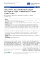
In both control and SSc neutrophils, Western blot analysis
identified two specific immunoreactive bands of 45 kDa
and 50 kDa, corresponding to A
2A
and A
2B
ARs, respec-
tively (Fig. 1). This confirmed the presence of both AR sub-
types in human neutrophils.
To characterize ARs, binding assays were conducted in
neutrophil membrane fractions. SSc patients were ran-
domly divided into two subgroups in order to obtain large
amounts of protein, as required by the experiments.
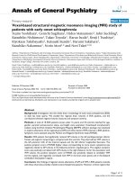
The selective A
2A
AR agonist [
3
H]CGS
21680
identified a
homogenous population of binding sites in control individ-
uals. Kd and B
max
values were 25 ± 1.3 nmol/l and 35 ± 2.4
fmol/mg protein, respectively (Fig. 2). Competition experi-
ments using [
3
H]CGS
21680
in combination with a variety of
A
2A
ligands revealed a pharmacological profile typical for
A
2A
ARs (R-PIA [R-N6-phenylisopropyladenosine] > teofil-
line > NECA > SCH
58261
; data not shown). Scatchard
analysis for SSc neutrophils revealed no significant differ-
ences in Kd and B
max
between patients (mean values: Kd =
23 ± 1.8 nmol/l, B
max
= 40 ± 3.2 fmol/mg protein) and
healthy control individuals (P > 0.05; Fig. 2), suggesting
that no alteration in A
2A
binding sites occurs in SSc. In
agreement with this, densitometric analysis of immunoblots
showed no significant changes in A
2A
AR immunoreactive
bands in SSc neutrophils relative to controls (optical den-
sity: 0.11 ± 0.03 for patients versus 0.15 ± 0.02 for
controls).
A
2B
AR binding sites were identified using [
3
H]NECA as
radioligand in the presence of 50 nmol/l CPA and 100
nmol/l SCH
58261
, to prevent nonspecific binding to A
1
and
A
2A
AR subtypes. We performed competition experiments
Arthritis Research & Therapy Vol 7 No 2 Bazzichi et al.
R192
using a wide range (50 nmol/l to 2 mmol/l) of [
3
H]NECA
concentrations to allow the identification of A
2B
AR low-
affinity binding sites. Data analysis revealed that the one-
site model produced a significantly better fit than the two-
site model (P < 0.05), both in control and SSc neutrophils.
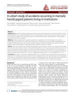
In our experimental conditions, control neutrophils exhib-
ited the presence of low-affinity binding sites with Kd and
B
max
values of 476 ± 34 nmol/l and 3696 ± 210 fmol/mg,
respectively (Fig. 3). Competition experiments using
[
3
H]NECA in combination with a variety of AR ligands
revealed a pharmacological profile typical for A
2B
ARs (R-
PIA > teofilline > SCH
58261
= MRS1220 > DPCPX > 2Cl-
adenosine > NECA > MRS1706; Table 1). Scatchard anal-
ysis for SSc neutrophils showed no significant differences
in Kd and B
max
between the two subgroups of patients.
However, a significant alteration in B
max
was found relative
to controls, whereas Kd values remained unaltered. Overall,
mean values for Kd and B
max
in SSc were 469 ± 35 nmol/l
and 1292 ± 98 fmol/mg protein, respectively (P < 0.05;
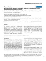
Fig. 3). Moreover, experiments conducted in individual
patients using a concentration of NECA of 500 nmol/l
showed similar specific binding values (expressed as fmol/
mg protein), confirming the homogeneity of A
2B
AR sites
between SSc subgroups (Fig. 4). The alteration in A
2B
AR
levels in SSc patients was confirmed by immunoblotting
assay. Densitometric analysis of immunoreactive bands
showed a reduction in A
2B
expression in SSc patients (opti-
cal density 0.22 ± 0.04) as compared with controls (optical
density 0.40 ± 0.06; P < 0.05; Fig. 1).
Functional coupling of A
2B
ARs to stimulatory G proteins in
neutrophil membranes was assessed by evaluating the
effects of the agonist NECA (in the presence of 100 nmol/
l SCH
58261
) on AC activity. NECA stimulated AC activity in
a concentration dependent manner. Dose-response curves
revealed significant differences between SSc patients
Figure 1
Immunoblotting analysis of A
2A
and A
2B
adenosine receptors (ARs) from systemic sclerosis (SSc) neutrophils and controlsImmunoblotting analysis of A
2A
and A
2B
adenosine receptors (ARs) from
systemic sclerosis (SSc) neutrophils and controls. Cells obtained from
26 healthy volunteers and 26 SSc patients were lysed as described in
the Methods section. Equal amounts of protein (50 µg) were separated
on polyacrylamide gel, blotted and probed with 0.1 µg/ml rabbit anti-
human A
2A
AR or A
2B
AR antibodies. Immunoreactive bands were visu-
alized according to electrogenerated chemiluminescence protocol. A
2A
and A
2B
AR antibodies recognized immunoreactive bands of 45 kDa
and 50 kDa, respectively. (a) Representative experiment performed on
neutrophils from one healthy volunteer and one SSc patient. (b) Densit-
ometric analysis of A
2A
and A
2B
AR immunoreactive bands from 26
healthy volunteers and 26 SSc patients. Graph bars: mean ± standard
error of band density, normalized to β-actin. White bars are controls;
grey bars are SSc patients.
Figure 2
Representative Scatchard plot of [
3
H]CGS
21680
saturation binding dataRepresentative Scatchard plot of [
3
H]CGS
21680
saturation binding
data. Empty circles indicate neutrophil membranes from healthy volun-
teers (affinity constant [Kd] = 25 ± 1.3 nmol/l; maximum number of
binding sites [B
max
] = 35 ± 2.4 fmol/mg); filled circles indicate neu-
trophil membranes from systemic sclerosis (SSc) patients overall (Kd =
23 ± 1.8 nmol/l; B
max
= 40 ± 3.2 fmol/mg). Assays were performed in
triplicate.
Available online />R193
(EC
50
= 373 ± 26 nmol/l; E
max
= 35 ± 2.9%) and controls
(EC
50
= 165 ± 9.3 nmol/l; E
max
= 43 ± 3.2%), suggesting
an alteration in A
2B
AR responsiveness in SSc (Fig. 5).
Discussion
In the present study we analyzed A
2A
and A
2B
AR subtypes
in neutrophils of patients affected by SSc, by means of
Western blot, radioligand binding techniques and func-
tional studies. This is the first report of use of Western blot
analysis to confirm the presence of A
2A
and A
2B
ARs in
human neutrophils.
A
2A
and A
2B
AR equilibrium binding parameters were meas-
ured using radioligand binding assays. Scatchard analysis
of [
3
H]CGS
21680
saturation binding to A
2A
AR showed no
significant difference in B
max
or Kd between SSc neu-
trophils and controls, suggesting that the A
2A
AR subtype
remained unaltered in SSc. Conversely, when A
2B
AR was
analyzed a reduction in B
max
(65%) was observed, with no
significant change in Kd values.
A
2B
ARs are known to be low-affinity adenosine binding
sites. Competition experiments using a variety of A
2B
AR
agonists and antagonists revealed a pharmacological pro-
file typical of A
2B
ARs, which is consistent with studies con-
ducted in transfected cell models. Our findings represent
Table 1
Specificity of [
3
H]NECA binding to A
2B
adenosine receptors in control neutrophil membranes
[
3
H]NECA Ki (µmol/l)
NECA 0.315 ± 0.028
2 Cl-adenosine 0.954 ± 0.600
R-PIA 1000 ± 86
SCH
58261
>10
Teofilline 47 ± 3.5
MRS1706 0.005 ± 0.0003
DPCPX 2 ± 0.12
MRS1220 >10
Competition experiments were performed, incubating aliquots of neutrophil membranes with 20 nmol/l [
3
H]NECA (plus 50 nmol/l CPA and 100
nmol/l SCH
58261
) in the presence of increasing ligand concentrations. Ki values are expressed as mean ± SEM of three separate experiments. Ki
values were calculated from IC
50
values (concentration of drug causing 50% inhibition of specific binding) using the Cheng and Prusoff equation.
Figure 3
Representative Scatchard plot of [
3
H]NECA saturation binding dataRepresentative Scatchard plot of [
3
H]NECA saturation binding data.
Competition binding experiments were performed, incubating aliquots
of neutrophil membranes with 20 nmol/l [
3
H]NECA and different NECA
concentrations (50 nmol/l to 2 mmol/l), in the presence of 50 nmol/l
CPA and 100 nmol/l SCH
58261
. Empty circles indicate neutrophil mem-
branes from healthy volunteers (affinity constant [Kd] = 476 ± 34 nmol/
l, maximum number of binding sites [B
max
] = 3696 ± 210 fmol/mg);
filled circles indicate neutrophil membranes from systemic sclerosis
(SSc) patients overall (Kd = 469 ± 35 nmol/l, B
max
= 1292 ± 98 fmol/
mg). Assays were performed in triplicate.
Figure 4
A
2B
adenosine receptor binding experiments performed in individual patients using NECA at 500 nmol/l concentrationA
2B
adenosine receptor binding experiments performed in individual
patients using NECA at 500 nmol/l concentration. Neutrophils were
obtained from healthy volunteers (n = 26) and systemic sclerosis (SSc)
patients (n = 26). Horizontal lines indicate the mean values.
Arthritis Research & Therapy Vol 7 No 2 Bazzichi et al.
R194
the first characterization of A
2B
ARs in neutrophils with
binding experiments.
In order to analyze a population of nonhomogenous
patients and to evaluate the impact of the disease on A
2
ARs, SSc patients were randomly divided into two sub-
groups. No difference was found when the two groups
were compared, suggesting that different degrees of dis-
ease severity and activity had no impact on the assays, but
that the disease per se is required to modulate levels and
functioning of A
2B
receptors.
Functional studies were performed to investigate whether
the decrease in level of A
2B
ARs was accompanied by alter-
ations in receptor responsiveness. An evaluation of the abil-
ity of NECA to increase AC activity revealed functional
coupling of A
2B
receptors to G proteins. In SSc patients a
significant reduction (by more than 50%) in NECA potency
was observed, without any effect on agonist efficacy.
Our findings suggest that a selective reduction in A
2B
AR
levels and responsiveness occurred in SSc. Alterations in
the expression and functionality of A
2B
ARs (low-affinity
ARs) in patients with SSc may be responsible for the
increase in free oxygen radicals, and consequent oxidative
damage, that characterizes SSc. This would account for
impaired control of hypoxic and inflammatory processes.
In neutrophils it has long been known that adenosine and
its analogues inhibit O
2
-
generation, phagocytosis and cell
adherence by occupying specific A
2
ARs. Because
hypoxia, ischaemia and inflammation can stimulate adenos-
ine production, A
2
AR regulation has been postulated to be
a self-protective mechanism for cells from activated neu-
trophils [24]. Eltzschig and coworkers [25] reported that
A
2B
ARs are selectively upregulated in endothelial cells by
hypoxia (more than fivefold increase in mRNA), which is
associated with ATP hydrolysis and release of adenosine.
Taken together, these findings show some coordination
between AR transcription and nucleoside signalling at the
vascular interface during hypoxia. We might speculate that
chronic inflammatory conditions in SSc patients impaired
regulatory mechanisms mediated by the anti-inflammatory
effects of adenosine via A
2B
AR activation. In addition, it
was reported by Visser and coworkers [26] that increases
in cAMP in activated neutrophils play an anti-inflammatory
role. The reduced activation of cAMP we observed in SSc
patients might be correlated with the inability of these
patients to control the inflammatory process.
It was no surprise to find an alteration in adenosinergic sys-
tem responsiveness in SSc. In fact, adenosine produces a
constellation of responses, including anti-inflammatory
actions and vasodilatation, mediated through interactions
with high-affinity receptor subtype A
2A
and low-affinity
receptor subtype A
2B
. Moreover, in SSc and related disor-
ders, alterations in adenosine metabolism have been sug-
gested. Indeed, purine analogue 2-chlorodeoxyadenosine,
which is utilized for the treatment of such chronic disorders
[27,28], appears to reduce the number of abnormal
fibroblasts.
A
2B
ARs were initially thought to be of lesser physiological
relevance because of their relatively low affinity for adenos-
ine, and it was only recently that important functions attrib-
utable to A
2B
ARs were discovered. A pivotal role for them
was postulated in inflammatory pathological conditions,
when adenosine is released at high levels (up to the micro-
molar range). In light of our findings, a closer examination of
A
2B
AR functions may be valuable because of the potential
therapeutic importance of these receptors as targets for
treatment with selective agents.
Conclusion
Our findings demonstrated a reduction in A
2
low-affinity
(A
2B
) AR density and functioning in neutrophils of patients
affected by SSc, suggesting an alteration in adenosinergic
system responsiveness. This reduction could relate to the
increased production of free oxygen radicals and conse-
quent oxidative damage that characterize SSc, highlighting
an impairment in the ability of neutrophils to control hypoxia
and inflammation.
No differences between two randomly selected subgroups
of SSc patients were found, thus suggesting that different
degrees of disease severity and activity had no impact on
Figure 5
A
2B
adenosine receptor (AR)-mediated stimulation of adenylyl cyclase activity in control (empty circles) and systemic sclerosis (SSc; filled cir-cles) neutrophil membranesA
2B
adenosine receptor (AR)-mediated stimulation of adenylyl cyclase
activity in control (empty circles) and systemic sclerosis (SSc; filled cir-
cles) neutrophil membranes. Membranes were incubated with different
NECA concentrations (ranging from 10 nmol/l to 100 µmol/l) and the
activity of adenylyl cyclase, expressed as pmol/min per mg protein, was
evaluated. Values are expressed as mean ± standard error of three
indipendent experiments. EC
50
values were 165 ± 9.3 for control ver-
sus 373 ± 26 nmol/l for SSc.
Available online />R195
the degree of A
2B
AR reduction. Consequently, the
functional status of A
2B
ARs may be considered a marker of
the disease, making it worthwhile to characterize a larger
cohort of patients, including their closest relatives and
patients with early SSc.
Competing interests
The author(s) declare that they have no competing
interests.
Authors' contributions
LB organized the study design and recruited the patients.
LT carried out the binding experiments and statistical anal-
ysis. AR participated in the immunoblotting experiments
and helped to draft the manuscript. FdF participated in the
collection of human samples. AL participated in the coordi-
nation of the study and helped with problem solving. SB
participated in the coordination of the study and in planning
the manuscript. CM participated in the coordination of the
study and designed the AC assay. All authors read and
approved the final manuscript.
References
1. Seibold JR: Scleroderma. In Kelley's Textbook of Rheumatology
Volume 2. 6th edition. Edited by: Ruddy S, Harris ED, Sledge CB.
Philadelphia: WB Saunders; 2000:1211-1239.
2. Medsger TA Jr: Systemic sclerosis (scleroderma): clinical
aspects. In Arthritis and Allied Conditions: Textbook of Rheuma-
tology Edited by: Koopman W. Philadelphia: Lippincott Williams &
Wilkins; 2000:1590-1624.
3. Murrel D: A radical proposal for the pathogenesis of
scleroderma. J Am Acad Dermatol 1993, 28:78-85.
4. Shingo M, Yoshioka K, Nobunaga M: Human vascular smooth
muscle cells and endothelial cells lack catalase activity and
are susceptible to hydrogen peroxide. Inflammation 1985,
9:309-320.
5. Schrier DJ, Imre KM: The effects of adenosine agonists on
human neutrophil function. J Immunol 1986, 137:3284-3289.
6. Rose FR, Hirschhorn R, Weissmann G, Cronstein BN: Adenosine
promotes neutrophil chemotaxis. J Exp Med 1988,
167:1186-1194.
7. Cronstein BN, Levin RI, Philips MR, Hirschhorn R, Abramson SB,
Weissman G: Neutrophil adherence to endothelium is
enhanced via adenosine A1 receptors and inhibited via adeno-
sine A2 receptors. J Immunol 1992, 148:2201-2206.
8. Cronstein BN, Kubersky SM, Weissman G, Hirschhorn R:
Engagement of adenosine receptors inhibits hydrogen perox-
ide (H
2
O
2
) release by activated human neutrophils. Clin Immu-
nol Immunopathol 1987, 42:76-85.
9. Cronstein BN, Levin RI, Belanoff J, Weissman G, Hirschhorn R:
Adenosine: an endogenous inhibitor of neutrophil-mediated
injury to endothelial cells. J Clin Invest 1986, 78:760-770.
10. Cronstein BN, Haines KA: Stimulus-response uncoupling in the
neutrophil. Adenosine A2-receptor occupancy inhibits the
sustained, but not the early, events of stimulus transduction in
human neutrophils by a mechanism independent of actin-fila-
ment formation. Biochem J 1992, 281:631-635.
11. Walker BA, Jacobson MA, Knight DA, Salvatore CA, Weir T, Zhou
D, Bai TR: Adenosine A3 receptor expression and function in
eosinophils. Am J Respir Cell Mol Biol 1997, 16:531-537.
12. Bouma MG, Jeunhomme TM, Boyle DL, Dentener MA, Voitenok
NN, Van den Wildenberg FA, Buurman WA: Adenosine inhibits
neutrophil degranulation in activated human whole blood:
involvement of adenosine A2 and A3. J Immunol 1997,
158:5400-5408.
13. Ezeamuzie CI: Involvement of A3 receptors in the potentiation
by adenosine of the inhibitory effect of theophylline on human
eosinophil degranulation: possible novel mechanism of the
anti-inflammatory action of theophylline. Biochem Pharmacol
2001, 61:1551-1559.
14. Ezeamuzie CI, Philips E: Positive coupling of atypical adenosine
A3 receptors on human eosinophils to adenylyl cyclase. Bio-
chem Biophys Res Commun 2003, 300:712-718.
15. Wakai A, Wang JH, Winter DC, Street JT, O'Sullivan RG, Red-
mond HP: Adenosine inhibits neutrophil vascular endothelial
growth factor release and transendothelial migration via A2B
receptor activation. Shock 2001, 15:297-301.
16. Meunier P, Filipe P, Emerit I, Freitas J, Guerra Rodrigo F, Manso C:
Adenosine deaminase in progressive systemic sclerosis. Acta
Derm Venereal 1995, 75:297-299.
17. Sasaki T, Nakajima H: Serum adenosine deaminase activity in
systemic sclerosis (scleroderma) and related disorders. J Am
Acad Dermatol 1992, 27:411-414.
18. Valentini G, Della Rossa A, Bombardieri S, Bencivelli W, Silman
AJ, D'Angelo S, Cerinic MM, Belch JF, Black CM, Bruhlmann P, et
al.: European multicentre study to define disease activity crite-
ria for systemic sclerosis. II. Identification of disease activity
variables and development of preliminary activity indexes. Ann
Rheum Dis 2001, 60:592-598.
19. Medsger TA Jr, Silman AJ, Steen VD, Black CM, Akesson A, Bacon
PA, Harris CA, Jablonska S, Jayson MI, Jimenez SA, et al.: A dis-
ease severity scale for systemic sclerosis: development and
testing. J Rheumatol 1999, 26:2159-2167.
20. Boyum A: Isolation of mononuclear cells and granulocytes
from human blood. Isolation of mononuclear cells by centrifu-
gation and sedimentation at 1 × g. Scand J Clin Lab Invest
1968, 21:77-89.
21. Feoktistov I, Biaggioni I: Pharmacological characterization of
adenosine A
2B
receptors. Studies in human mast cells co-
expressing A
2A
and A
2B
adenosine receptors subtypes. Bio-
chem Pharmacol 1998, 55:627-633.
22. Salomon Y: Adenylate cyclase assay. Adv Cyclic Nucleotide Res
1979, 10:35-55.
23. Johnson RA, Salomon Y: Determination of adenylyl cyclase cat-
alytitc activity using a single and double column procedure. In
Methods in Enzymology Volume 195. Edited by: Johnson RA,
Corbin JD. New York: Academic Press; 1994:1-21.
24. Martini C, Trincavelli L, Fiorini M, Nardi M, Bazzichi L, Lucacchini A:
Effect of FMLP stimulation on [
3
H] NECA binding to adenosine
receptors in neutrophil membranes. Adv Exp Med Biol 1998,
431:89-94.
25. Eltzschig HK, Ibla JC, Furuta GT, Leonard MO, Jacobson KA, Enjy-
oji K, Robson SC, Colgan SP: Coordinated adenosine nuclet-
oide phosphohydrolysis and nucleoside signaling in
posthypoxic endothelium: role of ectonucleotidases and ade-
nosine A
2B
receptors. J Exp Med 2003, 198:783-796.
26. Visser S, Theron AJ, Ramafi G, Ker JA, Anderson R: Apparent
involvement of the A
2A
subtype adenosine receptor in the anti-
inflammatory interactions of CGS 21680, cyclopentyladenos-
ine, and IB-MECA with human neutrophils. Biochem Pharmacol
2000, 60:993-999.
27. Davis LS, Sanal S, Sangueza OP: Treatment of scleromyxe-
dema with 2-chlorodeoxyadenosine. J Am Academy Dermatol
1996, 35:288-290.
28. Majewski S, Skopinska M, Blaszcyk M, Ryba M, Grieb P, Chorzel-
ski T, Jablonska S: Systemic administration of 2-chloro-2'-
deoxyadenosine (2-CdA) in patients with systemic
scleroderma. Arch Immunol Ther Exp (Warsz) 1994, 42:33-34.