Báo cáo y học: "Chemokine receptors in the rheumatoid synovium: upregulation of CXCR" potx
Bạn đang xem bản rút gọn của tài liệu. Xem và tải ngay bản đầy đủ của tài liệu tại đây (1.3 MB, 13 trang )
Open Access
Available online />R217
Vol 7 No 2
Research article
Chemokine receptors in the rheumatoid synovium: upregulation
of CXCR5
Caroline Schmutz
1
, Alison Hulme
1
, Angela Burman
2
, Mike Salmon
2
, Brian Ashton
1,3
,
Christopher Buckley
2
and Jim Middleton
1,3
1
Leopold Muller Arthritis Research Centre, Robert Jones and Agnes Hunt Orthopaedic Hospital, Oswestry, UK
2
Division of Immunity and Infection, Medical Research Council Centre for Immune Regulation, University of Birmingham, Edgbaston, UK
3
Institute for Science and Technology in Medicine, Medical School, Keele University, Stoke-on-Trent, UK
Corresponding author: Caroline Schmutz,
Received: 9 Aug 2004 Revisions requested: 23 Aug 2004 Revisions received: 7 Oct 2004 Accepted: 12 Nov 2004 Published: 16 Dec 2004
Arthritis Res Ther 2005, 7:R217-R229 (DOI 10.1186/ar1475)
http://arthr itis-research.com/conte nt/7/2/R217
© 2004 Schmutz et al., licensee BioMed Central Ltd.
This is an Open Access article distributed under the terms of the Creative Commons Attribution License ( />2.0), which permits unrestricted use, distribution, and reproduction in any medium, provided the original work is cited.
Abstract
In patients with rheumatoid arthritis (RA), chemokine and
chemokine receptor interactions play a central role in the
recruitment of leukocytes into inflamed joints. This study was
undertaken to characterize the expression of chemokine
receptors in the synovial tissue of RA and non-RA patients. RA
synovia (n = 8) were obtained from knee joint replacement
operations and control non-RA synovia (n = 9) were obtained
from arthroscopic knee biopsies sampled from patients with
recent meniscal or articular cartilage damage or degeneration.
The mRNA expression of chemokine receptors and their ligands
was determined using gene microarrays and PCR. The protein
expression of these genes was demonstrated by single-label
and double-label immunohistochemistry. Microarray analysis
showed the mRNA for CXCR5 to be more abundant in RA than
non-RA synovial tissue, and of the chemokine receptors studied
CXCR5 showed the greatest upregulation. PCR experiments
confirmed the differential expression of CXCR5. By
immunohistochemistry we were able to detect CXCR5 in all RA
and non-RA samples. In the RA samples the presence of
CXCR5 was observed on B cells and T cells in the infiltrates but
also on macrophages and endothelial cells. In the non-RA
samples the presence of CXCR5 was limited to macrophages
and endothelial cells. CXCR5 expression in synovial fluid
macrophages and peripheral blood monocytes from RA patients
was confirmed by PCR. The present study shows that CXCR5
is upregulated in RA synovial tissue and is expressed in a variety
of cell types. This receptor may be involved in the recruitment
and positioning of B cells, T cells and monocytes/macrophages
in the RA synovium. More importantly, the increased level of
CXCR5, a homeostatic chemokine receptor, in the RA synovium
suggests that non-inflammatory receptor–ligand pairs might
play an important role in the pathogenesis of RA.
Keywords: chemokine receptors, CXCR5, microarrays, rheumatoid synovium
Introduction
Rheumatoid arthritis (RA) is a chronic inflammatory condi-
tion that affects multiple joints, and it results in the accumu-
lation of leukocytes within the synovial tissue (ST) and
synovial fluid (SF). The inflammatory infiltrate consists pre-
dominantly of B lymphocytes, T lymphocytes and macro-
phages in the ST, whereas neutrophils are mainly found in
the SF. The lymphocyte infiltration is organized in lymphoid-
like microstructures in just under 50% of the RA patients;
however, the patients present germinal centre reactions in
only 20% of cases [1]. The pathogenesis of the RA is still
largely unknown but leukocytes and their products play an
important role in the development of inflammation, joint
destruction and pain [2,3]. The attraction of leukocytes into
the joints is controlled by chemokines, a family of small
chemotactic cytokine-like molecules that act as potent
mediators of inflammation [4].
Chemokine activity is dependent on the presence of and
interaction with chemokine receptors on the leukocyte sur-
face. Indeed, chemokines and their receptors are involved
together in the development and perpetuation of inflamma-
tion [5]. In vitro and in vivo experiments have indicated that
blocking chemokines or their receptors could potentially
DAB = 3,3'-diaminobenzidine; H&E = haematoxylin and eosin; PB = peripheral blood; PBS = phosphate-buffered saline; PCR = polymerase chain
reaction; RA = rheumatoid arthritis; RT = reverse transcription; SF = synovial fluid; ST = synovial tissue.
Arthritis Research & Therapy Vol 7 No 2 Schmutz et al.
R218
provide an effective treatment of inflammatory diseases
[5,6]. The 19 receptors so far identified belong to a super-
family of G-protein-coupled receptors with seven trans-
membrane domains [7]. Chemokine receptors have a reg-
ulatory effect on the maturation and traffic of leukocytes,
and they are implicated in several disease states [8]. There
have been several reports on chemokine receptor expres-
sion on T cells from RA ST, RA SF and RA peripheral blood
(PB) [9-13]. The expression of some chemokine receptors
on monocytes/macrophages, dendritic cells and neu-
trophils has also been reported [14-17], and the impor-
tance of the role of chemokine receptors in RA is emerging
[18,19].
CXCR5 is a chemokine receptor highly expressed in recir-
culating B cells, in subsets of CD4
+
and CD8
+
T cells and
monocytes [20,21]. It also has been identified on B-cell
infiltrates in Sjogren's syndrome [22,23]. CXCR5 is
involved in the immune-system homeostasis and in lym-
phoid organogenesis [24]. Several morphological and
functional studies suggest that lymphoid neogenesis takes
place in RA [1,25,26]. Furthermore, an important distur-
bance of follicle and germinal centre formation in the spleen
and Peyer's patches is observed in CXCR5-deficient mice
[27]. CXCL13, the unique ligand of CXCR5, is also
involved in follicular homing, as observed in CXCL13-defi-
cient mice [28].
In view of the role of chemokine receptors in leukocyte traf-
fic, the aim of the present study was to compare their
expression in inflamed and non-inflamed tissue to shed light
on which chemokine receptors may be involved in the
recruitment and retention of leukocytes in ST. We exam-
ined chemokine receptor expression in ST taken from RA
and non-RA patients using microarray technology, RT-PCR
and immunohistochemistry. The microarray and RT-PCR
experiments demonstrated the differential expression of
CXCR5, and immunohistochemistry showed that this
receptor is expressed in B-cell and T-cell infiltrates, on mac-
rophages and blood vessels. Our study identifies CXCR5
as a potentially interesting therapeutic target in RA and
points to the use of antagonists to this receptor as a treat-
ment strategy in the disease.
Materials and methods
Tissue and cell source
Tissue samples were obtained from patients with RA (n =
8) who fulfilled the American Rheumatism Association cri-
teria for RA (Table 1). The patients' mean age was 59 ±
14.8 years with a male to female ratio of 1:8. The disease
duration of six out of eight RA patients was over 10 years.
ST was taken from these subjects at the time of total knee
replacement. Non-RA patients (n = 9) had knee joint symp-
toms for suspected articular cartilage or meniscal damage
(Table 1). Their mean age was 47.6 ± 6.8 years with a male
to female ratio of 8:1. Except for one patient, the non-RA
patients had knee complaints for 1 year or less. ST biopsies
were obtained from these patients at the time of arthros-
copy. All samples were taken with informed consent and
ethical approval. The ST samples were taken from the
suprapatellar pouch and the medial gutter, which is
reported to provide representative sampling of synovial
membrane pathology [29]. Synovia were cut into approxi-
mately 4 mm
3
pieces and were either snap frozen in isopen-
tane and stored in liquid nitrogen or formalin fixed and
paraffin embedded.
Monocytes/macrophages were isolated from the PB and
SF of another four RA patients (Table 1). In brief, the blood
and hyaluronidase-treated SF were centrifuged over a ficoll
cushion (Amersham Biosciences, Chalfont St Giles, UK).
The macrophages were isolated from the buffy coat layer
(lymphocytes, macrophages) by adherence onto a glass
dish.
RNA extraction
Total RNA was extracted from frozen blocks of synovia or
from isolated monocytes/macrophages using TRIreagent
solution (Sigma, Poole, UK) according to the manufac-
turer's recommendation. The quantity recovered was deter-
mined by spectrophotometry and the integrity was
assessed by gel electrophoresis. For microarray experi-
ments, equal quantities (7 µg) of RNA from each RA or non-
RA patient were pooled and the messenger RNA was
extracted using the mRNA GeneElute Kit (Sigma). The
quantity recovered was determined by fluorometry using
SYBR Green II (Molecular Probes, Leiden, The Nether-
lands). RNA had to be pooled since only small biopsies
could be obtained from non-RA patients.
Microarray technology
The panorama human cytokine gene array (Sigma-Geno-
sys, Pampisford, UK) was used. This array contains 375 dif-
ferent cDNAs including 16 chemokine receptors and 33
chemokines, each printed in duplicate on nylon
membranes.
The probe labelling and hybridization were carried out
according to the manufacturer's instructions. Briefly,
33
P-
radiolabelled cDNA probes were prepared from 0.5 µg
mRNA (see earlier) using human cytokine cDNA labelling
primers (Sigma-Genosys) and AMV reverse transcriptase
at 42°C, and were purified on a Sephadex
®
G-25 spin col-
umn (Sigma-Genosys). The arrays were hybridized for 17–
18 hours at 65°C, washed and subjected to autoradiogra-
phy for various lengths of time using Kodak BioMax MR X-
ray film.
The intensity of hybridization signals was quantified using
the ArrayVision, version 6.0, software (Imaging Research
Available online />R219
Inc., Haverhill, UK). The intensity of each spot was cor-
rected for background levels using the 'corners between
spots' (set to 3 pixels) with or without 'segmentation' proto-
cols, and were normalized for differences in labelling using
the average values of seven housekeeping genes:
β
2
-
microglobulin,
β
-actin, cyclophilin A, glyceraldehyde-3-
phosphate dehydrogenase, HLA-A 0201 heavy chain,
human hypoxanthine phosphoribosyl transferase, and
α
-
tubulin. The remaining two housekeeping genes, L19 and
transferrin R, were excluded because of signal saturation
and differential expression, respectively. The software per-
forms the normalization automatically.
Reverse transcription-polymerase chain reaction
Total RNA aliquots from individual patients were reverse
transcribed using oligo(dT
18
) (MWG Biotech, Ebersberg,
Germany) and MMLV reverse transcriptase (Promega,
Southampton, UK) at 37°C for 1 hour. The reactions were
terminated at 70°C for 10 min and were diluted to 80 µl
with H
2
O. For two of the non-RA patients no more RNA
was available for RT-PCR following microarray analysis.
The PCR reactions were normalized against the ribosomal
RNA L27 using specific primers (MWG Biotech) (Table 2).
Appropriate cDNA dilutions were used subsequently for
the RT-PCR reactions using specific primers for CXCR5
(MWG Biotech) (Table 2). Specific primers were designed
from the published sequences. The number of cycles and
the annealing temperature were optimized for each primer
pair. The RT-PCR conditions were one cycle at 94°C for 3
min, 57°C for 1 min and 72°C for 1 min, X cycles at 94°C
for 1 min, 57°C for 1 min and 72°C for 1 min, and one cycle
at 94°C for 1 min, 57°C for 1 min and 72°C for 10 min. X
equals 34 cycles for CXCR5 and 24 cycles for L27.
Immunohistochemistry for CXCR5
The ST from the patients that had been examined at the
transcription level was also available for protein expression
analysis. Paraffin embedded sections were cut 4 µm thick
Table 1
Details of rheumatoid arthritis (RA) and non-RA patients
Patient (sex, age [years]) Diagnosis/pathology Disease duration (years) Medication
1 (male, 69) RA 37 Auranofin, NSAID
2 (female, 41) RA 23 NSAID
3 (female, 51) RA 10 NSAID, analgesic
4 (female, 79) RA 4 Methotrexate, NSAID, steroid
5 (female, 70) RA 8 Penicillamine, steroid, NSAID
6 (female, 63) RA + secondary osteoarthritis 39 Methotrexate, steroid, analgesic
7 (female, 33) juvenile chronic arthritis 20 NSAID
8 (female, 66) RA 38 NSAID, analgesic
1 (female, 50) Articular cartilage damage <1 NSAID, analgesic
2 (male, 44) Meniscal tear <1 -
3 (male, 42) Meniscal tear and articular cartilage damage 4 -
4 (male, 60) Meniscal degeneration <1 -
5 (male, 52) Articular cartilage degeneration 1 -
6 (male, 46) Meniscal tear <1 -
7 (male, 53) Meniscal degeneration <1 -
8 (male, 38) Meniscal tear 1 Steroid
9 (male, 43) Articular cartilage degeneration <1 -
1 (female, 91) RA <1 Analgesic
2 (female, 56) RA <1 NSAID, steroid, methotrexate
3 (male, 67) RA -
4 (male, 67) RA 4 Analgesic, NSAID, methotrexate
Synovia were obtained from eight RA patients and nine non-RA patients.
Monocytes/macrophages from peripheral blood/synovial fluid were obtained from the last four patients. NSAID, non-steroidal anti-inflammatory
drug.
Arthritis Research & Therapy Vol 7 No 2 Schmutz et al.
R220
on 3-aminopropyltriethoxysilane-coated slides. Sections
were deparaffinized and rehydrated before blocking endog-
enous peroxidase activity with H
2
O
2
(0.3%) in methanol.
The slides were rinsed with PBS and incubated with normal
serum (1:67 in PBS) for 10 min before applying anti-human
CXCR5 monoclonal antibody (1:666; R&D, Abingdon, UK)
and the respective IgG control (Dako, Ely, UK). The sec-
tions were rinsed with PBS and incubated with biotinylated
secondary antibody. The antibody binding was detected
using reagents in the Vectastain ABC Elite kit (Vector,
Peterborough, UK) and the chromogen 3,3'-diaminobenzi-
dine (DAB) (Vector). Sections were rinsed and counter
stained in Mayer's haematoxylin.
B cells and macrophages were localized using anti-human
CD20 antibodies (1:100; Dako) and CD68 antibodies
(clone PG-M1, 1:75; Dako), respectively. CD20 required
antigen demasking by 15 min microwaving in citrate buffer
(pH 6.0), but H
2
O
2
treatment was not necessary. CD68
antigen was demasked using 0.05% pronase in Tris-buff-
ered saline (pH 7.2) for 10 min.
Double immunohistochemistry was performed with anti-
human CD3 rabbit monoclonal antibodies (Labvision) and
CXCR5 antibodies. The slides were deparaffinized, rehy-
drated and microwaved for 15 min in citrate buffer pH 6.0
before being treated with H
2
O
2
in methanol. The slides
were incubated with 2.5% normal swine serum for 20 min
before applying CD3 diluted 1:60 in 2.5% serum for 30
min. The sections were rinsed with PBS and were incu-
bated with swine anti-rabbit antibody linked to alkaline
phosphatase (1:40; Dako). CD3 binding was detected
using Vector Red substrate (Vector). Sections were rinsed
and were either counter stained in Methyl Green (Vector) or
subjected to a second round of immunohistochemistry.
CXCR5 was used as for single immunohistochemistry
except that blocking and antibody dilutions were made in
2.5% normal horse serum and CXCR5 was revealed with
DAB-Nickel (Vector). No counter stain was performed for
double immunohistochemistry sections.
Results
Patients and tissue selection
Synovia were obtained from knee joints as this allowed the
use of arthroscopic samples of non-RA (normal) as controls
instead of osteoarthritic tissue, which can show more
enhanced inflammatory changes. The histology of H&E-
stained RA synovial sections demonstrated classic signs of
inflammation. Mononuclear cell infiltrates were visible in
seven out of eight patients and consisted of aggregate
structures; one of these seven patients also contained
more germinal-like centre structures. In addition one patient
revealed a diffuse infiltration. The synovium of the non-RA
patients showed minimal signs of inflammation. In eight out
of nine patients no mononuclear infiltrates were observed,
and in one case only a small infiltrate was seen. No
thickening of the intima was observed in the non-RA com-
pared with the RA samples.
Microarray analysis of chemokine receptor expression
To allow rapid preliminary screening of a large number of
chemokines and their receptors in RA ST and non-RA ST,
chemokine expression was investigated using microarrays.
A pair of human cytokine microarrays including 16 chemok-
ine receptors and 33 chemokines was hybridized with
labelled cDNA probes prepared from mRNAs obtained
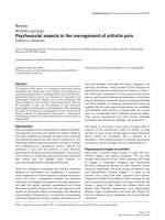
from RA and non-RA pools of synovial RNA. Figure 1
shows the results of hybridization of the RA and non-RA
probes to the array membranes. To reduce the bias that
could be introduced during the quantification, arrays show-
ing very similar signals for the housekeeping genes were
chosen and only non-saturated and non-regulated signals/
genes were used for normalization. The intensity of each
spot was corrected for background levels. The analysis
step was repeated eight times for each pair of
autoradiogram.
Of the 16 chemokine receptors present, the expression of
12 chemokine receptors was visible on the RA microarrays.
These were CCR1, CCR2a, CCR5, CCR7, CCR9,
CX3CR1, CXCR1, CXCR2, CXCR4, CXCR5, CXCR6
(STRL33) and Bob (Table 3). Expression of the same
receptors could be observed on the non-RA membranes
Table 2
Sequences of the primers used for RT-PCR
mRNA Product Sequence Size (base pairs) Accession number
L27 Forward 5'-GACGCAAAGCTGTCATCGTG-3' 344 BC007273
Reverse 5'-GCAGTTTCTGGAAGAACCAC-3'
CXCR5 Forward 5'-TGA CCT GAG GAA GCG TGA AG-3' 639 NM001716
Reverse 5'-CGT GAA GAC ACT CTC ACG TG-3'
Available online />R221
with the exception of Bob, CCR7 and CCR9. Bob/GPR15
is an orphan receptor that is a coreceptor for human and
simian immunodeficiency viruses, and its expression in the
RA synovium is a novel observation that might be worthy of
further investigation. The detection of CCR7 and CCR9 in
RA was only possible after extended exposure times, but at
the time points used for quantification no regulation was
demonstrated. Four chemokine receptors (CCR2b, CCR3,
CCR4 and CCR6) could not be detected in RA samples or
non-RA samples under our conditions.
The most obvious differences between RA samples and
non-RA samples were for the chemokine receptors CXCR5
and CXCR2, and to a lesser extent CXCR4, which gave
stronger signals in RA samples (Fig. 1). In order to quantify
the differential expression of these receptors the densities
of autoradiographic spots were measured using ArrayVi-
sion software (Table 3). The criteria we set for a gene to be
considered as upregulated or downregulated were a RA/
non-RA ratio higher than 3 or lower than 0.3, respectively,
and a 95% confidence interval below 10% (criteria as
[30]). In the present study the expression of CXCR5 and
CXCR4 was 22.6 ± 0.7-fold higher and 3.5 ± 0.1-fold
higher in RA tissue than in non-RA tissue, respectively.
These results indicated that, of the chemokine receptors
studied, CXCR5 was the most upregulated in RA (Table 3).
The upregulation of CXCR2 could not be calculated for
mathematical reasons because the signal intensity of
CXCR2 in non-RA tissue after correction for the back-
ground was zero. CXCR2 was only visible on the non-RA
autoradiogram upon prolonged exposure, at which point
the housekeeping genes were saturated and were there-
fore unsuitable for quantification purposes.
Out of the 33 chemokines present on the arrays, 29 of
these ligands were visible on the RA membranes and 21 on
the non-RA membranes (Fig. 1 and Table 3). These
included CXCL13, CXCL12, CXCL8, CXCL1-3 and
CXCL5, which are ligands for the chemokine receptors
CXCR5, CXCR4 and CXCR2. Several chemokines were
visible on RA microarrays but not on non-RA microarrays
(namely CXCL13, CCL21 and CCL24), suggesting that
these genes might be induced in the inflamed synovium. In
contrast, there were no chemokine signals that were
present on non-RA membranes and were absent on RA
membranes. Where chemokine signals were detectable on
RA and non-RA microarrays, it was possible to quantify the
degree of upregulation or downregulation using the criteria
described earlier for chemokine receptors. Of these chem-
okines, the following showed upregulation: CCL18 (4.5 ±
0.4-fold increase), CXCL9 (3.6 ± 0.1-fold increase),
CXCL5 (3.5 ± 0.3-fold increase), and CXCL8 (3.3 ± 0.2-
fold increase). No chemokines displayed a downregulation
with a RA/non-RA ratio less than 0.3. The upregulation of
CXCL9 in RA synovia is in agreement with the only micro-
array study of RA synovia, in which this chemokine was also
shown to be increased [31]. In our study only five chemok-
Figure 1
Microarray analysis of chemokine and chemokine receptor expression in the rheumatoid arthritis (RA) and non-RA synoviaMicroarray analysis of chemokine and chemokine receptor expression in the rheumatoid arthritis (RA) and non-RA synovia. A pair of human cytokine
array membranes were hybridized to
33
P-labelled cDNA probes prepared from pools of (a) RA mRNA (n = 8) and (b) non-RA mRNA (n = 9). The
membranes were washed and autoradiographed. (c) The position of the 33 chemokines (C), the 16 chemokine receptors (CR), the nine positive
control 'housekeeping genes' (HKG) and the six negative controls (NC). Each gene was printed in duplicate. The star indicates the position of the
genes CXCR1, CXCR2, CXCR4 and CXCR5 (reading top to bottom) and shows their differential expression in RA and non-RA synovia. Exposure
time was 7 days and 14 days for (a) and (b), respectively.
Arthritis Research & Therapy Vol 7 No 2 Schmutz et al.
R222
Table 3
Chemokine and chemokine receptor expression data analysis
Gene RA Non-RA Regulation (ratio RA/non-RA)
Receptors
Array column 3 (Fig. 1, C3)
CCR1 0.050 0.023 Up (2.2 ± 0.2)
CCR2a 0.031 0.012 ❍● Up (2.7 ± 0.2)
CCR2b 0.000 ❍ 0.000 ❍ Not visible (NA)
CCR3 0.000 ❍ 0.000 ❍ Not visible (NA)
CCR4 0.003 ❍ 0.000 ❍ Not visible (NA)
CCR5 0.042 0.000 ❍● Up (NA)
CCR6 0.001 ❍ 0.000 ❍ Not visible (NA)
CCR7 0.022 ❍● 0.011 ❍ Not visible (2.5 ± 0.9)
CCR9 0.001 ❍● 0.000 ❍ Not visible (NA)
CX3CR1 0.028 ❍● 0.019 ❍● Not visible (1.5 ± 0.1)
CXCR1 0.036 ❍● 0.008 ❍● Not visible (9.6 ± 5.6)
CXCR2 0.651 0.000 ❍● Up (NA)
CXCR4 0.190 0.055 Up (3.5 ± 0.1)
CXCR5 1.328 0.059 Up (22.6 ± 0.7)
CXCR6 (STRL33) 0.016 0.025 Not visible (0.7 ± 0.0)
Bob 0.034 0.000 ❍ Up (NA)
Chemokines
Array column 1 (Fig. 1, C1)
CCL21 (6Ckine) 0.030 0.019 ❍ Up (1.6 ± 0.1)
CXCL13 (BLC/BCA-1) 0.052 0.020 ❍ Up (2.8 ± 0.4)
CXCL10 (IP-10) 0.031 0.019 ❍● Up (1.6 ± 0.1)
CXCL5 (ENA-78) 0.050 0.015 ❍● Up (3.5 ± 0.3)
CCL11 (eotaxin) 0.050 0.052 Not visible (1.0 ± 0.0)
CCL24 (eotaxin-2) 0.032 0.013 ❍ Up (2.5 ± 0.3)
CX3CL1 (fractalkine) 0.030 ❍● 0.012 ❍ Not visible (2.5 ± 0.2)
CXCL1 (GRO-α) 0.090 0.057 Up (1.6 ± 0.0)
CXCL2 (GRO-β) 0.151 0.101 Up (1.5 ± 0.0)
CXCL3 (GRO-γ) 0.101 0.055 Up (1.8 ± 0.0)
CCL14 (HCC-1) 0.202 0.296 Down (0.7 ± 0.0)
CCL16 (HCC-4) 0.006 ❍● 0.000 ❍ Not visible (NA)
CCL1 (I-309) 0.007 ❍● 0.000 ❍ Not visible (NA)
CXCL8 (IL-8) 0.086 0.026 Up (3.3 ± 0.2)
CXCL7 (LDGF) 0.005 ❍ 0.000 ❍ Not visible (NA)
CCL15 (MIP-1δ) 0.009 ❍● 0.008 ❍ Not visible (11 ± 13)
XCL1 (lymphotactin) 0.048 0.031 Up (1.6 ± 0.0)
CCL2 (MCP-1) 0.482 0.545 Not visible (0.9 ± 0.0)
Available online />R223
ines (CXCL7, CCL13, CCL20, CCL17 and CCL25) could
not be detected at all, whether in RA or non-RA samples.
The rapid screening of several genes at once made array
technology a very attractive method. Its use has revealed
disadvantages, however, including the requirement for
large amounts of RNA (which are not always available from
human tissue biopsies), a susceptibility to experimental var-
iability and a lack of standard optimum methods for statisti-
cal analysis [32]. Arrays also present the risk of cross-
hybridization leading to false positive or negative results
[31]. However, the array approach remains a valuable tool
if the samples can be pooled and if it is used in conjunction
with alternative methods such as RT-PCR.
RT-PCR analysis of CXCR5
To confirm the array results and to examine individual
patients, RT-PCR was performed on the total RNA from
each patient sample (Fig. 2). PCR primers were run
through the BLAST program (available through the UK
MRC HGMP-RC website:
) to
ensure the gene specificity of the RT-PCR results and to
exclude the possibility of cross-hybridization with other
genes. Overall, CXCR5 RNA was more abundant in RA
patients than in non-RA patients, confirming the microarray
data. CXCR5 expression was detected in the synovia of all
eight RA patients and showed some degree of patient-to-
patient variation. The difference in CXCR5 expression
between RA patients and non-RA patients was unlikely to
be due to differences in the relative amount of cDNA pro-
duced by different RT reactions since the PCR reactions
were normalized using the ribosomal gene L27. RT-PCR
showed that the difference between RA patients and non-
RA patients was less marked for CXCR2 and CXCR4 than
for CXCR5 (data not shown).
CCL8 (MCP-2) 0.053 0.062 Not visible(0.9 ± 0.0)
CCL7 (MCP-3) 0.066 0.047 Up (1.4 ± 0.1)
CCL13 (MCP-4) 0.000 ❍ 0.002 ❍ Not visible (0.0)
CCL22 (MDC) 0.000 ❍● 0.008 ❍ Not visible (0.0)
Array column 2 (Fig. 1, C2)
Midkine 0.299 0.155 Up (1.9 ± 0.0)
CXCL9 (MIG) 0.166 0.047 Up (3.6 ± 0.1)
CCL3 (MIP-1α) 0.040 0.042 Not visible (1.0 ± 0.0)
CCL4 (MIP-1β) 0.023 0.018 ❍● Up (1.3 ± 0.2)
CCL20 (MIP-3α) 0.000 ❍ 0.013 ❍ Not visible(0.0)
CCL19 (MIP-3β) 0.023 0.042 Not visible (0.6 ± 0.0)
CCL23 (MPIF-1) 0.039 0.042 Up (0.9 ± 0.0)
CCL18 (PARC) 0.146 0.033 Up (4.5 ± 0.4)
CCL5 (RANTES) 0.037 0.031 Up (1.2 ± 0.1)
CXCL12 (SDF-1) 0.409 0.573 Down (0.7 ± 0.0)
CCL17 (TARC) 0.000 ❍ 0.000 ❍ Not visible (NA)
CCL25 (TECK) 0.000 ❍ 0.000 ❍ Not visible (NA)
Following hybridization to labelled mRNA extracted from rheumatoid arthritis (RA) and non-RA synovia, a pair of array membranes was
autoradiographed for varying lengths of time. The autoradiograms were scanned and analysed with the ArrayVision software (version 6.0; Imaging
Research Inc., Haverhill, UK). For each RA/non-RA pair the housekeeping genes on the membranes showed very similar intensities, were not
saturated and were used to normalize the data. The analysis measured the 'volume' of each spot (i.e. the density value of each spot multiplied by
its area). The background was measured using the 'corners between spots' protocol of the software and was deducted from the 'volumes'. The
ratio of RA synovia versus non-RA synovia was also calculated for each spot. The analysis was repeated eight times for each pair of
autoradiograms, providing 16 values for each gene (each gene is spotted in duplicate) on each pair. Figures in the columns RA, non-RA and ratio
RA/non-RA represent the average of 16 values. For each average ratio the 95% confidence level was calculated, and the results presented are
those from the autoradiogram pair giving the smallest variation. ❍, spot was not visible by eye on the corresponding autoradiogram; ●, spot was
visible after prolonged exposure. The mRNA regulation of RA versus non-RA as observed by eye at the time point used for quantification is
indicated by not visible, up or down. NA, ratio could not be calculated due to the presence of zero values. The recent systematic nomenclature of
chemokines is used, with the former names in parentheses. The order of the genes presented is the same as that appearing on the microarray in
Fig. 1.
Table 3 (Continued)
Chemokine and chemokine receptor expression data analysis
Arthritis Research & Therapy Vol 7 No 2 Schmutz et al.
R224
Immunohistochemistry
To identify the cell types expressing CXCR5, and since
RNA expression and protein expression do not always cor-
relate, the protein expression of this receptor and three
specific cell markers (CD20, CD3 and CD68) was investi-
gated by immunohistochemistry of paraffin-embedded
sections.
Seven out of eight RA patients presented substantial lym-
phoid follicles in their synovia. The specific cell markers
CD20 and CD3 confirmed the presence of B cells and T
cells, respectively, in these infiltrates. In every RA patient
where lymphoid follicles occurred, CXCR5
+
cells were
always present in these structures; this indicates a correla-
tion between the expression of CXCR5 and the occurrence
of lymphoid follicles. Serial sections indicated that CXCR5
was expressed by CD20
+
B cells (Fig. 3a,3c).
It was not possible to colocalize CXCR5 and CD3 in serial
sections, so a double-label immunohistochemistry tech-
nique was developed. Sections were treated with anti-CD3
followed by alkaline phosphatase and Vector red substrate.
Anti-CXCR5 was added to the same sections, and the col-
our developed using peroxidase and DAB-Nickel. CD3
expression alone gave a light red colour (Fig. 3e) and
CXCR5 expression alone produced a grey–black colour
(Fig. 3f). Where these two proteins colocalized a dark red
colour was obtained (Fig. 3f). Using this technique it was
evident that in the RA synovium there was a population of
CD3
+
T cells that expressed CXCR5 (Fig. 3f). These were
localized exclusively in lymphoid follicles in the synovia of
five out of the eight RA patients. The patient with diffuse
infiltration was negative for CXCR5
+
/CD3
+
cells. Serial
sections treated with anti-CXCR5 and the macrophage
marker anti-CD68 suggested that CXCR5
+
cells in the
intima included macrophages (Fig. 4a,4b). The endothelial
cells of synovial postcapillary venules were positive for
CXCR5 in the RA synovium (Fig. 4e).
In non-RA tissue, CXCR5 was localized in the intima and
endothelial cells (Fig. 5). Intimal cells were widely positive
for CXCR5 and serial sections indicated that these
included CD68
+
macrophage-like cells (Fig. 5a,5b). No
lymphocytic infiltrates were present in these synovial sam-
ples due to their non-inflamed nature. Sections treated with
CD20 and CD3 antibodies were negative, showing that no
B cells or T cells could be detected. In non-RA tissue and
RA tissue, fibroblasts were negative for CXCR5, as were
neutrophils in RA synovia, indicating selectivity in the cell
types expressing this receptor.
For all immunohistochemistry experiments in this study, the
use of isotype-matched immunoglobulin controls or sera
instead of primary antibodies resulted in negative staining
of RA sections and non-RA sections (Figs 3b,3d,3g,3h,
4c,4d,4f and 5c,5d,5f).
RT-PCR on isolated RA monocytes/macrophages
To further investigate whether macrophages themselves
are producing CXCR5 and to confirm the results of immu-
nohistochemistry, we performed RT-PCRs on monocytes/
macrophages isolated from the PB and SF of four addi-
tional RA patients (Fig. 6). CXCR5 was strongly expressed
in all four samples and there was little difference between
PB and SF.
Discussion
The major finding of the present study is that CXCR5 is
upregulated in the RA synovium. The cells expressing this
chemokine receptor are B lymphocytes, T lymphocytes,
macrophages and endothelial cells. The increased num-
bers of B lymphocytes, T lymphocytes and macrophages
producing CXCR5 in the RA synovium are probably
responsible for the increased expression of the receptor in
this chronically inflamed tissue. The majority (seven out of
eight) of the RA synovia included in this study contained
substantial lymphoid aggregates but only one out of nine
non-RA patients presented a very small infiltrate. These cell
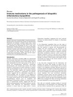
Figure 2
RT-PCR on rheumatoid arthritis and non-rheumatoid arthritis synovial tissueRT-PCR on rheumatoid arthritis and non-rheumatoid arthritis synovial tissue. CXCR5 RT-PCR products were separated on 0.8% agarose gels and
stained with ethidium bromide. The reactions were performed on the individual RNA samples that were applied to the microarray membranes. The
ribosomal RNA L27 was employed to normalize the amount of RNA used in each reaction.
Available online />R225
aggregates contained CD20
+
B cells that expressed
CXCR5. The expression of CXCR5 has been reported in
mature B cells and secondary lymphoid organs but as far
as the authors are aware this is the first report of a chemok-
ine receptor expressed by B cells in the RA synovium and
its ectopic lymphoid structures.
Our findings are particularly interesting in view of the func-
tional role of B cells in RA. This includes autoantibody pro-
duction, antigen presentation, a role in lymphoid follicle and
germinal centre formation, and the promising results of the
anti-CD20 treatment in RA [33,34]. The microarrays
showed that the mRNA for CXCL13, the only known ligand
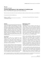
Figure 3
Immunohistochemistry of CXCR5 in lymphoid cell aggregates of rheu-matoid arthritis synoviaImmunohistochemistry of CXCR5 in lymphoid cell aggregates of rheu-
matoid arthritis synovia. Sections of rheumatoid synovium were treated
with (a) CXCR5 antibody or (b) isotype control. Serial sections were
treated with (c) anti-CD20 as a marker of B lymphocytes or (d) isotype
control. Arrows indicate B lymphocytes expressing CXCR5. (e) Rheu-
matoid synovium treated with the T-cell marker anti-CD3 followed by
alkaline phosphatase and Vector red substrate (methyl green counter-
stain). T cells stain a light red colour. (f) Serial section from the same
synovial sample as (e) treated with anti-CD3, alkaline phosphatase and
Vector red followed by anti-CXCR5, peroxidase and 3,3'-diaminobenzi-
dine (DAB)-Nickel substrate (no counterstain used). T cells that
express CXCR5 are stained dark red whereas cells expressing CXCR5
alone are grey–black in colour. (g) Control for (e), in which isotype-
matched rabbit immunoglobulin (Ig) was used instead of anti-CD3. (h)
Control for (f), in which isotype-matched rabbit and mouse Ig were
applied instead of CD3 and CXCR5 antibodies (no counterstain used).
Unless stated otherwise, DAB substrate was used. (a), (c) and (e)–(h)
Original magnification, 420 ×; isotype controls (b) and (d) original mag-
nification, 280 ×.
Figure 4
Immunohistochemistry of CXCR5 in the intima and postcapillary venules in rheumatoid arthritis synoviaImmunohistochemistry of CXCR5 in the intima and postcapillary
venules in rheumatoid arthritis synovia. (a) CD68
+
cells in the intima.
(b) Serial section to (a) stained for CXCR5. Note the colocalization of
CXCR5 and CD68 to the same group of cells. (c) and (d) Sections
from the same region as (a) and (b), treated with isotype-matched con-
trol immunoglobulin instead of CD68 and CXCR5 antibodies, respec-
tively. (e) Postcapillary venule positive for CXCR5 within a lymphoid
aggregate. Labelling was revealed using 3,3'-diaminobenzidine sub-
strate. (f) Isotype control for (e). (a), (b), (e) and (f) Original magnifica-
tion, 420 ×; (c) and (d) original magnification, 350 ×.
Arthritis Research & Therapy Vol 7 No 2 Schmutz et al.
R226
of CXCR5, was present in the RA synovium and not in the
non-RA synovium. Furthermore, other reports have shown
a CXCL13 message in RA synovia, together with its protein
that localizes to follicular dendritic cells, endothelial cells
and synovial fibroblasts, suggesting that these cells pro-
duce the chemokine [1,25]. Taken together with our data,
this indicates that CXCR5 on B cells may be important in
the recruitment of these cells into the RA synovium, in
addition to their positioning and retention within the syno-
vial infiltrates. In this regard, the role of CXCR5 on B cells
in secondary lymphoid organs has been well documented
[35,36]. CXCR5 guides B cells into the B-cell follicles and
also directly promotes the recruitment of these cells into
Peyer's patches via high endothelial venules [27,28,37,38].
In addition CXCR5-deficient mice exhibit impaired develop-
ment of lymph nodes and Peyer's patches, and the tissue
architecture of these organs is severely disturbed showing
a lack of B-cell follicles [27,28].
Our double immunohistochemistry data indicate that there
is a population of CXCR5
+
CD3
+
T cells present in the RA
synovium. CXCR5
+
T cells have been shown in secondary
lymphoid tissue where some of these cells localize within
germinal centres [20,39], and it is proposed that CXCR5
enables them to enter B-cell follicles guided by CXCL13
[36]. Within these follicles they may provide B-cell help and
have therefore been named follicular B helper T cells, since
purified human tonsillar CD4
+
CXCR5
+
T cells efficiently
stimulate the production of immunoglobulins by B cells
[39,40]. These follicular B helper T cells are CD57
+
,
whereas the majority of the CXCR5
+
T cells that are
present in interfollicular and T-cell areas of the lymphoid
tissue are CD57
-
and are poor B-cell helpers [41]. Since
lymphoid neogenesis occurs in the RA synovium it is pos-
sible that the CXCR5 expression on T cells as shown in the
present study is involved in the positioning of these cells
within the synovium and in providing B-cell help, although
further studies are required to characterize this synovial T-
cell population. Whether the two populations of
CXCR5
+
CD57
+
and CXCR5
+
CD57
-
T cells are present in
the RA synovium and what their role could be is still
unknown. However, CD57
+
T cells are reported to be
present in the RA synovium and SF, where levels of this
marker are elevated compared with controls [42,43]. Fur-
thermore, an involvement of CD57
+
T cells has been shown
in disease activity of RA [44].
Immunohistochemical experiments indicated that CD68
+
cells in the synovial intima express CXCR5. Intimal cells
comprise two cell types: macrophage-like cells and fibrob-
last-like cells. In RA the former macrophage-like cells are
numerous, comprising up to 80% of this cell layer [45]. It
Figure 5
Immunohistochemistry of CXCR5 in non-rheumatoid arthritis synoviaImmunohistochemistry of CXCR5 in non-rheumatoid arthritis synovia.
(a) CD68 staining in the intimal layer. (b) Serial section to (a) treated
with anti-CXCR5, showing that CXCR5
+
cells in the intimal layer
included those also positive for CD68. (c) and (d) Sections from the
same region as (a) and (b), treated with isotype-matched control immu-
noglobulin instead of CD68 and CXCR5 antibodies, respectively. (e)
Subintimal postcapillary venule stains for CXCR5 expression (arrow).
(f) Isotype-matched control for (e). Labelling was revealed using 3,3'-
diaminobenzidine substrate. (a), (b), (e) and (f) Original magnification,
420 ×; (c) and (d) original magnification, 350 ×.
Figure 6
RT-PCR on monocytes/macrophages from peripheral blood (PB) and synovial fluid (SF)RT-PCR on monocytes/macrophages from peripheral blood (PB) and
synovial fluid (SF). CXCR5 RT-PCR products were separated on 0.8%
agarose gels and stained with ethidium bromide. The reactions were
performed on four additional rheumatoid arthritis patients. The ribos-
omal RNA L27 was employed to normalize the amount of RNA used in
each reaction.
Available online />R227
has been reported that in the RA synovium anti-CD68
reacts strongly with intimal macrophages, but fibroblasts
also show some reactivity with this antibody [45].
Therefore, since macrophages are abundant in the RA
intima and because of their strong reactivity with anti-
CD68, it is likely that intimal macrophages are positive for
CXCR5. In the normal non-RA intima, macrophages are
positive for CD68 and fibroblasts are negative, making it
more certain that macrophages express CXCR5 in this cell
layer [45]. Consequently, RT-PCR was performed to verify
that RA macrophages/monocytes can express CXCR5.
The RT-PCR did indeed demonstrate CXCR5 mRNA in
macrophages from RA SF, as well as PB monocytes from
the same RA patients. Since the CXCR5 mRNA is
expressed at similar levels in RA PB and RA SF it is sug-
gested that the contribution of monocytes/macrophages to
the upregulation of CXCR5 in the RA synovium is due to
their increased number, rather than due to an increased
abundance of CXCR5 transcripts per cell. CXCR5 mRNA
has also been found in normal human PB monocytes by RT-
PCR [21]. Studies by ourselves and other workers have
shown that monocytes/macrophages express several other
CXC chemokine receptors in RA, including CXCR1,
CXCR2 and CXCR4 [15,16,46]. Furthermore, RA
monocytes/macrophages express CC chemokine recep-
tors such as CCR1, CCR2, CCR3 and CCR5 [14], illus-
trating their broad profile of chemokine receptor
expression.
Endothelial cells are another cell type expressing CXCR5
in the synovium. There have been several reports of
endothelial cells in the RA synovium expressing chemokine
receptors, including CXCR3 and CXCR4, in addition to the
Duffy antigen that is a non-signalling chemokine receptor
[18,47-49]. In the RA synovium there is increased turnover
of blood vessels with enhanced formation of new blood
vessels together with enhanced vascular regression
[50,51]. These mechanisms are regulated by the balance
of angiogenic and angiostatic factors, and these factors
include chemokines. Some chemokines are angiogenic
(e.g. CXCL8, CXCL12, CCL1 and CCL2) and other chem-
okines are angiostatic (e.g. CXCL9 and CXCL10), and
activation of their respective chemokine receptors results in
the stimulation of or inhibition of endothelial cell prolifera-
tion [47,52-57]. CXCL13 has been shown to have an angi-
ostatic function, inhibiting the angiogenic effects of FGF-2
on human umbilical vein endothelial cells [58]. In addition,
the presence of CXCR5 in a variety of cultured human
endothelial cells – from umbilical and saphenous veins, for
example – may mediate the angiostatic effects of CXCL13
[58,59]. Our data showing the presence of CXCR5 on
endothelial cells in the synovium and the presence of its lig-
and in this tissue [1,25]suggest that CXCR5 may play an
angiostatic role in RA pathophysiology, although the angi-
ostatic effects of CXCL13 could potentially be acting
through CXCR3, which is also expressed by the RA syno-
vial endothelium [48,60].
In the present study mRNA for other chemokine receptors
were detected in the RA synovium, such as CXCR1,
CXCR2, CXCR4, Bob, CCR1, CCR2a, CCR7, CCR9 and
CX3CR1 (CXCR3 was not on the microarray). All of these
showed variable degrees of increased mRNA expression in
RA, although the upregulation was less compared with that
of CXCR5. Several previous reports have shown the
expression of chemokine receptors by leukocytes from RA
joints. These have included CCR4–CCR6, CXCR3,
CXCR4 and CX3CR1 by T lymphocytes [9,12,13,19] and
CCR1–CCR5 and CXCR1–CXCR4 by monocytes/mac-
rophages [14-16,18]. Such reports mainly focused on
selected cell types and certain chemokine receptors. The
present study took a different approach. Ours was primarily
a whole-tissue study examining the mRNA expression of a
wide range of chemokine receptors in RA and control syn-
ovia. While our study is in general accord with previous
reports, differences may in part be due to the RA ST used.
This tissue was highly infiltrated and, in all but one sample,
had extensive lymphoid follicles bearing resemblance to
those of secondary lymphoid organs. This feature may be
responsible for the particular upregulation of the constitu-
tive chemokine receptor CXCR5. In addition, our RA
patients had long-standing disease (Table 1) and the
patient sample may also have influenced the types of chem-
okine receptors expressed.
Conclusion
Our study demonstrates the expression of CXCR5 on B
cells, on T cells, on monocytes/macrophages and on
endothelial cells in the RA synovium. The expression of a
marker shared by cells that are known to play a central role
in the process of chronic inflammation is of particular inter-
est and suggests that targeting CXCR5 could provide a
powerful new treatment for RA.
Competing interests
The author(s) declare that they have no competing
interests.
Authors' contributions
CS carried out the microarray work, the RT-PCR and the
double immunohistochemisty, and drafted the manuscript.
AH carried out the single colour immunohistochemistry. AB
isolated the peripheral blood and synovial fluid monocytes,
and isolated the RNA after adhesion. BA participated in the
design of the study. CB and MS collaborated on the study
or coordinated the collection of samples in Birmingham,
and contributed to the writing of the manuscript. JM con-
ceived the study, and participated in its design and in the
writing the manuscript. All authors read and approved the
final manuscript.
Arthritis Research & Therapy Vol 7 No 2 Schmutz et al.
R228
Acknowledgements
The authors are indebted to the patients who kindly agreed to take part
in this study. They thank Mr C McGeoch, Mr D Rees, Mr L van Niekerk
and Mr S White and the theatre teams for their help in obtaining synovial
tissue. They are also very grateful to P Evans, M Pritchard and N Har-
ness for their histological expertise and to J Menage for helpful immuno-
histochemistry discussion. Finally, the authors thankfully acknowledge
the Henry Smith Charity, Droitwich Medical Trust Ltd and the Orthopae-
dic Institute Ltd for their financial support.
References
1. Takemura S, Braun A, Crowson C, Kurtin PJ, Cofield RH, O'Fallon
WM, Goronzy JJ, Weyand CM: Lymphoid neogenesis in rheu-
matoid synovitis. J Immunol 2001, 167:1072-1080.
2. Szekanecz Z, Strieter RM, Kunkel SL, Koch AE: Chemokines in
rheumatoid arthritis. Springer Semin Immunopathol 1998,
20:115-132.
3. Feldmann M, Brennan FM, Maini RN: Rheumatoid arthritis. Cell
1996, 85:307-310.
4. Luster AD: Chemokines – chemotactic cytokines that mediate
inflammation. N Engl J Med 1998, 338:436-445.
5. Haringman JJ, Kraan MC, Smeets TJM, Zwinderman KH, Tak PP:
Chemokine blockade and chronic inflammatory disease: proof
of concept in patients with rheumatoid arthritis. Ann Rheum
Dis 2003, 62:715-721.
6. Podolin PL, Bolognese BJ, Foley JJ, Schmidt DB, Buckley PT, Wid-
dowson KL, Jin Q, White JR, Lee JM, Goodman RB, et al.: A potent
and selective nonpeptide antagonist of CXCR2 inhibits acute
and chronic models of arthritis in the rabbit. J Immunol 2002,
169:6435-6444.
7. D'Ambrosio D, Panina-Bordignon P, Sinigaglia F: Chemokine
receptors in inflammation: an overview. J Immunol Methods
2003, 273:3-13.
8. Murdoch C, Finn A: Chemokine receptors and their role in
inflammation and infectious diseases. Blood 2000,
95:3032-3043.
9. Buckley CD, Amft N, Bradfield PF, Pilling D, Ross E, Arenzana-
Seisdedos F, Amara A, Curnow SJ, Lord JM, Scheel-Toellner D,
Salmon M: Persistent induction of the chemokine receptor
CXCR4 by TGF-β1 on synovial T cells contributes to their accu-
mulation within the rheumatoid synovium. J Immunol 2000,
165:3423-3429.
10. Nanki T, Lipsky PE: Cytokine, activation marker, and chemokine
receptor expression by individual CD4
+
memory T cells in
rheumatoid arthritis synovium. Arthritis Res 2000, 2:415-423.
11. Shadidi KR, Thompson KM, Henriksen JE, Natvig JB, Aavak T:
Association of antigen specificity and migratory capacity of
memory T cells in rheumatoid arthritis. Scand J Immunol 2002,
55:274-283.
12. Ruth JH, Rottman JB, Katschke KJ Jr, Qin S, Wu L, LaRosa G,
Ponath P, Pope RM, Koch AE: Selective lymphocyte chemokine
receptor expression in the rheumatoid joint. Arthritis Rheum
2001, 44:2750-2760.
13. Ruth JH, Shahrara S, Park CC, Morel JC, Kumar P, Qin S, Koch
AE: Role of macrophage inflammatory protein-3α and its lig-
and CCR6 in rheumatoid arthritis. Lab Invest 2003, 83:579-588.
14. Katschke KJ Jr, Rottman JB, Ruth JH, Qin S, Wu L, LaRosa G,
Ponath P, Park CC, Pope RM, Koch AE: Differential expression
of chemokine receptors on peripheral blood, synovial fluid,
and synovial tissue monocytes/macrophages in rheumatoid
arthritis. Arthritis Rheum 2001, 44:1022-1032.
15. Brühl H, Wagner K, Kellner H, Schattenkirchner M, Schlöndorff D,
Mack M: Surface expression of CC- and CXC-chemokine
receptors on leucocytes subsets in inflammatory joint
diseases. Clin Exp Immunol 2001, 126:551-559.
16. Blades MC, Ingegnoli F, Wheller SK, Manzo A, Wahid S, Panayi
GS, Perretti M, Pitzalis C: Stromal cell-derived factor 1
(CXCL12) induces monocyte migration into human synovium
transplanted onto SCID mice. Arthritis Rheum 2002,
46:824-836.
17. Page G, Lebecque S, Miossec P: Anatomic localization of
immature and mature dendritic cells in an ectopic lymphoid
organ: correlation with selective chemokine expression in
rheumatoid synovium. J Immunol 2002, 168:5333-5341.
18. Shadidi KR: New drug targets in rheumatoid arthritis: focus on
chemokines. BioDrugs 2004, 18:181-187.
19. Szekanecz Z, Kim J, Koch AE: Chemokines and chemokine
receptors in rheumatoid arthritis. Semin Immunol 2003,
15:15-21.
20. Förster R, Emrich T, Kremmer E, Lipp M: Expression of the G-
protein-coupled receptor BLR1 defines mature, recirculating B
cells and a subset of T-helper memory cells. Blood 1994,
84:830-840.
21. Barella L, Loetscher M, Tobler A, Baggiolini M, Moser B:
Sequence variation of a novel heptahelical leucocyte receptor
through alternative transcript formation. Biochem J 1995,
309:773-779.
22. Amft N, Curnow SJ, Scheel-Toellner D, Devadas A, Oates J,
Crocker J, Hamburger J, Ainsworth J, Mathews J, Salmon M, Bow-
man SJ, Buckley CD: Ectopic expression of the B cell-attracting
chemokine BCA-1 (CXCL13) on endothelial cells and within
lymphoid follicles contributes to the establishment of germi-
nal center-like structure in Sjögren's syndrome. Arthritis
Rheum 2001, 44:2633-2641.
23. Salomonsson S, Larsson P, Tengnér P, Mellquist E, Hjelmström P,
Wahren-Herlenius M: Expression of the B cell-attracting chem-
okine CXCL13 in the target organ and autoantibody produc-
tion in ectopic lymphoid tissue in the chronic inflammatory
disease Sjögren's syndrome. Scand J Immunol 2002,
55:336-342.
24. Müller G, Höpken UE, Stein H, Lipp M: Systemic immunoregula-
tory and pathogenic functions of homeostatic chemokine
receptors. J Leukoc Biol 2002, 72:1-8.
25. Shi K, Hayashida K, Kaneko M, Hashimoto J, Tomita T, Lipsky PE,
Yoshikawa H, Ochi T: Lymphoid chemokine B-cell-attracting
chemokine-1 (CXCL13) is expressed in germinal center of
ectopic lymphoid follicles within the synovium of chronic
arthritis patients. J Immunol 2001, 166:650-655.
26. Hjelmström P: Lymphoid neogenesis: de novo formation of
lymphoid tissue in chronic inflammation through expression
of homing chemokines. J Leukoc Biol 2001, 69:331-339.
27. Förster R, Mattis AE, Kremmer E, Wolf E, Brem G, Lipp M: A puta-
tive chemokine receptor, BLR1, directs B cell migration to
defined lymphoid organs and specific anatomic compart-
ments of the spleen. Cell 1996, 87:1037-1047.
28. Ansel KM, Ngo VN, Hyman PL, Luther SA, Förster R, Sedgwick JD,
Browning JL, Lipp M, Cyster JG: A chemokine-driven positive
feedback loop organizes lymphoid follicles. Nature 2000,
406:309-314.
29. Kirkham B, Portek I, Lee CS, Stavros B, Lenarczyk A, Lassere M,
Edmonds J: Intraarticular variability of synovial membrane his-
tology, immunohistology, and cytokine mRNA expression in
patients with rheumatoid arthritis. J Rheumatol 1999,
26:777-784.
30. Lawrance IC, Fiocchi C, Chakravarti S: Ulcerative colitis and
Crohn's disease: distinctive gene expression profiles and
novel susceptibility candidate genes. Hum Mol Genet 2001,
10:445-456.
31. Ruschpler P, Lorenz P, Eichler W, Koczan D, Hänel C, Scholz R,
Melzer C, Thiesen H-J, Stiehl P: High CXCR3 expression in syn-
ovial mast cells associated with CXCL9 and CXCL10 expres-
sion in inflammatory synovial tissues of patients with
rheumatoid arthritis. Arthritis Res Ther 2003, 5:R241-R252.
32. Gu J, Märker-Hermann E, Baeten D, Tsai WC, Gladman D, Xiong
M, Deister H, Kuipers JG, Huang F, Song YW, et al.: A 588-gene
micorarray analysis of the peripheral blood mononuclear cells
of spondyloarthropathy patients. Rheumatology 2002,
41:759-766.
33. Weyand CM, Goronzy JJ, Takemura S, Kurtin PJ: Cell–cell inter-
actions in synovitis. Interactions between T cells and B cells in
rheumatoid arthritis. Arthritis Res Ther 2000, 2:457-463.
34. Goronzy JJ, Weyand CM: B cells as a therapeutic target in
autoimmune disease. Arthritis Res 2003, 5:131-135.
35. Campbell DJ, Kim CH, Butcher EC: Chemokines in the systemic
organization of immunity. Immunol Rev 2003, 195:58-71.
36. Müller G, Lipp M: Shaping up adaptive immunity: the impact of
CCR7 and CXCR5 on lymphocyte trafficking. Microcirculation
2003, 10:325-334.
37. Okada T, Ngo VN, Ekland EH, Förster R, Lipp M, Littman DR,
Cyster JG: Chemokine requirements for B cell entry to lymph
nodes and Peyer's patches. J Exp Med 2002, 196:65-75.
Available online />R229
38. Ebisuno Y, Tanaka T, Kanemitsu N, Kanda H, Yamaguchi K, Kaisho
T, Akira S, Miyasaka M: Cutting edge: the B cell chemokine CXC
chemokine ligand 13/B lymphocyte chemoattractant is
expressed in the high endothelial venules of lymph nodes and
Peyer's patches and affects B cell trafficking across high
endothelial venules. J Immunol 2003, 171:1642-1646.
39. Schaerli P, Willimann K, Lang AB, Lipp M, Loetscher P, Moser B:
CXC chemokine receptor 5 expression defines follicular hom-
ing T cells with B cell helper function. J Exp Med 2000,
192:1553-1562.
40. Breitfeld D, Ohl Lars, Kremmer E, Ellwart J, Sallusto F, Lipp M,
Förster R: Follicular B helper T cells express CXC chemokine
receptor 5, localize to B cell follicles, and support immu-
noglobulin production. J Exp Med 2000, 192:1545-1552.
41. Kim CH, Rott LS, Clark-Lewis I, Campbell DJ, Wu L, Butcher EC:
Subspecialization of CXCR5
+
T cells: B helper activity is
focused in a germinal center-localized subset of CXCR5
+
T
cells. J Exp Med 2001, 193:1373-1381.
42. Dupuy d'Angeac A, Monier S, Jorgensen C, Gao Q, Travaglio-Enci-
noza A, Bologna C, Combe B, Sany J, Reme T: Increased per-
centage of CD3
+
, CD57
+
lymphocytes in patients with
rheumatoid arthritis. Correlation with duration of disease.
Arthritis Rheum 1993, 36:608-612.
43. Arai K, Yamamura S, Seki S, Hanyu T, Takahashi HE, Abo T:
Increase of CD57
+
T cells in knee joints and adjacent bone
marrow of rheumatoid arthritis (RA) patients: implication for
an anti-inflammatory role. Clin Exp Immunol 1998,
111:345-352.
44. Maeda T, Yamada H, Nagamine R, Shuto T, Nakashima Y, Hirata
G, Iwamoto Y: Involvement of CD4
+
, CD57
+
T cells in the dis-
ease activity of rheumatoid arthritis. Arthritis Rheum 2002,
46:379-384.
45. Edwards JCW: The synovium. In Rheumatology Edited by: Klip-
pel JH, Dieppe PA. London: Mosby; 1998:6.1-6.8.
46. Patterson AM, Schmutz C, Davis S, Gardner L, Ashton BA, Middle-
ton J: Differential binding of chemokines to macrophages and
neutrophils in the human inflamed synovium. Arthritis Res
2002, 4:209-214.
47. Pablos JL, Santiago B, Galindo M, Torres C, Brehmer MT, Blanco
FJ, García-Lázaro FJ: Synoviocyte-derived CXCL12 is displayed
on endothelium and induces angiogenesis in rheumatoid
arthritis. J Immunol 2003, 170:2147-2152.
48. García-López MA, Sánchez-Madrid F, Rodríguez-Frade JM, Mel-
lado M, Acevedo A, García MI: CXCR3 chemokine receptor dis-
tribution in normal and inflamed tissues: expression on
activated lymphocytes, endothelial cells, and dendritic cells.
Lab Invest 2001, 81:409-418.
49. Patterson AM, Siddall H, Chamberlain G, Gardner L, Middleton J:
Expression of the duffy antigen/receptor for chemokine
(DARC) by the inflamed synovial endothelium. J Pathol 2002,
197:108-116.
50. Walsh DA, Wade M, Mapp PI, Blake DR: Focally regulated
endothelial proliferation and cell death in human synovium.
Am J Pathol 1998, 152:691-702.
51. Paleolog EM: Angiogenesis in rheumatoid arthritis. Arthritis Res
2002, 4:S81-S90.
52. Addison CL, Daniel TO, Burdick MD, Liu H, Ehlert JE, Xue YY,
Buechi L, Walz A, Richmond A, Strieter RM: The CXC chemokine
receptor 2, CXCR2, is the putative receptor for ELR
+
CXC
chemokine-induced angiogenic activity. J Immunol 2000,
165:5269-5277.
53. Bernardini G, Spinetti G, Ribatti D, Camarda G, Morbidelli L, Ziche
M, Santoni A, Capogrossi MC, Napolitano M: I-309 binds to and
activates endothelial cell function and acts as an angiogenic
molecule in vivo. Blood 2000, 96:4039-4045.
54. Salcedo R, Ponce ML, Young HA, Wasserman K, Ward JM, Klein-
man HK, Oppenheim JJ, Murphy WJ: Human endothelial cells
express CCR2 and respond to MCP-1: direct role of MCP-1 in
angiogenesis and tumor progression. Blood 2000, 96:34-40.
55. Koch AE, Volin MV, Woods JM, Kunkel SL, Connors MA, Harlow
LA, Woodruff DC, Burdick MD, Strieter RM: Regulation of angio-
genesis by C-X-C chemokines interleukin-8 and epithelial
neutrophil activating peptide 78 in the rheumatoid joint. Arthri-
tis Rheum 2001, 44:31-40.
56. Romagnani P, Annunziato F, Lasagni L, Lazzeri E, Beltrame C,
Francalanci M, Uguccioni M, Galli G, Cosmi L, Maurenzig L, et al.:
Cell cycle-dependent expression of CXC chemokine receptor
3 by endothelial cells mediates angiostatic activity. J Clin
Invest 2001, 107:53-63.
57. Salcedo R, Zhang X, Young HA, Michael N, Wasserman K, Ma W-
H, Martins-Green M, Murphy WJ, Oppenheim J: Angiogenic
effects of prostaglandin E2 are mediated by up-regulation of
CXCR4 on human microvascular endothelial cells. Blood 2003,
102:1966-1977.
58. Spinetti G, Camarda G, Bernardini G, Romano Di Peppe S, Cap-
ogrossi MC, Napolitano M: The chemokine CXCL13 (BCA-1)
inhibits FGF-2 effects on endothelial cells. Biochem Biophys
Res Commun 2001, 289:19-24.
59. Hillyer P, Mordelet E, Flynn G, Male D: Chemokines, chemokine
receptors and adhesion molecules on different human
endothelia: discriminating the tissue-specific functions that
affect leucocyte migration. Clin Exp Immunol 2003,
134:431-441.
60. Jenh C-H, Cox MA, Hipkin W, Lu T, Pugliese-Sivo C, Gonsiorek W,
Chou C-C, Narula SK, Zavodny PJ: Human B cell-attracting
chemokine 1 (BCA-1; CXCL13) is an agonist for the human
CXCR3 receptor. Cytokine 2001, 15:113-121.