Báo cáo y học: "CXCR3/CXCL10 expression in the synovium of children with juvenile idiopathic arthritis" pot
Bạn đang xem bản rút gọn của tài liệu. Xem và tải ngay bản đầy đủ của tài liệu tại đây (828.28 KB, 9 trang )
Open Access
Available online />R241
Vol 7 No 2
Research article
CXCR3/CXCL10 expression in the synovium of children with
juvenile idiopathic arthritis
Georgia Martini
1
, Francesco Zulian
1
, Fiorella Calabrese
2
, Marta Bortoli
3
, Monica Facco
3
,
Anna Cabrelle
3
, Marialuisa Valente
2
, Franco Zacchello
1
and Carlo Agostini
3
1
Department of Paediatrics, Padua University School of Medicine, Italy
2
Pathology Institute, Padua University School of Medicine, Italy
3
Department of Clinical and Experimental Medicine, Padua University School of Medicine, Italy
Corresponding author: Georgia Martini,
Received: 14 Apr 2004 Revisions requested: 26 May 2004 Revisions received: 16 Nov 2004 Accepted: 22 Nov 2004 Published: 7 Jan 2005
Arthritis Res Ther 2005, 7:R241-R249 (DOI 10.1186/ar1481)
http://arthr itis-research.com/conte nt/7/2/R241
© 2005 Martini et al.; licensee BioMed Central Ltd.
This is an Open Access article distributed under the terms of the Creative Commons Attribution License ( />2.0), which permits unrestricted use, distribution, and reproduction in any medium, provided the original work is cited.
Abstract
The accumulation of T cells in the synovial membrane is the
crucial step in the pathophysiology of the inflammatory
processes characterizing juvenile idiopathic arthritis (JIA). In this
study, we evaluated the expression and the pathogenetic role in
oligoarticular JIA of a CXC chemokine involved in the directional
migration of activated T cells, i.e. IFNγ-inducible protein 10
(CXCL10) and its receptor, CXCR3. Immunochemistry with an
antihuman CXCL10 showed that synovial macrophages,
epithelial cells, and endothelial cells bear the chemokine. By
flow cytometry and immunochemistry, it has been shown that
CXCR3 is expressed at high density by virtually all T
lymphocytes isolated from synovial fluid (SF) and infiltrating the
synovial membrane. Particularly strongly stained CXCR3
+
T
cells can be observed close to the luminal space and in the
perivascular area. Furthermore, densitometric analysis has
revealed that the mRNA levels for CXCR3 are significantly
higher in JIA patients than in controls. T cells purified from SF
exhibit a definite migratory capability in response to CXCL10.
Furthermore, SF exerts significant chemotactic activity on the
CXCR3
+
T-cell line, and this activity is inhibited by the addition
of an anti-CXCL10 neutralizing antibody. Taken together, these
data suggest that CXCR3/CXCL10 interactions are involved in
the pathophysiology of JIA-associated inflammatory processes,
regulating both the activation of T cells and their recruitment into
the inflamed synovium.
Keywords: chemokines, CXCL10, juvenile idiopathic arthritis, pathogenesis
Introduction
The trafficking and accumulation of immunocompetent
cells are essential components in the pathophysiology of
the inflammatory processes. A number of recent data sug-
gest that most of these events are regulated by chemok-
ines, a superfamily of 8–10 kDa molecules that has been
divided into four branches (C, CC, CXC, and CXXXC)
according to variations in a shared cysteine [1,2]. The cur-
rent roster approaches more than 50 related proteins.
Structural variations of chemokines have been associated
with differences in their ability to regulate the trafficking of
immune cells during inflammatory disorders. The biological
activity of chemokines is mediated by seven-transmem-
brane-domain, G-protein-coupled receptors classified as
C, CC, CXC, or CXXXC chemokine receptors according to
the type of chemokine bound. Chemokine receptors are
constitutively expressed on some cells, whereas they are
inducible on others [3].
Three CXC chemokines (IP-10/CXCL10, Mig/CXCL9, and
I-TAC/CXCL11) that are produced in response to IFNγ
allow for the accumulation of activated lymphocytes by
interacting with a specific receptor (CXCR3) [2]. Although
the interactions of chemokine receptors are often charac-
terized by considerable promiscuity, CXCR3 is selective in
the recruitment of Th1 cells, B cells, and NK (natural killer)
cells but not of nonlymphoid cells. Juvenile idiopathic arthri-
tis (JIA) is characterized by chronic inflammation of the
cDNA = complementary DNA; GAPDH = glyceraldehyde-3-phosphate dehydrogenase; IFNγ = interferon γ ; IL = interleukin; JIA = juvenile idiopathic
arthritis; PB = peripheral blood; PBS = phosphate-buffered saline; PCR = polymerase chain reaction; RT-PCR = reverse transcriptase PCR; SF =
synovial fluid; TCR = T-cell receptor; Th1 = T helper cell type 1.
Arthritis Research & Therapy Vol 7 No 2 Martini et al.
R242
synovium in multiple joints. Early studies of the synovial
membrane in JIA have shown the presence of a dense infil-
trate of activated T cells clustered around activated den-
dritic cells, suggesting that lymphocyte recruitment is
crucial in the pathogenesis of the disease [4,5]. There is
also strong evidence of an up-regulation of IFNγ expression
in synovial tissue relative to that in peripheral blood of
patients with JIA [6,7], indicating a Th1 type polarization of
local inflammatory response. Taken together, these data
suggest that lymphocyte-specific CXC chemokines could
be involved in the mechanisms promoting the development
of inflammatory events in JIA patients.
In this study, using immunohistochemical and molecular
studies of tissue sections and flow cytometry evaluation of
cells recovered from synovial fluid, we evaluated the role of
CXCR3/CXCL10 interactions in the regulation of T-cell
migration into the joints of patients with JIA. We have dem-
onstrated the presence of IP-10/CXCL10 in the synovial
tissue and its release into the synovial fluid, where it exerts
chemotactic activity toward activated CXCR3
+
T cells.
Taken together, our data suggest that the local production
of CXCL10 is involved in the pathophysiology of JIA-asso-
ciated inflammatory processes.
Materials and methods
Study populations
We analyzed synovial tissue from nine patients with oligoar-
ticular JIA who were undergoing arthroscopic synovec-
tomy. All the patients fulfilled the revised criteria for JIA
according to the International League of Associations for
Rheumatology (ILAR) classification [8] and were managed
at the Pediatric Rheumatology Unit of Padua University.
The procedure was performed in the case of persistently
inflamed joints that did not respond either to systemic anti-
inflammatory therapy or to intra-articular steroid injections.
In all these patients, gadolinium-enhanced MRI showed
marked thickening of the synovial membrane throughout
the joint. The patients' mean age at onset of the disease
was 70.6 months (range 34–156); the average disease
duration at synovectomy was 29.5 months (range 2–60).
As controls, three synovial tissue specimens obtained from
children with noninflammatory arthropathy were analyzed
by immunochemistry. These subjects had presented with
either hexadactylism, bone dysplasia, or bone fracture.
Paired samples of peripheral blood (PB) and synovial fluid
(SF) from 20 consecutive patients undergoing intra-articu-
lar steroid injection were examined. These patients' mean
age at onset of the disease was 77 months (range 13–
264) and the mean disease duration was 17 months (range
2–108). Patients who were having systemic anti-inflamma-
tory treatment at the time were excluded from the study.
Since the local ethics committee was not established yet at
the beginning of the study, institutional review board
approval was not requested, but informed consent was
obtained from the parents of all the children included in this
study.
Phenotypic evaluation of lymphocytes from peripheral
blood and synovial fluid
The commercially available conjugated or unconjugated
monoclonal antibodies used were from the Becton Dickin-
son (Sunnyvale, CA, USA) and PharMingen (San Diego,
CA, USA) series and included CD3, CD4, CD8, CD45R0,
CD45RA, and isotype-matched controls. Fluorescein-iso-
thiocyanate-labelled mouse antihuman CXCR3 (R&D Sys-
tems Inc, Minneapolis, MN, USA) was also used, and the
frequency of PB and SF cells positive for this reagent was
determined by flow cytometry as previously reported [9].
Specifically, cells were scored using a FACSCalibur ana-
lyzer (Becton Dickinson) and data were processed using
the Macintosh CELLQuest software program (Becton
Dickinson).
For immunofluorescence analysis, control mouse IgG
1
and
IgG
2a
were obtained from Becton Dickinson.
Chemotactic activity of synovial fluid
The CXCR3-positive cell line 300-19 (kindly provided by Dr
B Moser, Theodor-Kocher Institute, University of Bern,
Switzerland) was used to evaluate the chemotactic activity
of SF. The cells were grown in RPMI 1640 medium supple-
mented with 1% glutamine, 5% human serum, 1% kanamy-
cin, and 100 U/ml human recombinant IL-2. Cells were
periodically expanded by restimulation with phytohemag-
glutinin (1 µg/ml) in the presence of irradiated blood mono-
nuclear cells (10:1 ratio of feeder cells : 300-19 cells) and
were used for experiments after a culture period of 10 to 14
days.
Cell migration was measured in a 48-well modified Boyden
chamber (AC48, Neuro Probe Inc, Gaithersburg, MD,
USA). The chamber contains two sections. Chemotactic
stimuli were loaded in the bottom section, and cells were
put into the top compartment. Polyvinylpyrrolidone-free
polycarbonate membranes with 3- to 5-µm pores and
coated with fibronectin were placed between the two
chamber parts. Only the bottom face of filters was pre-
treated with fibronectin; this treatment maximizes attach-
ment of migrating cells to filters, increasing their
adherence. SF samples or control medium (30 µl) was
added to the bottom wells, and 50 µl of 300-19 cells resus-
pended in RPMI 1640 medium was added to the top wells.
The chamber was incubated at 37°C with 5% CO
2
for 2
hours. The membranes were then removed, washed with
PBS on the upper side, fixed, and stained with DiffQuik
(Dade AG, Düdingen, Switzerland). Cells were counted in
Available online />R243
three fields per well at magnification ×800. All assays were
performed in triplicate. In blocking experiments, cell sus-
pensions were preincubated before chemotaxis assay for
30 min at 4°C with antihuman IP-10 antibodies at 20 µg/
ml. In a few experiments, T cells purified from SF were eval-
uated for their migratory capability in response to CXCL10
(20 ng/ml and 200 ng/ml, R&D Systems).
Data are expressed as a migration index, which is the ratio
between the number of migrating cells in the presence of
the stimulus and that in medium alone.
Immunohistochemical analysis
Expression of CXCR3 and CXCL10 was detected by
immunohistochemistry with anti-CXCR3 and anti-IP-10
antibodies, respectively (R&D Systems). Paraffin-embed-
ded sections (4 µm thick) from patients and controls were
used for immunostaining with the standard avidin–biotin
complex method (Vectastain ABC kit; Vector Laboratories,
Burlingame, CA, USA), as previously described [10].
Briefly, for the microwave antigen-retrieval procedure,
slides were placed in a 2-L glass beaker containing 0.01
mol/L citrate buffer, pH 5.9, and microwaved at full power
(800 W for 5 min, three times) before cooling and equilibra-
tion in PBS.
To neutralize endogenous peroxidase activity, we pre-
treated slides with 3% hydrogen peroxide for 5 min. Pri-
mary antibodies were applied at a concentrations of 1:100
for both antibodies (anti-hCXCR3 monoclonal antibody
and anti-hIP-10/CXCL10 polyclonal antibody) for 1 hour in
a humidified chamber at 37°C. Immunoreactivity was
detected using biotinylated secondary antibodies (1:50
rabbit antigoat and 1:1000 goat antirabbit antibodies
diluted in PBS–bovine serum albumin buffer) incubated for
45 min, followed by a 30-min incubation with avidin–perox-
idase (1:200) and visualized by a 7-min incubation with the
use of 0.1% 3,3'-diaminobenzidene tetrahydrochloride as
the chromogen. Thereafter the slides were rinsed and
washed with PBS for 5 min, and the sections were coun-
terstained with Mayer's hematoxylin. The last steps were
performed at room temperature. Control slides were incu-
bated with Tris-buffered saline containing isotype-matched
antibodies instead of the primary antibody; they were invar-
iably negative (data not shown). The intensity of antibody
staining was classified as strong, moderate, weak, and neg-
ative. Parallel control slides were prepared either lacking
primary antibody or lacking primary and secondary antibod-
ies, or were stained with normal sera to control for back-
ground reactivity.
Immunohistochemistry for the characterization of inflamma-
tory infiltrate, endothelial cells, and synovial cells was car-
ried out using the following monoclonal antibodies CD45
(1:20), CD45RO (1:100), CD20 (1:100), CD68 (1:50),
CD4 (1:100), CD8 (1:100), CD31 (1:30) (all from Dako
Glostrup, Denmark), and cytokeratin–CAM 5.2 (1:1 Bec-
ton Dickinson). The immunoreaction products were devel-
oped using the avidin–biotin–peroxidase complex method
as described above.
Confocal microscopy
In order to evaluate the expression of CXCL10 by synovial
macrophages, confocal microscopy experiments were per-
formed in three patients with JIA. Paraffined sections were
prepared for immunofluorescent labelling. Briefly, primary
antibodies against CD68 and IP-10 (diluted 1:50 and 1:1
00, respectively, in PBS with 5 g/L bovine serum albumin
and 1 g/L gelatin, respectively) and secondary antibodies
(goat antimouse IgG and donkey antigoat IgG) conjugated
with Texas red or Alexa 488 (Sigma, Milan, Italy) were used.
Double labelling using both antibodies on the same section
was performed. Primary antibodies and secondary antibod-
ies were incubated for 1 hour at room temperature. Nuclear
staining was carried out with DAPI (4' 6-diamidino-2-
phenyindole; Sigma) in PBS. Slides were stored at 4°C and
analyzed within 24 hours. As a control, the primary antibody
was omitted.
Immunofluorescence was observed with a Leica TCS SL
spectral confocal and multiphoton system (Leica, Heidel-
berg, Germany). We used an argon laser at 488 nm in com-
bination with a helium neon laser at 543 nm to excite the
green (CD68) and red (IP-10) fluorochromes simultane-
ously. Emitted fluorescence was detected with a 505–530-
nm band-pass filter for the green signal and a 560-nm long-
pass filter for the red signal.
RT-PCR
RNA was extracted from the tissues using TRIzol reagent
(Invitrogen, San Giuliano Milanese, Milan, Italy). The con-
centration of RNA was estimated by spectrophotometer.
The RNA was treated with DNase I (Invitrogen) to remove
any genomic DNA that might contaminate the RNA prepa-
rations. Complementary DNA (cDNA) was prepared using
a synthesis kit (SuperScript II DNA Preamplification Sys-
tem; Invitrogen). A cDNA reaction mixture from 0.1 µg of
RNA was used for DNA amplification by PCR. A typical
amplification reaction included 2 units of Taq polymerase
(Takara, Shiga, Japan), 20 pmol of sense and antisense oli-
gonucleotide primers, and 200 µM each of dATP, dCTP,
dGTP, and dTTP. Amplification was carried out for 30
cycles: 1 min at 92°C, 1 min at 55°C, and 1 min at 72°C.
The amplified DNA was electrophoresed on a 2% agarose
gel (Invitrogen), stained with ethidium bromide, visualized
under ultraviolet light, and photographed.
The primer sequences used were as follows: for glyceral-
dehyde-3-phosphate dehydrogenase (GAPDH), 5'-TCC-
Arthritis Research & Therapy Vol 7 No 2 Martini et al.
R244
ATG-ACA-ACT-TTG-GTA-TCG-3' (sense) and 5'-GTC-
GCT-GTT-GAA-GTC-AGA-GGA-3' (antisense); for
CXCR3, 5'-TTG-ACC-GCT-ACC-TGA-ACA-TA-3' (sense)
and 5'-ACG-TCT-ACC-CTG-CTT-TCT-CG-3'. The
expected sizes for the cDNA amplicons were as follows:
376 bp for GAPDH, 377 bp for IP-10, and 456 bp for
CXCR3. All assays were performed in triplicate.
The number of cycles (30) was chosen to ensure that the
amount of products synthesized was proportional to the
amount of specific mRNA in the original preparation.
After PCR amplification, PCR products (15 µl) were sub-
jected to electrophoresis on 2% agarose gels containing
0.03 µg/ml ethidium bromide. The quantification of tran-
script level was carried out by scanning photographs of
gels and analyzing the area under peaks, using Quantity
one Biorad software. Levels of mRNA expression were nor-
malized by calculating them as a percentage of 3GAPDH
mRNA expression levels [11]. The band intensity for
3GAPDH did not differ significantly between experiments.
Statistical analysis
Data were analyzed with the assistance of the Statistical
Analysis System. Data are expressed as means ± standard
deviation. Mean values were compared using the ANOVA
test.
Results
Immunohistochemical analysis of the expression and
cellular distribution of CXCL10 in the synovial
membrane during JIA
Immunohistochemical analysis was used to investigate the
pattern of expression of this chemokine in synovial mem-
branes from nine children with JIA and three age-matched
controls. All the JIA synovial tissues showed moderate or
strong staining for CXCL10 (Table 1). As shown in Fig. 1a
and, at higher magnification, in Fig. 1b, CXCL10 was dem-
onstrated on the surface of three types of cells, specifically
macrophages, epithelial cells, and endothelial cells, as
determined by cell morphology. Most of the IP-10-express-
ing cells were macrophages. Matched controls revealed no
CXCL10 staining (Fig. 1c,d). In order to verify whether
macrophages express CXCL10 morphology, data were
confirmed by the use of confocal microscopy. As shown in
Fig. 2, double staining with CD68 and CXCL10 clearly
demonstrated that CD68
+
macrophages showed an
intense coexpression of the chemokine.
CXCL10 is present in synovial fluid from patients with JIA
and mediates chemotactic activity
To evaluate if CXCL10 is released into the SF and is capa-
ble of inducing T-cell migration, the chemotactic activity of
supernatants from the SF of four patients with JIA was
tested on a T-cell clone expressing high levels of CXCR3
(300-19). As shown in Fig. 3, SF of all the patients we stud-
ied exerted significant chemotactic activity on the CXCR3
+
T-cell line. The addition of an anti-CXCL10 neutralizing anti-
Table 1
CXCR3 and CXCL10 expression in patients with juvenile idiopathic arthritis and controls
Subject no. Sex Age at onset (months) CXCR3 CXCL10
Patients
1F54++++
2 F 34 +++ +++
3 F 84 +++ ++
4 F 70 +++ ++
5 F 65 +++ ++
6 F 156 +++ ++
7 F 141 +++ +++
8 M 70 +++ ++
9 F 42 +++ +++
Controls
1F72++
2F36++
3M24 +
+++, strong; ++, moderate; +, weak; , negative.
Available online />R245
body (α CXCL10) but not of a control antibody inhibited
chemotactic activities, suggesting the presence of IP-10/
CXCL10 in SF and its responsibility in the chemotaxis of
CXCR3
+
cells. In a second set of experiments, T cells puri-
fied from SF exhibited a definite migratory capability per se,
which was significantly enhanced in response to CXCL10.
Two representative experiments are represented in Fig. 4.
Immunohistochemical and flow cytometry analysis of
the expression of CXCR3 by PB, SF, and synovial-tissue
T lymphocytes in JIA
The possibility that CXCL10 in synovial fluid and mem-
brane might account for the recruitment of CXCR3
+
T lym-
phocytes from the bloodstream to the synovium was
investigated by immunohistochemical analysis of the
expression of this chemokine receptor. All the JIA patients
showed CXCR3-expressing lymphocytes infiltrating the
synovium, with strong or moderate staining intensities (see
Table 1). Particularly strongly stained cells were observed
close to the perivascular area (as in Fig. 5a,b, showing two
different magnifications of the same slide). In a few cases,
a follicular pattern of strongly marked lymphocytes was vis-
ible close to the luminal space (Fig. 6). The control synovial
tissues revealed no CXCR3 staining (Fig. 5c,d).
Densitometric analysis showed that CXCR3 mRNA levels
were significantly higher in patients with JIA than in controls
(CXCR3:GADPH ratio 2.25 ± 1.8 vs 0.6 ± 0.49, P < 0.05)
(Fig. 7).
Flow cytometry analysis confirmed the selective recruit-
ment of CXCR3
+
lymphocytes into the synovium. We ana-
lyzed paired samples of PB and SF from 20 children with
JIA, and in 18 of these patients, T lymphocytes isolated
from the SF showed greater expression of CXCR3 with
than did those from PB, both in terms of percentage of pos-
itive cells and of the MFI (P = 0.01) (Table 2). Flow cytom-
etry profiles for one representative patient are shown in Fig.
8. Taken together, these results strongly suggest a role for
the CXCL10 released into the synovial compartment in the
accumulation of its selective CXCR3-receptor expressing
T cells.
Discussion
JIA is characterized by a persistent accumulation in the syn-
ovial membrane of T lymphocytes most of which express
surface markers indicative of activation, such as CD45RO,
and a type-1 cytokine profile [4,5]. The cellular infiltrate is
defined largely by the composition of locally produced
chemokines as well as by the diversity of circulating leuko-
cytes expressing the relevant receptors. Our principal find-
ings are that in JIA, CXCL10/IP-10 is strongly expressed in
synovial membranes and is released into synovial fluid (SF),
where it exerts a definite chemotactic activity on CXCR3
+
T-cell clones and on T cells purified from SF; and that there
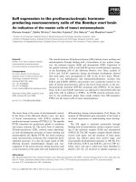
Figure 1
IP-10/CXCL10 expression in the synovium of a patient with juvenile idiopathic arthritisIP-10/CXCL10 expression in the synovium of a patient with juvenile idiopathic arthritis. Few inflammatory cells showing moderate staining; original
magnification ×50 (a), ×100 (b). Negative staining in control patient: panoramic view (c) (original magnification ×25) and particular view (d) (original
magnification ×50).
(a)
(b)
(c) (d)
2 µm
4 µm
1 µm
2 µm
Arthritis Research & Therapy Vol 7 No 2 Martini et al.
R246
is an accumulation of CXCR3 expressing T lymphocytes
from the bloodstream to the synovial fluid and membrane.
These findings suggest a role for CXCL10 in the mecha-
nism of T-cell activation and recruitment into the inflamed
synovium.
The high expression of CXCR3 by T cells retrieved from the
synovia of patients with JIA might be considered a by-prod-
uct of the in vivo cell hyperactivity of the tissue T-cell com-
partment in this disease. In fact, recent data clearly indicate
Figure 2
Expression of IP-10/CXCL10 in the synovium of a representative patient with JIAExpression of IP-10/CXCL10 in the synovium of a representative
patient with JIA. Immunofluorencence confocal laser scanning micros-
copy indicates the presence of chemokine IP-10 (red) (a); (b) the same
cells are shown to be synovial macrophages, as they are marked with
CD68 (green). (c) The co-localization of IP10 and CD68 by macro-
phages (brown) is clearly visible. Original magnification ×1000.
(a)
(b)
(c)
20 µm
Figure 3
Chemotactic activity of 300-19 cells in the presence of synovial fluid alone (grey bar), synovial fluid with an anti-CXCL10 neutralizing anti-body (αCXCL10) (black bar), and synovial fluid with a control antibody (white bar) from four representative patients with juvenile idiopathic arthritisChemotactic activity of 300-19 cells in the presence of synovial fluid
alone (grey bar), synovial fluid with an anti-CXCL10 neutralizing anti-
body (αCXCL10) (black bar), and synovial fluid with a control antibody
(white bar) from four representative patients with juvenile idiopathic
arthritis.
Figure 4
Chemotactic activity migration indices of T cells from synovial fluids of two representative patients with juvenile idiopathic arthritis in the pres-ence of RPMI 1640 medium alone or medium containing CXCL10 at 20 ng/ml or at 200 ng/mlChemotactic activity migration indices of T cells from synovial fluids of
two representative patients with juvenile idiopathic arthritis in the pres-
ence of RPMI 1640 medium alone or medium containing CXCL10 at
20 ng/ml or at 200 ng/ml.
migrating cell number/field
synovial fluid + αCXCL10
synovial
fluid
synovial fluid + unrelated mAb
Patient no. 1
Patient no. 2
Patient no. 3
Patient no. 4
20 40 6010 30 500
medium
CXCL10
20 ng/ml
0.0 1.51.0 2.0 2.5 3.0 3.5 4.0 4.5 5.00.5
CXCL10
200 ng/ml
migration index
Patient no. 1
Patient no. 2
Available online />R247
that CXCR3 and its ligands become functional on recently
activated T cells [12]. After antigenic challenge or in
response to stimulation through the T-cell receptor (TCR),
T cells express CXCR3, respond with chemotaxis to
CXCR3 ligands, and produce IFNγ. Furthermore, in the
presence of persistent antigenic stimulations, CXCR3
expression is maintained and poised for rapid up-regulation
with reactivation. We and other authors have previously
shown that CXCR3/CXCL10 interaction is involved in the
pathogenesis of other Th1-mediated processes, such as
Crohn's disease and sarcoidosis [13,14]. A similar
sequence of events could take place in the synovia of chil-
dren with JIA. In fact, as previously reported [15], the eval-
uation of the molecular organization of the TCR revealed
that T cells proliferating in children with JIA show a
preferential usage of definite TCR gene regions, indicating
Figure 5
CXCR3 expression in the synovium of a patient with juvenile idiopathic arthritisCXCR3 expression in the synovium of a patient with juvenile idiopathic arthritis. Note the marked staining of inflammatory cell infiltrate in the perivas-
cular area [original magnification ×50 (a), ×100 (b)]. Negative staining in control patient: panoramic view (c) (original magnification ×25) and partic-
ular view (d) (original magnification ×50).
(a) (b)
(c)
(d)
2 µm
2 µm
2 µm
4 µm
Figure 6
CXCR3 expression in juvenile idiopathic arthritis synoviumCXCR3 expression in juvenile idiopathic arthritis synovium. A follicular
pattern of strongly marked lymphocytes is visible close to the lumen
surface. Original magnification ×25.
4 µm
Figure 7
Semiquantitative RT-PCR determination of CXCR3 expression in patients and controlsSemiquantitative RT-PCR determination of CXCR3 expression in
patients and controls. Unnumbered frame: DNA marker. Representative
results of agarose-gel electrophoresis of RT-PCR products of CXCR3
mRNA (456 bp) and glyceraldehyde-3-phosphate dehydrogenase (234
bp) for nine patients (frames 1–9) and three controls (frames 10–12).
D
N
AM
a
r
k
e
r
12 3 4 5 6 7 89 101112
CXCR3
GAPDH
Arthritis Research & Therapy Vol 7 No 2 Martini et al.
R248
an ordered immune response in which a specific TCR has
been triggered and CXCR3 expression is induced [16].
CXCL10 was expressed by macrophages in synovial mem-
brane of patients with JIA but not of controls. This finding
suggests that CXCL10 is part of the matrix of cytokines
that regulates the accessory activity of macrophages at
sites of inflammatory lesions in the synovial
microenvironment. Since large amounts of type 1 inflamma-
tory cytokines, such as IFNγ, tumor necrosis factor α, IL-15,
and IL-18, have been detected in JIA synovium [7], it is
likely that these cytokines act in concert, sustaining the
local proinflammatory responses and up-regulating
CXCL10 expression. In turn, since CXCL10 is known to be
capable of up-regulating cytokine synthesis in human Th1
cells, it is likely that macrophage-derived chemokines as IL-
18 and IL-15 could participate in the maintenance of the
default Th1/Tc1 polarization seen during JIA inflammation.
It should be noted that, as shown in Fig. 2, anti-IP-10
almost completely inhibited the migration of the CXCR3
+
300-19 T cells in response to synovial fluid. Given the abil-
ity of I-ITAC/CXCL11 and Mig/CXCL10 to favor T-cell
recruitment [17], we are currently investigating whether this
non-ERL chemokine may influence entry of T cells into the
JIA synovia.
It remains to be established whether synovial endothelial
cells express CXCL10 (Fig. 1a). In a previous report it has
been shown that human umbilical-vein-derived endothelial
cell monolayers stimulated with IFNγ and tumor necrosis
factor α produce IP-10/CXCL10, retaining it on their sur-
face, and that this leads to a rapid adhesion of T lym-
phocytes. This effect was drastically reduced by anti-
CXCR3 monoclonal antibody [18]. Furthermore, it is known
that unstimulated human umbilical-vein-derived endothelial
cells are able to retain IP-10 added exogenously, through
binding to cell-surface proteoglycans [19]. Finally, recent
data have definitively demonstrated that human endothelial
cells may express a previously unrecognized receptor for
CXC chemokines named CXCR3B and derived from an
alternative splicing of the CXCR3 gene [20]. This receptor
shows higher affinity for CXCL10 than classic CXCR3,
mediates the inhibition of endothelial-cell growth, and
accounts for the known angiostatic capability of CXCL10.
Thus, it is possible that nonspecific binding of IP-10 may be
responsible for the CXCL10 positivity we observed on
endothelial cells. Further studies are in progress to deter-
mine whether synovial endothelial cells express CXCR3B
in vivo and, if this be the case, to determine the putative
role of CXCR3B/IP-10 interactions on the balance of ang-
iogenic/angiostatic events in the JIA synovia.
Previous studies on chemokines and their receptors in
modulating the recruitment of leukocytes at the sites of
inflammation suggested that targeting these molecules
with engineered agents might have therapeutic utility in
down-modulating inflammatory responses. Results of
CXCR3 or IP-10/CXCL10 blockade have already been
reported in animal models. Recently, some authors have
shown a rapid and marked improvement of adjuvant-
Table 2
CXCR3 expression in peripheral blood (PB) and synovial fluid (SF) lymphocytes in five representative patients with juvenile
idiopathic arthritis
Mean fluorescence of CXCR3
a
Patient no. In PB cells In SF cells D/s
b
1 35.79 55.8 36.5
2 23.02 77.13 36
3 20.27 48.88 22.4
4 15.84 27.75 34
5 16.44 20.59 20.2
a
P ≤ 0.001 in every case.
b
On the Kolmogorov–Smirnov test; D/s values >10, and P values <0.05 were considered significant. D/s is calculated
as a function of the number of data; it ranged from 0.5 to 100 and is a measure of the significance of the difference between two distributions.
Figure 8
Flow cytometry profile of CXCR3 expression in peripheral blood (PB) and synovial fluid (SF) lymphocytes from patient 3 and a control subjectFlow cytometry profile of CXCR3 expression in peripheral blood (PB)
and synovial fluid (SF) lymphocytes from patient 3 and a control
subject.
r
e
l
a
t
i
v
e
c
e
l
l
n
u
m
b
e
r
SF lymphocytes
control
PB lymphocytes
log. fluorescence intensity
Available online />R249
induced arthritis in rats treated with IP-10 DNA vaccine
[21]. Moreover, anti-mCXCR3 neutralizing antibodies were
found to inhibit Th1 lymphocyte recruitment to peripheral
inflammatory sites in a mouse model [22]. Further studies
are needed in animal models to explore the therapeutic
potential of CXCR3- or CXCL10-antagonists, with the ulti-
mate goal of offering new clues for immune intervention in
Th1-mediated diseases such as JIA and rheumatoid
arthritis.
Conclusion
Our results provide the first evidence of the functional role
of CXCR3/CXCL10 interactions in mediating recruitment
of T cells at sites of synovial inflammation in JIA. An in-depth
molecular study of mechanisms regulating overexpression
of CXCR3/CXCL10 might help in defining the role of these
molecules in synovial inflammatory responses, offering new
insights into elements controlling the immune response
within joints.
Competing interests
The author(s) declare that they have no competing
interests.
Authors' contributions
GM conceived and coordinated the study and drafted the
manuscript. FZ participated in the design of the study. FC
performed the immunohistochemistry and helped to draft
the manuscript. MB and MF carried out the chemotaxis. AC
performed the flow cytometry experiments. MV participated
in the immunohistochemistry. FZ participated in the design
of the study. CA conceived the study and helped in the
draft of the manuscript. All authors read and approved the
final manuscript
Acknowledgements
This work was supported by a grant from the Regione Veneto (Venice,
Italy) and COFIN MIUR 2002 (No. 2002068787002).
References
1. Luster AD: Chemokines – Chemotactic cytokines that mediate
inflammation. N Engl J Med 1998, 338:437-445.
2. Rollins BJ: Chemokines. Blood 1997, 90:909-928.
3. Baggiolini M: Chemokines and leukocyte traffic. Nature 1998,
392:565-568.
4. Silverman ED, Isacovics B, Pesche D, Laxer RM: Synovial fluid
cells in juvenile rheumatoid arthritis: evidence of selective T
cell migration to inflamed tissue. Clin Exp Immunol 1993,
91:90-95.
5. Murray KJ, Luyrink L, Grom AA, Passo MH, Emery H, Witte D,
Glass DN: Immunohistological characteristics of T cell infil-
trates in different forms of childhood onset chronic arthritis. J
Rheumatol 1996, 23:2116-2124.
6. Ozen S, Tucker LB, Miller LC: Identification of Th subsets in
juvenile rheumatoid arthritis confirmed by intracellular
cytokine staining. J Rheumatol 1998, 25:1651-1653.
7. Scola MP, Thompson SD, Brunner HI, Tsoras MK, Witte D, van
Dijk MA, Grom AI, Passo MH, Glass DN: Interferon-γ: interleukin-
4 ratios and associated type 1 cytokine expression in juvenile
rheumatoid arthritis synovial tissue. J Rheumatol 2002,
29:369-378.
8. Petty RE, Southwood TR, Baum J, Bhettay E, Glass DN, Manners
P, Maldonado-Cocco J, Suarez-Almazor M, Orozco-Alcala J, Pieur
AM: Revision of proposed classification criteria for Juvenile
Idiopathic Arthritis: Durban, 1997. J Rheumatol 1998,
25:1991-1994.
9. Agostini C, Trentin L, Facco M, Sancetta R, Cerutti A, Tassinari C,
Cimarosto L, Adami F, Cipriani A, Zambello R, Semenzato G: Role
of IL-15, IL-2 and their receptors in the development of T cell
alveolitis in pulmonary sarcoidosis. J Immunol 1996,
157:910-918.
10. Agostini C, Calabrese F, Rea F, Facco M, Tosoni A, Loy M, Binotto
G, Valente M, Trentin L, Semenzato G: CXCR3 and its ligand
CXCL10 are expressed by inflammatory cells infiltrating lung
allografts and mediate chemotaxis of T cells at sites of
rejection. Am J Pathol 2001, 158:1703-1711.
11. Horikoshi T, Sakakibara M: Quantification of relative mRNA
expression in the rat brain using simple RT-PCR and ethidium
bromide staining. J Neurosci Methods 2000, 99:45-51.
12. Haringman JJ, Ludikhuize J, Tak PP: Chemokines in joint disease:
the key to inflammation? Ann Rheum Dis 2004, 63:1186-1194.
13. Singh UP, Singh S, Iqbal N, Weaver CT, McGhee JR, Lillard JW Jr:
IFN-gamma-inducible chemokines enhance adaptive immu-
nity and colitis. J Interferon Cytokine Res 2003, 23:591-600.
14. Agostini C, Cassatella M, Zambello R, Trentin L, Gasperini S, Perin
A, Piazza F, Siviero M, Facco M, Dziejman M, et al.: Involvement
of the IP-10 chemokine in sarcoid granulomatous reactions. J
Immunol 1998, 161:6413-6420.
15. Thompson SD, Murray KJ, Grom AA, Passo MH, Choi E, Glass DN:
Comparative sequence analysis of the human T cell receptor
beta chain in juvenile rheumatoid arthritis and juvenile spond-
yloarthropathies: evidence for antigenic selection of T cells in
the synovium. Arthritis Rheum 1998, 41:482-497.
16. Wedderburn LR, Robinson N, Patel A, Varsani H, Woo P: Selec-
tive recruitment of polarized T cells expressing CCR5 and
CXCR3 to the inflamed joints of children with juvenile idio-
pathic arthritis. Arthritis Rheum 2000, 43:765-774.
17. Farber JM: HuMig: a new human member of the chemokine
family of cytokines. Biochem Biophys Res Commun 1993,
192:223-230.
18. Piali L, Weber C, LaRosa G, Mackay CR, Springer TA, Clark-Lewis
I, Moser B: The chemokine receptor CXCR3 mediates rapid
and shear-resistant adhesion-induction of effector T lym-
phocytes by the chemokines IP10 and Mig. Eur J Immunol
1998, 28:961-972.
19. Luster AD, Greenberg SM, Leder P: The IP-10 chemokine binds
to a specific cell surface heparin sulfate site shared with plate-
let factor 4 and inhibits endothelial cells proliferation. J Exp
Med 1995, 182:219-231.
20. Lasagni L, Francalanci M, Annunziato F, Lazzeri E, Giannini S,
Cosmi L, Sagrinati C, Mazzinghi B, Orlando C, Maggi E, et al.: An
alternatively spliced variant of CXCR3 mediates the inhibition
of endothelial cell growth induced by IP-10, Mig, and I-TAC,
and acts as functional receptor for platelet factor 4. J Exp Med
2003, 197:1537-1549.
21. Salomon I, Netzer N, Wildbaum G, Schif-Zuck S, Maor G, Karin N:
Targeting the function of IFN-γ-inducible protein 10 sup-
presses ongoing adjuvant arthritis. J Immunol 2002,
169:2685-2693.
22. Xie JH, Nomura N, Lu M, Chen SL, Koch GE, Weng Y, Rosa R, Di
Salvo J, Mudgett J, Peterson LB, et al.: Antibody-mediated block-
ade of the CXCR3 chemokine receptor results in diminished
recruitment of T helper 1 cells into sites of inflammation. J Leu-
koc Biol 2003, 73:771-780.