Báo cáo y học: "Inhibition of antithrombin by hyaluronic acid may be involved in the pathogenesis of rheumatoid arthritis" potx
Bạn đang xem bản rút gọn của tài liệu. Xem và tải ngay bản đầy đủ của tài liệu tại đây (603.11 KB, 6 trang )
Open Access
Available online />R268
Vol 7 No 2
Research article
Inhibition of antithrombin by hyaluronic acid may be involved in
the pathogenesis of rheumatoid arthritis
Xiaotian Chang
1
, Ryo Yamada
1
and Kazuhiko Yamamoto
1,2
1
Laboratory for Rheumatic Diseases, SNP Research Center, The Institute of Physical and Chemical Research (RIKEN), Kanagawa, Japan
2
Department of Allergy and Rheumatology, Graduate School of Medicine, University of Tokyo, Tokyo, Japan
Corresponding author: Xiaotian Chang,
Received: 10 Jul 2004 Revisions requested: 27 Sep 2004 Revisions received: 26 Nov 2004 Accepted: 1
Dec 2004 Published: 11 Jan 2005
Arth
ritis Res Ther 2005, 7:R268-R273 (DOI 10.1186/ar1487)
http://arthr itis-research.com/conte nt/7/2/R268
© 2005 Chang et al., licensee BioMed Central Ltd.
This is an Open Access article distributed under the terms of the Creative Commons Attribution License ( />2.0), which permits unrestricted use, distribution, and reproduction in any medium, provided the original work is cited.
Abstract
Thrombin is a key factor in the stimulation of fibrin deposition,
angiogenesis, proinflammatory processes, and proliferation of
fibroblast-like cells. Abnormalities in these processes are
primary features of rheumatoid arthritis (RA) in synovial tissues.
Tissue destruction in joints causes the accumulation of large
quantities of free hyaluronic acid (HA) in RA synovial fluid. The
present study was conducted to investigate the effects of HA
and several other glycosaminoglycans on antithrombin, a
plasma inhibitor of thrombin. Various glycosaminoglycans,
including HA, chondroitin sulfate, keratan sulfate, heparin, and
heparan, were incubated with human antithrombin III in vitro. The
residual activity of antithrombin was determined using a
thrombin-specific chromogenic assay. HA concentrations
ranging from 250 to 1000 µg/ml significantly blocked the ability
of antithrombin to inhibit thrombin in the presence of Ca
2+
or
Fe
3+
, and chondroitin A, B and C also reduced this ability under
the same conditions but to a lesser extent. Our study suggests
that the high concentration of free HA in RA synovium may block
antithrombin locally, thereby deregulating thrombin activity to
drive the pathogenic process of RA under physiological
conditions. The study also helps to explain why RA occurs and
develops in joint tissue, because the inflamed RA synovium is
uniquely rich in free HA along with extracellular matrix
degeneration. Our findings are consistent with those of others
regarding increased coagulation activity in RA synovium.
Keywords: antithrombin, glycosaminoglycan, hyaluronic acid, rheumatoid arthritis, thrombin
Introduction
Thrombin is a multifunctional protease that can activate
hemostasis and coagulation through the cleavage of fibrin-
ogen to form fibrin clots. Increasing fibrin deposition is a
predominant feature of rheumatoid arthritis (RA) in synovial
tissue, which contributes to chronic inflammation and pro-
gressive tissue abnormalities [1]. Thrombin also acts as a
mitogen to stimulate the abnormal proliferation of synovial
cells during RA pathogenesis. In this regard, thrombin can
elevate the expression of nuclear factor-κB, interleukin-6,
and granulocyte colony-stimulating factor in fibroblast-like
cells of the RA synovium [2,3]. By a similar mechanism,
thrombin can upregulate the transcription of vascular
endothelial growth factor receptor and thereby induce the
permeability, proliferation, and migration of capillary
endothelial cells or their progenitors during angiogenesis
[4-6]. Thrombin also plays an important role in the proin-
flammatory process by stimulating neutrophil adhesion to
vessel walls and releasing prostacyclin [7]. Thus, thrombin
is essential for enhancing synovial thickness and inflamma-
tion during the pathogenesis of RA.
The principal plasma inhibitor of thrombin is antithrombin, a
single-chain 51 kDa glycoprotein that is synthesized in liver.
The inhibitory activity of antithrombin on thrombin is signifi-
cantly enhanced by heparin, a type of glycosaminoglycan
(GAG) [8]. The GAG family comprises large anionic
polysaccharides with similar disaccharide repeats of uronic
acid and hexosamine. Physiologically important GAGs
include hyaluronic acid (HA), chondroitin sulfates, keratan
sulfate (KS), heparin, and heparan, which are the major
components of joint cartilage, synovial fluid, and other soft
connective tissues [9,10]. Along with the destruction of RA
joint tissue, a remarkable quantity of various GAG
CS = chondroitin sulfate; GAG = glycosaminoglycan; HA = hyaluronic acid; KS = keratan sulfate; PADI = peptidylarginine deiminase; RA = rheuma-
toid arthritis.
Arthritis Research & Therapy Vol 7 No 2 Chang et al.
R269
molecules, especially HA, are released from the extracellu-
lar matrix of the synovium [9,10], which is a key feature of
RA progression. Because GAGs and heparin share a simi-
lar molecular structure, we investigated how HA and other
GAGs affect antithrombin activity.
Methods
Highly purified HA, chondroitin sulfate A (CSA), chondroi-
tin sulfate B (CSB), chondroitin sulfate C (CSC), KS,
heparin, or heparan (Seikagaku, Tokyo, Japan) were incu-
bated for 24 hours with human antithrombin III at 150 µg/
ml (Sigma, St. Louis, MO, USA) at 37°C in working buffer
(100 mmol/l Tris-HCl, pH 7.5) containing 5 mmol/l CaCl
2
or FeCl
3
. The concentration of antithrombin was deter-
mined according to its physiologic level in synovial fluid
[11,12]. The reaction was stopped with EDTA. Residual
activity of antithrombin was analyzed using the chromoge-
nic Actichrome AT III (American Diagnostica, Greenwich,
CT, USA) kit, which quantifies antithrombin III activity as fol-
lows. After exposure to GAGs, antithrombin was incubated
with the thrombin reagent provided with the kit and residual
thrombin activity was determined by incubation with the
thrombin-specific chromogenic substrate in the kit. Absorb-
ance was measured at a wavelength of 405 nm. Hence, the
inhibitory ability of antithrombin on thrombin was inversely
proportional to the residual thrombin activity. This assay
method is usually used in the clinical setting. We prepared
a series of control tests in which HA, CSA, CSB, CSC, and
KS were digested in 0.1 mol/l phosphate buffer (prepare
100 ml of the buffer with 94 ml of 0.1 M KH
2
PO
4
and 6 ml
of 0.1 M K
2
HPO
4
, pH 6.2) at 37°C for 2 hours with 0.1
units/ml hyaluronidase (Seikagaku, Japan) before incuba-
tion with antithrombin. Hyaluronidase preferentially digests
HA rather than other GAGs.
To determine whether HA can prevent heparin from stimu-
lating antithrombin, we simultaneously incubated heparin
(10 µg/ml) and various concentrations of HA with anti-
thrombin (150 µg/ml) at 37°C for 24 hours in the presence
of 5 mmol/l CaCl
2
. To investigate the effect of HA on anti-
thrombin in the presence of other metal ions, we incubated
HA (1 mg/ml) and human antithrombin III (150 µg/ml) at
37°C for 24 hours in the presence of CaCl
2
, FeCl
3
, KCl,
MgCl
2
, and NaCl at various concentrations. Residual anti-
thrombin activity was measured as described above.
Results
In the absence of heparin, antithrombin partly inhibited
thrombin activity. Low concentrations of HA did not signifi-
cantly affect antithrombin activity, regardless of the pres-
ence or absence of Ca
2+
or Fe
3+
. However, HA
concentrations above 250 µg/ml considerably suppressed
the inhibitory ability of antithrombin against thrombin in the
presence of Ca
2+
or Fe
3+
, and 1 mg/ml HA completely
blocked antithrombin activity under the same conditions.
Consequently, thrombin activity was gradually elevated by
increasing HA concentrations between 250 and 1000 µg/
ml. However, HA at concentrations above 1000 µg/ml pro-
gressively lost the ability to prevent inhibition of thrombin
activity by antithrombin. Furthermore, HA after digestion
with hyaluronidase inhibited antithrombin activity at rela-
tively low concentrations (100 µg/ml) in the presence of
Ca
2+
. This observation indicated that the inhibitory effect of
HA on antithrombin was not caused by impurities in the rea-
gent. The control without antithrombin indicated that HA
does not directly affect thrombin (Fig. 1).
CSA, CSB, and CSC also inhibited the antithrombin effect
in the presence of Ca
2+
but to a lesser extent than did HA
(Fig. 2). KS did not significantly affect antithrombin activity.
Exposing CSs and KS to hyaluronidase did not clearly
change this effect, indicating that CSs themselves inhibit
antithrombin (data not shown). In contrast to HA, heparin
and heparan clearly stimulated thrombin inhibition by anti-
thrombin (Fig. 2). However, the stimulatory effect of heparin
was considerably decreased in the presence of HA and
Ca
2+
. Moreover, the ability of HA to prevent heparin activity
was progressively strengthened with increased concentra-
tions of HA within the range 250–1000 µg/ml (Fig. 3).
Other metal ions, including K
+
, Mg
2+
, and Na
+
, did alter the
effect of HA on antithrombin (Fig. 4).
Discussion
The destruction of joint tissue is a primary feature of RA. In
the inflamed RA synovium, proliferating macrophages and
colonizing lymphocytes, together with persistent angiogen-
esis, produce large amounts of matrix metalloproteinases
that destroy the surrounding cartilage and extracellular
matrix of connective tissue [13]. Because GAGs are the
basic structural components of joint cartilage, synovial fluid,
and soft tissues [9,10], the RA synovium produces an
abundance of free GAGs during tissue destruction. Among
these, HA is a predominant component of the articular sur-
face and synovial fluid, in which the HA concentration is
between 1500 and 2500 µg/ml [14,15]. Pitsillides and
coworkers [14] found that the ratio of free HA to bound HA
was significantly increased in the RA (4.53 ± 0.40) as com-
pared with the healthy (1.87 ± 0.42) synovium, although
the total concentration of hyaluronan was not increased in
the rheumatoid synovium. Their histochemical staining also
showed that hyaluronan was concentrated in the lining
layer of noninflamed synovial membrane but was more uni-
formly distributed throughout rheumatoid samples. On the
other hand, the HA level is very low among various other tis-
sues. For example, the concentration of serum HA from
healthy individuals averages 16 ng/ml, which is 1 × 10
5
fold
lower than that in synovial fluid [16,17].
The present study found that HA at concentrations
between 250 and 1000 µg/ml significantly blocked the
Available online />R270
ability of antithrombin to inhibit thrombin. This finding helps
to explain why RA occurs and develops in joint tissue,
because the inflamed RA synovium is uniquely rich in free
HA and other GAGs, along with extracellular matrix degen-
eration. Although the HA levels are higher in RA than in
healthy sera [18], we demonstrated that the relatively low
levels of HA do not prevent antithrombin activity and thus
cannot cause blood clots in the circulation. Hence, only the
conditions in the RA synovium can drive the pathogenesis
of thrombin-related RA, which includes abnormal angio-
genesis, extreme proliferation of fibroblast-like cells, exces-
sive fibrin deposition, and proinflammatory processes.
Thus, thrombin-related RA worsens because of the snow-
ball effect of HA release in inflamed joints.
Our notion is supported by many other studies. Jones and
coworkers [11] found that antithrombin activity is selec-
tively depressed in RA synovial fluid as compared with that
in osteoarthritis, although the concentration of the anti-
thrombin–thrombin complex was significantly increased.
Ohba and coworkers [12] also found high levels of
thrombin activity in RA synovial fluid. These findings sup-
port the notion that inhibiting antithrombin activity plays an
essential role in RA pathogenesis. Wang and coworkers
[10] recently constructed a model of arthritis by injecting
various GAGs into mice. We postulate that the injected
GAGs significantly disrupted the inhibition of thrombin by
antithrombin, which therefore caused connective tissue
disease through abnormally activated angiogenesis, proin-
flammatory processes, and fibrin deposition. On the other
hand, heparan, which has an almost identical structure to
that of heparin but contains fewer sulfates, stimulated anti-
thrombin activity in a similar manner to heparin. These
observations indicate that the diverse effects of GAGs on
antithrombin are due to differences in their molecular con-
figurations. Heparin pentasaccharide can form complexes
with antithrombin and expose a reactive proteinase binding
loop on the protein surface [19,20]. Because the molecular
structure of HA is analogous to that of heparin, HA might
exert its effect by binding to the heparin-binding region of
antithrombin. However, such binding did not stimulate the
activity of antithrombin as did heparin and heparan; in fact,
it blocked the ability of antithrombin to inhibit thrombin. In
the present study, the stimulatory effect of heparin on anti-
thrombin was considerably decreased in the presence of
HA, supporting the notion that HA could compete with
heparin for the heparin-binding region of antithrombin.
Remarkably, HA affected the inhibition by antithrombin only
within the range 250–1000 µg/ml. At concentrations
above 2000 µg/ml, HA either lost its inhibitory effect or ele-
vated the ability of antithrombin to inhibit thrombin. The
physiologic level of free HA in the RA synovium is just within
the range 500–1000 µg/ml [14]. Some clinical studies
have shown that injecting HA into articular rheumatoid
joints can ameliorate inflammation [21,22]. Although further
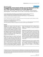
Figure 1
Effect of hyaluronic acid (HA) on antithrombin (AT)Effect of hyaluronic acid (HA) on antithrombin (AT). Various concentrations of HA, digested or not with hyaluronidase, were incubated with anti-
thrombin in the presence of 5 mmol/l CaCl
2
or FeCl
3
. Thrombin activity in the absence of both HA and antithrombin (blank) was considered as 1 and
the activities of the other tests were normalized based on comparisons with blank. Values are expressed as mean ± standard deviation of data from
triplicate experiments.
Arthritis Research & Therapy Vol 7 No 2 Chang et al.
R271
investigation is required to elucidate the exact mechanism
by which HA inhibits antithrombin, the results of the present
study do not refute the notion that optimal proteoglycan
uptake can improve overall articular function in patients
with arthritis.
Why HA inhibited antithrombin more after than before
hyaluronidase digestion remains obscure. Perhaps the
small HA molecule can easily bind and thus exert a more
inhibitory role on antithrombin. Nagaya and coworkers [23]
found high hyaluronidase activity in the synovial fluid and
serum of RA patients, implying an abundance of small HA
molecules in the RA synovium. Maneirio and coworkers
[24] reported that HA at various molecular weights had dif-
ferent effects on the interleukin-1 induced synthesis of both
nitric oxide and prostaglandin E
2
in chondrocytes. How
Ca
2+
and Fe
3+
are involved in inhibiting antithrombin by HA
is also poorly understood. Some investigators found that
Ca
2+
dramatically promotes the ability of heparin to drive
antithrombin activity [8,25,26]. Thus, both Ca
2+
and Fe
3+
ions might play similar roles in HA-induced changes in the
configuration of antithrombin.
Synovial fluid from RA patients contains a far greater abun-
dance of free iron than that from patients with osteoarthritis
[27,28]. It was reported that Fe
3+
stored in the RA syn-
ovium perpetuates inflammation by supporting the produc-
tion of oxygen radicals and by promoting hyaluronic acid
degradation, as well as the release of lysosomal enzymes
[29]. Telfer and coworkers [30] recently found that proin-
flammatory cytokines produced in the RA synovium
increased the accumulation of iron in synovial fluid. On
other hand, Davies and coworkers [31] reported that
neutrophils from synovial fluid and the circulation of RA
patients could increase the release of free Ca
2+
at inflam-
matory sites. Caruthers and coworkers [32] also showed
that calcium signaling is altered in T lymphocytes from RA
patients.
Genome-wide single nucleotide polymorphism analysis has
shown that peptidylarginine deiminase (PADI4), an enzyme
that post-translationally catalyzes peptidyl arginine to citrul-
line, is closely associated with RA [33]. We recently found
that recombinant human PADI4 protein inactivated human
antithrombin III via citrullination in vitro. We also detected
Figure 2
Effects of various glycosaminoglycans (GAGs) on antithrombin (AT)Effects of various glycosaminoglycans (GAGs) on antithrombin (AT).
Hyaluronic acid (HA), chondroitin sulfate A (CSA), chondroitin sulfate B
(CSB), chondroitin sulfate C (CSC), keratan sulfate (KS), heparin, or
heparan (500 µg/ml) was incubated with 150 µg/ml antithrombin and 5
mmol/l CaCl
2
. Controls consisted of only GAG or AT and blank (work-
ing buffer only). Thrombin activity of blank was considered as 1 and the
activities of other tests were normalized based on comparisons with
blank. Values are expressed as mean ± standard deviation of data from
triplicate experiments.
Figure 3
Heparin stimulates antithrombin (AT) activity in the presence of hyaluronic acid (HA)Heparin stimulates antithrombin (AT) activity in the presence of
hyaluronic acid (HA). Heparin (10 µg/ml) and various concentrations of
HA were incubated with 150 µg/ml antithrombin in presence of 5
mmol/l CaCl
2
. Thrombin activity of blank (reaction buffer only) was con-
sidered as 1 and the activities of other tests were normalized based on
comparisons with blank. Values are expressed as mean ± standard
deviation of data from triplicate experiments.
Available online />R272
an increased level of citrullinated antithrombin in the
plasma of RA patients [34]. PADI4 is extensively expressed
in RA synovial tissue [35,36]. Thus, we suggested that the
citrullination of antithrombin is one potential pathway
through which PADI4 contributes to the pathogenesis of
RA [34]. This notion does not contradict the current find-
ings. We postulate that the genetic, single nucleotide poly-
morphism-associated disorder of PADI4 and its excessive
citrullination of antithrombin play important roles in initiating
the RA pathogenic process, whereas inhibition of anti-
thrombin by HA contributes to the development of RA
rather than its initiation, because free HA in the synovium
achieves high concentrations along with RA progression.
Because of abundant Fe
3+
and altered Ca
2+
metabolism
together with significant hyaluronidase activity in the RA
synovium, thrombin-related RA specifically worsens in joint
tissue as a result of antithrombin inactivation by local
PADI4 and free HA (Fig. 5).
HA is an important component of the extracellular matrix.
Thrombin and antithrombin play key roles in hemostasis
and are involved in the pathogenic processes of many
diseases [6,37,38]. The findings presented here should
also be useful in investigating the nature of other diseases.
Conclusion
At concentrations of 250–1000 µg/ml in vitro, HA blocked
the thrombin-inhibitory ability of antithrombin in the pres-
ence of Ca
2+
and Fe
3+
. This finding suggested that the high
concentration of free HA in diseased RA synovium locally
blocks antithrombin under physiologic conditions and
thereby deregulates the activity of thrombin. These proc-
esses in turn drive the thrombin-related pathogenesis of
RA, which includes extensive fibrin deposition, extreme
angiogenesis, and abnormal fibroblast-like cell proliferation.
Our findings are consistent with those of previous reports
regarding increased coagulation activity in the RA
synovium.
Competing interests
The author(s) declare that they have no competing
interests.
Authors' contributions
XC designed and executed the study and prepared the
manuscript. RY and KY supervised the project, evaluated
data, and assisted in preparing the manuscript.
Acknowledgements
We thank every member of the Rheumatology Diseases Laboratory of
Riken for their general contribution to making this study possible.
References
1. Carmassi F, de Negri F, Morale M, Song KY, Chung SI: Fibrin deg-
radation in the synovial fluid of rheumatoid arthritis patients: a
model for extravascular fibrinolysis. Semin Thromb Hemost
1996, 22:489-496.
Figure 4
Effects of various metal ions on ability of hyaluronic acid (HA) to inhibit the activity of antithrombin (AT)Effects of various metal ions on ability of hyaluronic acid (HA) to inhibit
the activity of antithrombin (AT). HA (1000 µg/ml) and antithrombin
(150 µg/ml) were incubated with various concentrations of CaCl
2
,
FeCl
3
, KCl, MgCl
2
, or NaCl. Thrombin activity of blank (reaction buffer
only) was considered as 1 and the activities of other tests were normal-
ized based on comparisons with blank. Values are expressed as mean
± standard deviation of data from triplicate experiments.
Figure 5
Proposed mechanism of involvement of hyaluronic acid (HA) and pepti-dylarginine deiminase (PADI4) in the pathogenesis of rheumatoid arthritisProposed mechanism of involvement of hyaluronic acid (HA) and pepti-
dylarginine deiminase (PADI4) in the pathogenesis of rheumatoid arthri-
tis. VEGF, vascular endothelial growth factor.
Arthritis Research & Therapy Vol 7 No 2 Chang et al.
R273
2. Shin H, Kitajima I, Nakajima T, Shao Q, Tokioka T, Takasaki I, Hanyu
N, Kubo T, Maruyama I: Thrombin receptor mediated signals
induce expressions of interleukin 6 and granulocyte colony
stimulating factor via NF-kappa B activation in synovial
fibroblasts. Ann Rheum Dis 1999, 58:55-60.
3. Shin H, Nakajima T, Kitajima I, Shigeta K, Abeyama K, Imamura T,
Okano T, Kawahara K, Nakamura T, Maruyama I: Thrombin recep-
tor-mediated synovial proliferation in patients with rheuma-
toid arthritis. Clin Immunol Immunopathol 1995, 76:225-233.
4. Tsopanoglou NE, Maragoudakis MEJ: On the mechanism of
thrombin-induced angiogenesis. Potentiation of vascular
endothelial growth factor activity on endothelial cells by up-
regulation of its receptors. J Biol Chem 1999,
274:23969-23976.
5. Maragoudakis ME, Tsopanoglou NE, Andriopoulou P: Mechanism
of Thrombin-induced angiogenesis. Biochem Soc Trans 2002,
30:173-177.
6. Narayanan S: Multifunctional roles of thrombin. Ann Clin Lab
Sci 1999, 29:275-280.
7. Morris R, Winyard PG, Brass LF, Blake DR, Morris CJ: Thrombin
in inflammation and healing' relevance to rheumatoid arthritis.
Ann Rheum Dis 1994, 53:72-79.
8. Wiebe EM, Stafford AR, Fredenburgh JC, Weitz JI: Mechanism of
catalysis of inhibition of factor IXa by antithrombin in the pres-
ence of heparin or pentasaccharide. J Biol Chem 2003,
278:35767-35774.
9. Lozzo RV: Matrix proteoglycans: from molecular design to cel-
lular function. Annu Rev Biochem 1998, 67:609-652.
10. Wang JY, Roehrl MH: Glycosaminoglycans are a potential
cause of rheumatoid arthritis. Proc Natl Acad Sci USA 2002,
99:14362-14367.
11. Jones HW, Bailey R, Zhang Z, Dunne KA, Blake DR, Cox NL, Mor-
ris CJ, Winyard PG: Inactivation of antithrombin III in synovial
fluid from patients with rheumatoid arthritis. Ann Rheum Dis
1998, 57:162-165.
12. Ohba T, Takase Y, Ohhara M, Kasukawa R: Thrombin in synovial
fluid of paients with rheumatoid arthritis mediates prolifera-
tion of synovial fibroblast-like cells by induction of plate
derived growth factor. J Rheumatol 1996, 23:1505-1511.
13. Jain A, Nanchahal J, Troeberg L, Green P, Brennan F: Production
of cytokines, vascular endothelial growth factor, matrix metal-
loproteinases, and tissue inhibitor of metalloproteinases 1 by
tenosynovium demonstrates its potential for tendon destruc-
tion in rheumatoid arthritis. Arthritis Rheum 2001,
44:1754-1760.
14. Pitsillides AA, Worrall JG, Wilkinson LS, Bayliss MT, Edwards JC:
Hyaluronan concentration in non-inflamed and rheumatoid
synovium. Br J Rheumatol 1994, 33:5-10.
15. Nakayama Y, Shirai Y, Yoshihara K, Uesaka S: Evaluation of gly-
cosaminoglycans levels in normal joint fluid of the knee. J Nip-
pon Med Sch 2000, 67:92-95.
16. Takei YG, Honma T, Ito A: Quantitation of hyaluronic acid in
serum with automated microparticle photometric agglutina-
tion assay. J Immunoassay Immunochem 2002, 23:85-94.
17. Wyatt HA, Dhawan A, Cheeseman P, Mieli-Vergani G, Price JF:
Serum hyaluronic acid concentrations are increased in cystic
fibrosis patients with liver disease. Arch Dis Child 2002,
86:190-193.
18. Partsch G, Leeb B, Stancikova M, Raffayova H, Eberl G, Hitzel-
hammer H, Smolen JS: Low serum hyaluronan in psoriatic
arthritis patients in comparison to rheumatoid arthritis
patients. Clin Exp Rheumatol 1996, 14:381-386.
19. Skinner R, Abrahams JP, Whisstock JC, Lesk AM, Carrell RW,
Wardell MR: The 2.6 A structure of antithrombin indicates a
conformational change at the heparin binding site. J Mol Biol
1997, 266:601-609.
20. Jin L, Abrahams JP, Skinner R, Petitou M, Pike RN, Carrell RW:
The anticoagulant activation of antithrombin by heparin. Proc
Natl Acad Sci USA 1997, 94:14683-14688.
21. Kobayashi K, Matsuzaka S, Yoshida Y, Miyauchi S, Wada Y, Moriya
H: The effects of intraarticularly injected sodium hyaluronate
on levels of intact aggrecan and nitric oxide in the joint fluid of
patients with knee osteoarthritis. Osteoarthritis Cartilage 2004,
12:536-542.
22. Moreland LW: Intra-articular hyaluronan (hyaluronic acid) and
hylans for the treatment of osteoarthritis: mechanisms of
action. Arthritis Res Ther 2003, 5:54-67.
23. Nagaya H, Yamagata T, Yamagata S, Iyoda K, Ito H, Hasegawa Y,
Iwata H: Examination of synovial fluid and serum hyaluroni-
dase activity as a joint marker in rheumatoid arthritis and oste-
oarthritis patients (by zymography). Ann Rheum Dis 1999,
58:186-188.
24. Maneiro E, de Andres MC, Fernandez-Sueiro JL, Galdo F, Blanco
FJ: The biological action of hyaluronan on human osteoartritic
articular chondrocytes: the importance of molecular weight.
Clin Exp Rheumatol 2004, 22:307-312.
25. Rezaie AR: Calcium enhances heparin catalysis of the anti-
thrombin-factor Xa reaction by a template mechanism. Evi-
dence that calcium alleviates Gla domain antagonism of
heparin binding to factor Xa. J Biol Chem 1998,
273:16824-16827.
26. Bedsted T, Swanson R, Chuang YJ, Bock PE, Bjork I, Olson ST:
Heparin and calcium ions dramatically enhance antithrombin
reactivity with factor IXa by generating new interaction
exosites. Biochemistry 2003, 42:8143-8152.
27. Blake DR, Gallagher PJ, Potter AR, Bell MJ, Bacon PA: The effect
of synovial iron on the progression of rheumatoid disease. A
histologic assessment of patients with early rheumatoid
synovitis. Arthritis Rheum 1984, 27:495-501.
28. Ahmadzadeh N, Shingu M, Nobunaga M: Iron-binding proteins
and free iron in synovial fluids of rheumatoid arthritis patients.
Clin Rheumatol 1989, 8:345-351.
29. Morris CJ, Blake DR, Wainwright AC, Steven MM: Relationship
between iron deposits and tissue damage in the synovium: an
ultrastructural study. Ann Rheum Dis 1986, 45:21-26.
30. Telfer JF, Brock JH: Proinflammatory cytokines increase iron
uptake into human monocytes and synovial fibroblasts from
patients with rheumatoid arthritis. Med Sci Monit 2004,
10:BR91-BR95.
31. Davies EV, Williams BD, Whiston RJ, Cooper AM, Campbell AK,
Hallett : Altered Ca
2+
signalling in human neutrophils from
inflammatory sites. Ann Rheum Dis 1994, 53:446-449.
32. Carruthers DM, Arrol HP, Bacon PA, Young SP: Dysregulated
intracellular Ca
2+
stores and Ca
2+
signaling in synovial fluid T
lymphocytes from patients with chronic inflammatory arthritis.
Arthritis Rheum 2000, 43:1257-1265.
33. Suzuki A, Yamada R, Chang X, Tokuhiro S, Sawada T, Suzuki M,
Nagasaki M, Nakayama-Hamada M, Kawaida R, Ono M, et al.:
Functional haplotypes of PADI4, encoding citrullinating
enzyme peptidylarginine deiminase 4, are associated with
rheumatoid arthritis. Nat Genet 2003, 34:395-402.
34. Chang X, Yamada R, Sawada T, Suzuki A, Yamamoto K: The inhi-
bition of antithrombin by peptidylarginine deiminase 4 may
contribute to pathogenesis of rheumatoid arthritis. Rheumatol-
ogy (Oxford) 2004 in press.
35. Vossenaar ER, Radstake TR, Van Der Heijden A, Van Mansum MA,
Dieteren C, De Rooij DJ, Barrera P, Zendman AJ, Van Venrooij WJ:
Expression and activity of citrullinating peptidylarginine deim-
inase enzymes in monocytes and macrophages. Ann Rheum
Dis 2004, 63:373-381.
36. Chang X, Yamada R, Suzuki A, Sawada T, Yoshino S, Tokuhiro S,
Yamamoto K: Localization of peptidylarginine deiminase 4
(PADI4) and citrullinated protein in synovial tissue of rheuma-
toid arthritis. Rheumatology (Oxford) 2004 in press.
37. van Boven HH, Lane DA: Antithrombin and its inherited defi-
ciency states. Semin Hematol 1997, 34:188-204.
38. Ishiguro K, Kojima T, Kadomatsu K, Nakayama Y, Takagi A, Suzuki
M, Takeda N, Ito M, Yamamoto K, Matsushita T, et al.: Complete
antithrombin deficiency in mice results in embryonic lethality.
J Clin Invest 2000, 106:873-878.