Báo cáo y học: "Amplification of autoimmune disease by infection" potx
Bạn đang xem bản rút gọn của tài liệu. Xem và tải ngay bản đầy đủ của tài liệu tại đây (106.7 KB, 11 trang )
74
CMV = cytomegalovirus; EAE = experimental autoimmune encephalomyelitis; EBV = Epstein–Barr virus; HHV = human herpesvirus; IFN = inter-
feron; MHC = major histocompatibility complex; MHV = murine herpesvirus; PCR = polymerase chain reaction; RA = rheumatoid arthritis; SLE =
systemic lupus erythematosus; TCR = T-cell receptor; TNF = tumor necrosis factor.
Arthritis Research & Therapy Vol 7 No 2 Posnett and Yarilin
Abstract
Reports of infection with certain chronic persistent microbes
(herpesviruses or Chlamydiae) in human autoimmune diseases are
consistent with the hypothesis that these microbes are reactivated
in the setting of immunodeficiency and often target the site of auto-
immune inflammation. New experimental animal models demon-
strate the principle. A herpesvirus or Chlamydia species can be
used to infect mice with induced transient autoimmune diseases.
This results in increased disease severity and even relapse. The
evidence suggests that the organisms are specifically imported to
the inflammatory sites and cause further tissue destruction,
especially when the host is immunosuppressed. We review the
evidence for the amplification of autoimmune inflammatory disease
by microbial infection, which may be a general mechanism
applicable to many human diseases. We suggest that patients with
autoimmune disorders receiving immunosuppressing drugs should
benefit from preventive antiviral therapy.
What do herpesviruses, Chlamydiae and
parvovirus B19 have in common?
The question of how infectious organisms contribute to
autoimmunity has continued to be of interest to clinical
rheumatologists and basic immunologists. Recent reviews
have considered the possible contributions of different
non-mutually exclusive mechanisms, including molecular
mimicry, bystander activation, cryptic antigens, and
epitope spreading [1–3]. However, current understanding,
as reflected by these reviews, does not account for the
skewed list of infectious organisms often quoted as being
associated with various autoimmune disorders. As
outlined in Table 1, certain organisms are repeatedly
mentioned as being linked to different autoimmune
disorders. These are human herpesviruses (HHVs), in
particular the non-neurotropic herpesviruses such as
Epstein–Barr virus (EBV), cytomegalovirus (CMV) and
HHV6 (the group also includes HHV7 and HHV8),
Chlamydiae and parvovirus B19. As these organisms are
mentioned in the context of so many different diseases it is
unlikely that they would have specific etiologic roles.
Moreover, there is a large, controversial and often
contradictory literature on these associations, which
suggests that pathogenic mechanisms might be
redundant and non-specific. New data demonstrate a role
for such microbes in augmenting disease expression in
several experimental mouse models [4–6].
One approach is to examine relevant similarities between
Chlamydiae, parvovirus B19 and non-neurotropic
herpesviruses. Although, superficially, they have nothing in
common, they may share cell tropisms (Table 2) in that
they have a predilection for hematopoietic cells and
endothelial cells. The ability of these organisms to ‘hitch a
ride’ and get around in hematopoietic cells might actually
serve a vital function. For instance, the infectious life-cycle
of herpesviruses includes three functions for infected host
cells: first, initial viral replication; second, a long-term
latency reservoir; and third, the production of infectious
virus at a convenient mucosal or skin site. The initial host
cell for productive lytic infection, for example with EBV,
may be an oral mucosa epithelial cell [7], but it is quickly
replaced by the major target cell, the B lymphocyte, during
acute infectious mononucleosis. For EBV the same cell
serves as the latency reservoir. Conveniently, herpesvirus
latency is frequently established in circulating hemato-
poietic cells. To complete the infectious life cycle, virus
must be produced and transmitted to uninfected
individuals. This occurs at mucosal sites: salivary glands,
buccal mucosa and urogenital mucosa [8–10]. It is
assumed that, at intervals, productive infection occurs in
mucosal epithelial cells even in normal individuals and that
these mucosal cells are infected in turn via circulating
hematopoietic cells after local reactivation of latent virus.
For this purpose EBV-infected B cells may use the CD48
molecule to bind heparan sulfate on epithelial cells
[11,12]. This may occur at sites of chronic or intermittent
inflammation. Indeed, the lymphoid organs of Waldeyer’s
ring, where EBV is thought to reactivate, are sites of
Review
Amplification of autoimmune disease by infection
David N Posnett
1,2
and Dmitry Yarilin
1,2
1
Immunology Program, Graduate School of Medical Sciences, Weill Medical College, Cornell University, Ithaca, NY, USA
2
Department of Medicine, Division of Hematology-Oncology, Weill Medical College, Cornell University, Ithaca, NY, USA
Corresponding author: David N Posnett,
Published: 10 February 2005
Arthritis Res Ther 2005, 7:74-84 (DOI 10.1186/ar1691)
© 2005 BioMed Central Ltd
75
Available online />physiologic chronic inflammation. Other such sites of
physiologic inflammation include the gastrointestinal
mucosa and certain types of urogenital mucosa such as
the cervical transitional mucosa [12].
Low-grade histological inflammation of the prostate may
be more common than is generally thought [13], and was
noted in 66% of autopsies of men over the age 40 in one
study [14] and in all men with benign prostate hypertrophy
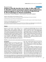
Table 1
Disease associations
Chlamydia Chlamydia
Disease EBV CMV HHV6 HHV7 HHV8 trachomatis pneumoniae PV B19 References
SLE +
a
+
b
+ + + [127–133]
RA ++ ++ + ++ ++ ++ [64,95,130,134]
Sjoegren’s disease ++ ++ [135,136]
Myocarditis + + + ++ [137–139]
MS + + ++ ++ [91,140–142]
T1DM
c
++ [143]
IgA nephritis ++ ++ [144,145]
Guillain–Barré syndrome + + [146,147]
Uveitis ++ + [148,149]
Reiter’s syndrome + + ++ [64,89]
Polymyositis dermatomyositis + + [130]
Aplastic anemia ++ [66]
ITP + + + [150,151]
Vasculitis + + + ++ [130]
Behcet’s disease + [130]
Giant cell arteritis ++? + [152,153]
Scleroderma + + [154,155]
Glomerulonephritis ++ ++ [144,145,156–158]
Autoimmune infertility + [159,160]
Psoriais + [161]
Pityriasis rosea ++ ++ [162]
Atherosclerosis + + ++ [98]
Leprosy + + [102,103,163]
After transplant ++
d
++
d
+ + + + + [123,124,164–173]
a
Associations that include some form of documented presence (by culture, electron microscopy, immunohistochemistry, PCR or in situ
hybridization) of the microbe in autoimmune target tissues are indicated by ++. Other associations are indicated by +. Note that the references are
not comprehensive and omit most of the contradictory literature; the purpose was to look for evidence of microbial presence specifically in the
autoimmune target tissues.
b
CMV in SLE is often a complication from immunosuppressive therapy causing colitis, ileitis, retinitis, pneumonitis or vasculitis, but infection can
also occur before therapy. It is unclear whether infection occurs on top of a pre-existing autoimmune lesion in an autoimmune tissue (for example
skin or kidney). In settings of viral reactivation due to immunosuppression, the virus may be expressed ubiquitously and we were therefore more
interested in reports in which expression was limited to an autoimmune target tissue.
c
A recent review lists up to six viruses associated with type I diabetes mellitus (T1DM), but we focus here only on those mentioned repeatedly in
association with a wide variety of autoimmune disorders.
d
PTLD (post-transfusion lymphoproliferative disease) represents a spectrum of disorders in which lymphocytes (predominantly B cells) infiltrate the
allo-transplant organ. PTLD can evolve from a condition that is reversible upon cessation of immunosuppression, to an irreversible monoclonal
lymphoma. Productive herpesvirus infections, especially EBV and CMV, occur in situ in allotransplants. By contrast, EBV is not usually present in
rejected transplant tissues. Chlamydiae can cause infectious complications in severely immunodeficient transplant patients but do not directly
infiltrate the transplanted tissues.
CMV, cytomegalovirus; EBV, Epstein–Barr virus; HHV, human herpesvirus; ITP, immune thrombocytopenia; MS, multiple sclerosis; PV, parvovirus;
RA, rheumatoid arthritis; SLE, systemic lupus erythematosus.
76
Arthritis Research & Therapy Vol 7 No 2 Posnett and Yarilin
[15]. Discrete focal inflammation of clinically normal
salivary glands has also been noted [16]. Finally, asympto-
matic airway inflammation is common and can be elicited
by ubiquitous stimuli such as smoke or smog [17,18].
Herpesviruses must have evolved a way of migrating to
such mucosal sites, perhaps by taking advantage of inflam-
matory cells that go there naturally. A possible unintended
sequel is that inflammatory cells may also migrate to
internal sites of inflammation, such as the synovium of an
arthritic patient. Reactivation of virus at these sites does
not serve the purpose of the virus but may aggravate the
disease process. The prediction from this model is that any
organism that uses hematopoietic inflammatory cells to
migrate to a site of inflammation can be reactivated in
autoimmune target tissues. Thus, there need not be a
specific organism associated with a specific disease.
Herpesviruses
How well does this model fit for the organisms listed in
Table 1? EBV (HHV4) is well known to infect resting B
lymphocytes. CD27
–
, CD5
–
, IgD
–
memory B cells later
provide a latency reservoir [8,19]. There are estimates that
1 in 10
5
to 1 in 10
6
B cells carry latent EBV in normal
adults [20]. Upon reactivation of EBV in the lymphoid
tissue of Waldeyer’s ring [8], shedding occurs from the
oral mucosa. Although not yet proven, it is possible that
mucosal epithelial cells adjacent to these lymphoid organs
produce infectious virus [11,21]. Indeed, EBV can infect
several cell types other than B cells, including endothelial
cells [22], follicular dendritic cell lines [23], T lymphoma
cells in hemophagocytic syndrome [24], smooth muscle
tumor cells in immunosuppressed hosts [25] and synovio-
cytes from patients with rheumatoid arthritis (RA) [26–28].
Acute lytic infection with CMV (HHV5) occurs in
monocytes in the blood, and a latency reservoir is
established in circulating myelomonocytic cells and their
CD33
+
CD34
+
bone marrow progenitors [29–31]. About
0.01 to 0.004% of mononuclear cells from peripheral
blood donors, who had received granulocyte colony-
stimulating factor mobilization for transplant purposes,
contained the viral genome [32]. CMV can also infect
dendritic cells [33,34] and endothelial cells, and may
establish a latency reservoir in these cells too [30]. Lytic
infection can involve epithelial cells, fibroblasts, stromal
cells, neuronal cells, smooth muscle cells and hepatocytes
in infected target tissues. CMV seems to be reactivated
from latency by allostimulation [29,35]. Perhaps
reactivation also occurs by immune stimulation at a
mucosal site where CMV is excreted, such as the salivary
glands, the lactating mammary glands or the urogenital
tract [10,36,37], allowing both horizontal sexual trans-
mission and vertical transmission to the newborn infant.
Tumor necrosis factor-α (TNF-α) can substitute for
allostimulation in inducing expression of the CMV IE-1
gene [29,38], but for complete CMV reactivation it is likely
that other checkpoints must be overcome [39], perhaps
regulated by other cytokines such as interleukin-13 and
granulocyte/macrophage colony-stimulating factor, which
are known to promote the replication of human CMV [38].
Moreover, CMV has evolved its own specialized CC-
chemokine gene, MCK-2. The presence of MCK-2 results
in greater inflammatory responses and enables CMV
shedding in the salivary glands [40,41].
HHV6 infects cells of the myelomonocytic lineage both
acutely and then latently. This includes bone marrow
progenitors and myelomonocytic cells in peripheral blood
[42–45]. HHV6 also has tropism for T cells, B cells,
natural killer cells (viral subgroup A) and dendritic cells
[45]. Finally, lytic infection can occur in many other cell
types including neurons, muscle cells and epithelial cells.
The last of these probably allow productive infection at a
mucosal site, such as the salivary glands [46–48].
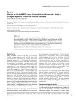
Table 2
Characteristics of human herpesviruses, Chlamydiae and parvovirus
Main Proposed Other
Organism Receptors cellular tropism latency cell tropism References
EBV CD21, MHC-II, α5β1 integrin B B EPC, EC [8]
CMV EGFR M/M M/M; EC N, EPC, EC [174]
HHV6 CD46+ M/M, T, B M/M N, EPC [175]
HHV7 CD4
+
heparan sulfate receptor T M/M N, EPC, EC [176,177]
HHV8 Heparan sulfate receptor, EGFR EC, M/M, B, T B N, EPC [174,178]
Chlamydia pneumoniae Heparan sulfate receptor M/M EC,EPC [179]
Chlamydia trachomatis M/M EC,EPC
Parvovirus B19 Erythrocyte P antigen Erythroid precursors EC [66,67]
B, B cells; CMV, cytomegalovirus; EBV, Epstein–Barr virus; EC, endothelial cells; EGFR, epidermal growth factor receptor; EPC, epithelial cells;
HHV, human herpesvirus; MHC, major histocompatibility complex; M/M, myelomonocytic cells; N, neural cells; T, T cells.
77
HHV7 may infect predominantly T cells but also
myelomonocytic cells [49–51]. Like other herpesviruses it
can infect epithelial and endothelial cells. Salivary glands
are a major site of production of HHV7 [9,52]. HHV6 and
HHV7 antigenemia occurs in the setting of CMV
reactivation in transplant patients [53].
Finally, HHV8 targets myelomonocytic cells, lymphocytes
and endothelial cells [54,55]. There may be a latency
reservoir in B cells and circulating monocytes. Epithelial
cells can also be infected and HHV8 is detected in the
saliva of asymptomatic persons [9,52,56].
The cellular receptors used for herpesviral entry and fusion
are often expressed ubiquitously (Table 2) and do not
completely explain the targeted cell types. Just because a
receptor is known does not mean it is the only one. CD21
and major histocompatibility complex (MHC) class II are
known EBV receptors on B cells but α
5
β
1
integrin is a
receptor for entry into polarized tongue and nasopharyn-
geal epithelial cells [7]. Nevertheless, there is a recurrent
pattern in that these β- and γ-herpesviruses establish
latency in hematopoietic cells and are reactivated for
production of infectious virus at a suitable mucosal site. To
some extent this may also apply to α-herpesviruses,
although their distinguishing feature is tropism for, and
latency in, neuronal cells and host-to-host transmission
through skin lesions.
Chlamydiae
Chlamydiae are bacteria that live within vacuoles in
eukaryotic cells. Acute infections target mucosal cell
surfaces (lung, genital tract or eye). Persistence for many
years is common, and studies in mouse models have
shown that quiescent organisms can be reactivated
[57,58]. Host cells include endothelial cells (Chlamydia
pneumoniae) and epithelial cells (Chlamydia trachomatis).
Circulating monocytes also carry Chlamydiae and may
serve to disseminate the organism [59,60]. In vitro, small
amounts of interferon-γ (IFN-γ) arrest chlamydial develop-
ment and promote a morphologically distinguishable
persistent form. This is reversible in the presence of
excess tryptophan [57,61]. Thus, it is thought that IFN-γ
limits available intracellular pools of tryptophan for the
bacteria without affecting their viability and that this
occurs via the tryptophan decyclizing enzyme indoleamine
2,3-dioxygenase. However, not all Chlamydiae are
dependent on exogenous tryptophan: serovars D–K of
Chlamydia trachomatis, with urogenital rather than ocular
tropism, possess trpRBA, a tryptophan synthase gene
cluster, and can synthesize tryptophan from indole
substrates produced by vaginal microbial flora [62]. In
IFN-γ knockout mice, and even more so in mice with
severe combined immunodeficiency, C. trachomatis
(strain MoPn) disseminates to various tissues from the
genital tract and infection fails to resolve [63]. Thus, as
with the Herpesviruses, the host inflammatory response
can control the persistence of Chlamydiae, although the
mechanistic details differ. The proinflammatory cytokine
mix present in the arthritic synovium may promote the local
persistence of Chlamydiae [57,61,64,65].
Parvovirus B19
With parvovirus B19, acute infection occurs in the upper
respiratory tract [66,67]. At least 50% of the general
population have been exposed and have detectable IgG
antibodies. There are three clinical syndromes: fifth
disease (erythema infectiosum), hydrops fetalis, and
transient aplastic crisis/pure red cell aplasia. The latter led
to the discovery that parvovirus B19 has exquisite cell
tropism for early erythroid cells and progenitors, resulting
in a cytopathic effect in giant pro-normoblasts [66].
However, anemia develops primarily when red cell turn-
over is increased, as in patients with chronic hemolysis.
The virus uses globoside or erythrocyte P antigen to gain
entry to these cells. Although the receptor is present on
other cells, including megakaryocytes and endothelial
cells, productive infection is restricted to pronormoblasts
[66]. Parvoviruses of other animal species infect lympho-
cytes and monocytes, but this has not been shown for
B19 in humans. A reticuloendothelial site for B19 infection
remains a possibility (N. Young, personal communication).
Parvovirus B19 is a single-stranded DNA virus that does not
enter typical latency or become integrated in the host cell
genome. However, persistence of the organism does occur.
In the original description [68], viremia was described in
healthy asymptomatic blood donors. By nested PCR,
parvovirus DNA was found in bone marrow from 4 of 45
random cadavers [69]. It is also known that the virus can
be transmitted by blood products [70]. Virus can ‘persist’
in normal and immunodeficient patients without clinical
evidence of disease [70,71]. Patients with congenital
immunodeficiency, children with leukemia during or after
chemotherapy, patients with AIDS, and transplant
recipients may suffer persistent parvovirus B19 infection
and the viral DNA load can be as high as in acute infection
[66]. Cryptic infection with low-grade viral replication in
normal hosts [72] may explain why B19 DNA is found in
the bone marrow of patients with arthritis [73], some of
whom may have B19 DNA in the synovium and the
synovial fluid [74–76] and occasionally viral DNA is
widespread including in the serum and skin [77].
The pathogenic role of viral DNA in the synovium is
debated because control samples from osteoarthritis
patients, or patients with recent joint trauma, also
contained B19 DNA. While transgenic expression of
nonstructural protein-1 (NS1) of parvovirus B19 in C57Bl/6
mice did not result in spontaneous arthritis, it did render
mice of a resistant genetic background susceptible to
collagen-induced arthritis [78]. In these mice NS1 was
Available online />78
expressed in the synovium after arthritis induction. There
are further associations where B19 DNA has been found
in the relevant tissues, for example hepatitis, myocarditis
and various types of vasculitis [67].
Perhaps cryptic infection is normally contained in the
presence of neutralizing antibodies, which are present in
many sera and can be administered therapeutically in the
form of intravenous immunoglobulin to immunodeficient
patients [66]. It is not known whether this virus uses
hematopoietic cells for dissemination within the body, nor
is it known where or how the virus is excreted for
dissemination to new hosts. Data are also lacking on
whether the inflammatory milieu might influence viral
replication. Autoimmunity associated with parvovirus B12
infection (Table 1) is thought to be due to immune
complexes, cross-reactive antibodies, immune dysregulation
or the production of inflammatory cytokines [79–82].
Overall, the data on this virus are not as strong as those
for herpesviruses and Chlamydiae in support of the
hypothesis proposed herein.
Circumstantial evidence for the hypothesis
In summary, it is possible that both herpesviruses and
Chlamydiae gain access to sites of chronic tissue
inflammation through a Trojan horse mechanism, because
the influx of inflammatory hematopoietic cells will include a
small number of cells that carry these organisms in
dormant forms. There is some circumstantial evidence for
this hypothesis. First, several studies aimed at discovering
the autoantigen in human autoimmune diseases have used
TCR repertoire analysis. In several instances, expanded
CD4 and CD8 clones were found. Although investigators
had invariably been hunting for autoantigen-reactive
clones, the only specificities that have been uncovered are
herpesvirus antigens! For example, CD4 clones from RA
synovia examined by Li and colleagues were ‘auto-
reactive’ with EBV-transformed B cell lines [83]. CD8
clones in psoriatic arthritis bore the signature TCR BV
CDRIII region of T cells specific for BMLF1 of EBV [84].
CD8 clones from RA synovia characterized by Bonneville
and colleagues in a series of elegant studies were reactive
with latent and lytic viral antigens, including BZLF1 and
BMLF1 [85,86]. Curiously, the EBV antigens identified
were often lytic gene products. This implied that productive
viral infection might have occurred in the synovium.
These results were corroborated by using MHC class I
tetramers, specifically EBV and CMV peptides bound to
HLA-A2. Synovial T cells specific for herpesvirus antigens
were found enriched in the synovium in comparison with
blood obtained at the same time from the same patient
[87,88]. Finally, these studies revealed that the
concentration of herpes-specific T cells in the inflam-
matory synovium was not disease specific. This pheno-
menon was observed in RA, in psoriatic arthritis, in
ankylosing spondylitis, in uveitis, and in multiple sclerosis,
where target tissues were also enriched in CMV-specific
and EBV-specific T cells [89]. In this context the much
touted association of a disease such as multiple sclerosis
with HHV-6 or Chlamydia [90,91] is less puzzling. As with
CMV and EBV, these organisms may reactivate within the
autoimmune target tissue.
Whether herpesviruses are produced in situ in autoimmune
target tissues has been examined in several studies [26–28].
Koide and colleagues were able to culture infectious EBV
from RA synoviocytes obtained ex vivo [26]. Takeda and
colleagues provided immunohistological and in situ
hybridization studies in support of productive viral infection
in RA synovia [27]. Many studies have provided serological
evidence of productive EBV infection in RA, and also for
HHV6 and CMV [92,93]. Productive infection by EBV in
the oral mucosa is significantly increased in RA in
comparison with normal subjects [92]. Finally, PCR studies
for viral DNA and RNA in RA synovia have yielded
contradictory results [28,94,95]. However, negative results
can easily be explained by the rapid and efficient clearance
of virus-infected cells by a competent immune system.
Some samples that were negative by PCR were
nevertheless enriched for EBV-specific CD8 cells [94].
As discussed, T cells specific for lytic viral antigens can
accumulate in the inflammatory target tissues in several
autoimmune diseases. However, this is not specific to
autoimmunity. It might also occur in other inflammatory
lesions, including atherosclerotic plaques for example
[96–98]. The association between herpesvirus infection of
the arterial wall and atherosclerosis is striking for Marek’s
disease in chickens [99]. Infection of apoE
–/–
mice with a
murine γ-herpesvirus accelerates atherosclerosis, and viral
mRNA is present in the aorta [100]. There may be other
examples, as suggested by unusual reports such as the
detection of EBV by PCR and immunohistochemistry in
fibroadenomas of the breast in immunosuppressed hosts
[101], and the association of EBV with leprosy [102,103].
The key question is whether this matters for disease
progression. If these microbes aggravate disease by
superimposed infection, antimicrobial therapy would be
predicted to halt disease progression. This question has
now been addressed in animal models.
Murine models to test the hypothesis
Murine herpesvirus (MHV)-68 is a mouse gamma
herpesvirus. It most closely resembles EBV and HHV8
and is a natural pathogen of small rodents. This virus has
now been used to infect mice with transiently induced
arthritis [4] using serum transferred from K/BxN mice
[104]. Normally, a clinically severe but transient inflam-
matory arthritis develops within 2 days and resolves after 3
to 4 weeks.
Arthritis Research & Therapy Vol 7 No 2 Posnett and Yarilin
79
The course of this transient arthritis was aggravated and
prolonged by infection with MHV-68 given 2 to 5 days
after arthritis induction [4]. In immunocompetent mice the
disease remained transient, but in severely immuno-
compromised mice a relapse of arthritis was observed.
The relapse was due to lytic viral infection in synovial
tissues of recovering arthritic, but not normal, joints in the
same animal. Infection was demonstrated by PCR,
immunohistochemistry and electron microscopy. Virus-
specific T cells were enriched in the affected joints.
Clinical relapse of arthritis could be inhibited with an
antiviral drug, cidofovir, known to be active against
MHV-68. Latent infection could be reactivated in the
synovium when normal mice, latently infected with
MHV-68, were treated with Cytoxan. This was associated
with increased arthritis and viral antigens in the synovium
by immunohistochemistry. These data strongly suggest
that a herpesvirus infection can be imported to the
inflammatory site of an autoimmune target tissue. Genuine
viral infection is established, and this alters the course of
the autoimmune disease.
MHV-68 infection is also known to exacerbate experi-
mental autoimmune encephalomyelitis (EAE) in mice, an
experimental mouse model for multiple sclerosis [5]. The
mechanism by which the virus altered disease expression
was not uncovered in this study. Although viral DNA was
not detected in the diseased spinal cords, this might have
been due to insufficient sensitivity of the assays. In an
immunocompetent host, as in these mice, virus-infected
cells are instantly removed and only the telltale viral
antigen-specific T cells remain as proof of what has
happened.
C. pneumoniae was used to infect mice (intraperitoneally)
on day 7 of EAE induction. C. pneumoniae, but not C.
trachomatis, resulted in more severe neurological disease
[6]. C. pneumoniae, usually present only in spleen and
lungs, was found in the central nervous system by reverse
transcriptase PCR and by immunohistochemical staining
associated with perivascular lymphocytic infiltrates. In
conclusion, several animal models, using herpesviruses or
Chlamydiae, support our hypothesis.
Mechanisms of amplification of
autoimmunity
Imported infection as described above can theoretically
have one of three effects: first, it can exacerbate ongoing
disease leading to greater severity and duration; second, it
can induce a relapse; or third, it can lead to chronic
progressive disease. In the KxN arthritis model using the γ-
herpesvirus MHV-68 [4], exacerbation of transient arthritis
was observed in immunocompetent mice. Disease was
also exacerbated in Cytoxan-treated immunodeficient
mice, and in severely immunocompromised RAG1
–/–
mice
a relapse due to lytic viral infection in the synovia was
observed. In EAE the same virus (MHV-68) exacerbated
disease [5]. Only immunocompetent mice were examined
and the observation period was not long enough to assess
relapse or chronicity. These authors did not find lytic viral
infection in the central nervous system by viral plaque
assays or by PCR. For C. pneumoniae and EAE [6],
exacerbation was also noted in immunocompetent mice,
but relapse or chronicity was not examined. In that paper,
in vitro responses to myelin basic protein, such as T cell
proliferation and γ-IFN production, were measured. Mice
with EAE plus C. pneumoniae infection had larger
responses to myelin basic protein than mice with EAE
alone, suggesting that autoimmune responses were
amplified by the infection.
Our data from immune-suppressed mice showed
extensive viral infection, with MHV-68 in the synovium
involving all cell types including fibroblasts and synovial
lining cells [4]. By electron microscopy many of these cells
were lytically destroyed, extracellular free viral particles
were abundant and polymorphonuclear cells ingesting
viral particles were seen. This picture suggests lytic viral
infection. In an immunocompetent mouse, this infection
would presumably be contained by a cellular and a
humoral immune response. A local antiviral immune
response would no doubt contribute to autoimmune
inflammation. Cytotoxic tissue damage, whether induced
by cytotoxic T cells or due to lytic viral infection, would
result in a proinflammatory milieu. Cytokines and
chemokines could contribute to inflammation in a non-
specific way. However, infection might also release
sequestered autoantigens and thus spread the repertoire
of targeted autoantigens.
Indeed, Horwitz and colleagues have demonstrated
bystander tissue destruction by Coxsackie virus in
autoimmune diabetes [105]. As a result, sequestered
autoantigen was released, which re-stimulated resting
auto-reactive T cells in TCR transgenic mice, containing
an overabundance of such T cells specific for an islet
autoantigen. Both Coxsackie virus and the drug
streptozotocin, an islet-damaging agent, had this effect
[106]. Coxsackie virus is not a persistent or latent virus of
hematopoietic cells. Mechanisms pertaining to the
amplification of autoimmunity by MHV-68 or Chlamydiae
might therefore differ and have not yet been rigorously
examined.
In RA, studies need to be performed to examine whether
viral infection with herpesviruses contributes to the
emergence of new autoimmune responses. Of interest are
responses to the following: collagen type II, proteoglycans
and chondrocyte glycoprotein 39; nuclear lamins,
topoisomerase II and RA33 antigen (heterogeneous
nuclear ribonucleoprotein A2); cytoplasmic antigens such
as anti-neutrophil cytoplasmic antibodies; extracellular
Available online />80
antigens such as keratin and IgG, the target of typical
rheumatoid factors; and apoptosis-related proteins such
as annexin V, calpastatin, vimentin and filaggrin
[107–115]. For the last two antigens, arginine is replaced
by citrulline, a process that occurs during apoptosis and is
catalyzed by peptidyl arginine deiminase [110]. One
indication that immunosuppressive therapy, with potential
reactivation of endogenous herpesviruses, is associated
with the emergence of new antibody specificities, has
been published. In patients with RA (726 paired samples),
initial drug therapy (often methotrexate) was associated
with a change from a negative to a positive antinuclear
antibody test in 12.5% [116].
Antimicrobials for autoimmunity?
The implication from these studies is that it may be time to
design trials of antimicrobial drugs for selected patients
with autoimmune diseases such as RA. It is already
common practice to treat transplant patients and cancer
patients receiving strong immunosuppressive drugs with
acyclovir or valacyclovir, to prevent the reactivation of
CMV and EBV. Whether patients with autoimmune
diseases, such as RA and systemic lupus erythematosus
(SLE), on immunosuppressive drugs such as metho-
trexate, azathioprine or cytoxan could also benefit from
antiviral drugs need to evaluated. The occurrence of EBV-
related lymphomas in methotrexate-treated patients with
RA [117,118] suggests that EBV-specific immuno-
surveillance is deficient [119]. EBV genomic DNA,
measured by real-time PCR, was increased in the
peripheral blood mononuclear cells of patients with RA by
about 1 log over controls [120]. However, fluctuations of
EBV DNA in the blood mononuclear cells were not
correlated with immunosuppressive therapy (either metho-
trexate alone or methotrexate plus anti-TNF-α) in small
groups of patients. EBV DNA in the affected joints was
not measured. Whether those patients with higher viral
load did worse than others was also not reported.
The use of antimicrobials for autoimmunity is not without
precedent, and successes have been reported. In most
cases antibiotics have been used for their non-
antimicrobial effects. Dapsone (which inhibits neutrophil
function), tetracyclines (which block collagenase) and
chloroquine (which blocks antigen presentation and
cytokine secretion) have all been used in treating RA and
SLE [121]. However, organisms like Chlamydiae are
susceptible to antibiotics including tetracyclines, raising
the possibility that some of these drugs might have been
beneficial as a result of antimicrobial activity.
To optimize chances of therapeutic success, we suggest
that patients first be screened for reactivated herpes-
viruses, parvovirus B12 and persistent Chlamydiae.
Screening for CMV or EBV reactivation by quantitative
PCR is standard practice in bone marrow transplant
patients. This helps to guide the clinical use of antiviral
drugs, which are now often used for prophylaxis [122-
125]. These include acyclovir, gancyclovir and the oral
prodrugs valacyclovir and valgancyclovir. We propose the
same approach for autoimmunity. Depending on the
organism(s) present in the analyzed autoimmune tissues,
antiviral drugs for EBV or CMV, tetracycline or other
antibiotics for Chlamydiae, or intravenous immunoglobulin
for parvovirus could be tried. Note that there are few data
on the efficacy of antibiotics for chronic Chlamydia
infections [126]. Careful monitoring for the presence of
the microbial organism in the relevant tissue (synovial fluid
in RA) will be desirable to monitor the effectiveness of the
drug. For example, quantitative PCR assays for
herpesviruses, parvovirus and Chlamydiae could be used.
Cultures might also be helpful. Finally, prophylactic
antiviral therapy in patients receiving immunosuppressive
drugs such as low-dose methotrexate in RA should be
considered.
Competing interests
The author(s) declare that they have no competing interests.
Acknowledgements
We thank the following colleagues for their critical input: D Thorley-
Lawson, WA Muller, RL Nachman, L Ivashkiv, M Kuntz-Crow and A
Asch. This work was supported in part by an Arthritis Foundation grant.
References
1. Olson JK, Croxford JL, Miller SD: Virus-induced autoimmunity:
potential role of viruses in initiation, perpetuation, and pro-
gression of T-cell-mediated autoimmune disease. Viral
Immunol 2001, 14:227-250.
2 Benoist C, Mathis D: Autoimmunity provoked by infection: how
good is the case for T cell epitope mimicry? Nat Immunol
2001, 2:797-801.
3 Hafler DA: The distinction blurs between an autoimmune
versus microbial hypothesis in multiple sclerosis. J Clin Invest
1999, 104:527-529.
4 Yarilin DA, Valiando J, Posnett DN, A mouse Herpesvirus
induces relapse of experimental autoimmune arthritis. J
Immunol 2004, 173:5238-5246.
5 Peacock JW, Elsawa SF, Petty CC, Hickey WF, Bost KL: Exacer-
bation of experimental autoimmune encephalomyelitis in
rodents infected with murine gammaherpesvirus-68. J
Immunol 2003, 33:1849-1858.
6 Du C, Yao SY, Ljunggren-Rose A, Sriram S: Chlamydia pneumo-
niae infection of the central nervous system worsens experi-
mental allergic encephalitis. J Exp Med 2002, 196:1639-1644.
7 Tugizov SM, Berline JW, Palefsky JM: Epstein–Barr virus infec-
tion of polarized tongue and nasopharyngeal epithelial cells.
Nat Med 2003, 9:307-314.
8 Thorley-Lawson DA: Epstein–Barr virus: exploiting the immune
system. Nat Rev Immunol 2001, 1:75-82.
9 Lucht E, Brytting M, Bjerregaard L, Julander I, Linde A: Shedding
of cytomegalovirus and herpesviruses 6, 7, and 8 in saliva of
human immunodeficiency virus type 1-infected patients and
healthy controls. Clin Infect Dis 1998, 27:137-141.
10 Gautheret-Dejean A, Aubin JT, Poirel L, Huraux JM, Nicolas JC,
Rozenbaum W, Agut H: Detection of human Betaherpesvirinae
in saliva and urine from immunocompromised and immuno-
competent subjects. J Clin Microbiol 1997, 35:1600-1603.
11 Ianelli CJ, De Lellis R, Thorley-Lawson DA: CD48 binds to
heparan sulfate on the surface of epithelial cells. J Biol Chem
1998, 273:23367-23375.
12 Johansson EL, Rudin A, Wassen L, Holmgren J: Distribution of
lymphocytes and adhesion molecules in human cervix and
vagina. Immunology 1999, 96:272-277.
Arthritis Research & Therapy Vol 7 No 2 Posnett and Yarilin
81
13 Krieger JN, Ross SO, Riley DE: Chronic prostatitis: epidemiol-
ogy and role of infection. Urology 2002, 60:8-12.
14 Billis A, Magna LA: Inflammatory atrophy of the prostate.
Prevalence and significance. Arch Pathol Lab Med 2003, 127:
840-844.
15 Nickel JC, Downey J, Young I, Boag S: Asymptomatic inflamma-
tion and/or infection in benign prostatic hyperplasia. BJU Int
1999, 84:976-981.
16 Harrison JD, Epivatianos A, Bhatia SN: Role of microliths in the
aetiology of chronic submandibular sialadenitis: a clinico-
pathological investigation of 154 cases. Histopathology 1997,
31:237-251.
17 Roth MD, Arora A, Barsky SH, Kleerup EC, Simmons M, Tashkin
DP: Airway inflammation in young marijuana and tobacco
smokers. Am J Respir Crit Care Med 1998, 157:928-937.
18 Sherwin RP, Richters V, Everson RB, Richters A: Chronic glan-
dular bronchitis in young individuals residing in a metropoli-
tan area. Virchows Arch 1998, 433:341-348.
19 Joseph AM, Babcock GJ, Thorley-Lawson DA: EBV persistence
involves strict selection of latently infected B cells. J Immunol
2000, 165:2975-2981.
20. Decker LL, Klaman LD, Thorley-Lawson DA: Detection of the
latent form of Epstein–Barr virus DNA in the peripheral blood
of healthy individuals. J Virol 1996, 70:3286-3289.
21. Deacon EM, Matthews JB, Potts AJ, Hamburger J, Bevan IS,
Young LS: Detection of Epstein–Barr virus antigens and DNA
in major and minor salivary glands using immunocytochem-
istry and polymerase chain reaction: possible relationship
with Sjogren’s syndrome. J Pathol 1991, 163:351-360.
22. Jones K, Rivera C, Sgadari C, Franklin J, Max EE, Bhatia K, Tosato
G: Infection of human endothelial cells with Epstein–Barr
virus. J Exp Med 1995, 182:1213-1221.
23. Lindhout E, Lakeman A, Mevissen ML, de Groot C: Functionally
active Epstein–Barr virus-transformed follicular dendritic cell-
like cell lines. J Exp Med 1994, 179:1173-1184.
24. Lay JD, Tsao CJ, Chen JY, Kadin ME, Su IJ: Upregulation of
tumor necrosis factor-alpha gene by Epstein–Barr virus and
activation of macrophages in Epstein–Barr virus-infected T
cells in the pathogenesis of hemophagocytic syndrome. J Clin
Invest 1997, 100:1969-1979.
25. McClain KL, Leach CT, Jenson HB, Joshi VV, Pollock BH, Parmley
RT, Di Carlo FJ, Chadwick EG, Murphy SB: Association of
Epstein–Barr virus with leiomyosarcomas in children with
AIDS. N Engl J Med 1995, 332:12-18.
26. Koide J, Takada K, Sugiura M, Sekine H, Ito T, Saito K, Mori S,
Takeuchi T, Uchida S, Abe T: Spontaneous establishement of
an Epstein–Barr virus infected fibroblast line from the syn-
ovial tissue of a rheumatoid arthritis patient. J Virol 1997, 71:
2478-2481.
27. Takeda T, Mizugaki Y, Matsubara L, Imai S, Koike T, Takada K:
Lytic Epstein–Barr virus infection in the synovial tissue of
patients with rheumatoid arthritis. Arthritis Rheum 2000, 43:
1218-1225.
28. Saal JG, Krimmel M, Steidle M, Gerneth F, Wagner S, Fritz P,
Koch S, Zacher J, Sell S, Einsele H, et al.: Synovial Epstein–Barr
virus infection increases the risk of rheumatoid arthritis in
individuals with the shared HLA-DR4 epitope. Arthritis Rheum
1999, 42:1485-1496.
29. Soderberg-Naucler C, Fish KN, Nelson JA: Reactivation of latent
human cytomegalovirus by allogeneic stimulation of blood
cells from healthy donors. Cell 1997, 91:119-126.
30. Jarvis MA, Nelson JA: Human cytomegalovirus persistence and
latency in endothelial cells and macrophages. Curr Opin
Microbiol 2002, 5:403-407.
31. Kondo K, Xu J, Mocarski ES: Human cytomegalovirus latent
gene expression in granulocyte-macrophage progenitors in
culture and in seropositive individuals. Proc Natl Acad Sci
USA 1996, 93:11137-11142.
32. Slobedman B, Mocarski ES: Quantitative analysis of latent
human cytomegalovirus. J Virol 1999, 73:4806-4812.
33. Halary F, Amara A, Lortat-Jacob H, Messerle M, Delaunay T,
Houles C, Fieschi F, Arenzana-Seisdedos F, Moreau JF,
Dechanet-Merville J: Human cytomegalovirus binding to DC-
SIGN is required for dendritic cell infection and target cell
trans-infection. Immunity 2002, 17:653-664.
34. Raftery MJ, Schwab M, Eibert SM, Samstag Y, Walczak H,
Schonrich G: Targeting the function of mature dendritic cells
by human cytomegalovirus: a multilayered viral defense strat-
egy. Immunity 2001, 15:997-1009.
35. Hummel M, Abecassis MM: A model for reactivation of CMV
from latency. J Clin Virol 2002, Suppl 2:123-136.
36. Forbes BA: Acquisition of cytomegalovirus infection: an
update. Clin Microbiol Rev 1989, 2:204-216.
37. Ho M: Epidemiology of cytomegalovirus infections. Rev Infect
Dis 1990, 12 (Suppl 7):701-710.
38. Streblow DN, Nelson JA: Models of HCMV latency and reactiva-
tion. Trends Microbiol 2003, 11:293-295.
39. Reddehase MJ, Podlech J, Grzimek NK: Mouse models of
cytomegalovirus latency: overview. J Clin Virol 2002, Suppl 2:
23-36.
40. Saederup N, Mocarski ES Jr: Fatal attraction: cytomegalovirus-
encoded chemokine homologs. Curr Top Microbiol Immunol
2002, 269:235-256.
41. Saederup N, Aguirre SA, Sparer TE, Bouley DM, Mocarski ES:
Murine cytomegalovirus CC chemokine homolog MCK-2
(m131-129) is a determinant of dissemination that increases
inflammation at initial sites of infection. J Virol 2001, 75:9966-
9976.
42. Kondo K, Kondo T, Okuno T, Takahashi M, Yamanishi K: Latent
human herpesvirus 6 infection of human monocytes/
macrophages. J Gen Virol 1991, 72:1401-1408.
43. Kondo K, Kondo T, Shimada K, Amo K, Miyagawa H, Yamanishi K:
Strong interaction between human herpesvirus 6 and periph-
eral blood monocytes/macrophages during acute infection. J
Med Virol 2002, 67:364-369.
44. Luppi M, Barozzi P, Morris C, Maiorana A, Garber R, Bonacorsi G,
Donelli A, Marasca R, Tabilio A, Torelli G: Human herpesvirus 6
latently infects early bone marrow progenitors in vivo. J Virol
1999, 73:754-759.
45. Lusso P: Human herpesvirus 6 (HHV-6). Antiviral Res 1996, 31:
1-21.
46. Levy JA, Ferro F, Greenspan D, Lennette ET: Frequent isolation
of HHV-6 from saliva and high seroprevalence of the virus in
the population. Lancet 1990, 335:1047-1050.
47. Fox JD, Briggs M, Ward PA, Tedder RS: Human herpesvirus 6
in salivary glands. Lancet 1990, 336:590-593.
48. Di Luca D, Mirandola P, Ravaioli T, Dolcetti R, Frigatti A, Bovenzi
P, Sighinolfi L, Monini P, Cassai E: Human herpesviruses 6 and
7 in salivary glands and shedding in saliva of healthy and
human immunodeficiency virus positive individuals. J Med
Virol 1995, 45:462-468.
49. Black JB, Pellett PE: Human herpesvirus 7. Rev Med Virol 1999,
9:245-262.
50. Mirandola P, Secchiero P, Pierpaoli S, Visani G, Zamai L, Vitale M,
Capitani S, Zauli G: Infection of CD34
+
hematopoietic progenitor
cells by human herpesvirus 7 (HHV-7). Blood 2000, 96:126-131.
51. Kempf W, Adams V, Wey N, Moos R, Schmid M, Avitabile E,
Campadelli-Fiume G: CD68+ cells of monocyte/macrophage
lineage in the environment of AIDS-associated and classic-
sporadic Kaposi sarcoma are singly or doubly infected with
human herpesviruses 7 and 6B. Proc Natl Acad Sci USA 1997,
94:7600-7605.
52. Sada E, Yasukawa M, Ito C, Takeda A, Shiosaka T, Tanioka H,
Fujita S: Detection of human herpesvirus 6 and human her-
pesvirus 7 in the submandibular gland, parotid gland, and lip
salivary gland by PCR. J Clin Microbiol 1996, 34:2320-2321.
53. Lautenschlager I, Lappalainen M, Linnavuori K, Suni J, Hockerst-
edt K: CMV infection is usually associated with concurrent
HHV-6 and HHV-7 antigenemia in liver transplant patients. J
Clin Virol 2002, Suppl 2:S57-S61.
54. Monini P, Colombini S, Sturzl M, Goletti D, Cafaro A, Sgadari C,
Butto S, Franco M, Leone P, Fais S, et al.: Reactivation and per-
sistence of human herpesvirus-8 infection in B cells and
monocytes by Th-1 cytokines increased in Kaposi’s sarcoma.
Blood 1999, 93:4044-4058.
55. Blasig C, Zietz C, Haar B, Neipel F, Esser S, Brockmeyer NH,
Tschachler E, Colombini S, Ensoli B, Sturzl M: Monocytes in
Kaposi’s sarcoma lesions are productively infected by human
herpesvirus 8. J Virol 1997, 71:7963-7968.
56. Pauk J, Huang ML, Brodie SJ, Wald A, Koelle DM, Schacker T,
Celum C, Selke S, Corey L: Mucosal shedding of human her-
pesvirus 8 in men. N Engl J Med 2000, 343:1369-1377.
57. Morrison RP: New insights into a persistent problem – chlamy-
dial infections. J Clin Invest 2003, 111:1647-1649.
Available online />82
58. Cotter TW, Miranpuri GS, Ramsey KH, Poulsen CE, Byrne GI:
Reactivation of chlamydial genital tract infection in mice. Infect
Immun 1997, 65:2067-2073.
59. Koehler L, Nettelnbreker E, Hudson AP, Ott N, Gerard HC, Brani-
gan PJ, Schumacher HR, Drommer W, Zeidler H: Ultrastructural
and molecular analyses of the persistence of Chlamydia tra-
chomatis (serovar K) in human monocytes. Microb Pathog
1997, 22:133-142.
60. Villareal C, Whittum-Hudson JA, Hudson AP: Persistent Chlamy-
diae and chronic arthritis. Arthritis Res 2002, 4:5-9.
61. Rottenberg ME, Gigliotti-Rothfuchs A, Wigzell H: The role of
IFN-gamma in the outcome of chlamydial infection. Curr Opin
Immunol 2002, 14:444-451.
62. Caldwell HD, Wood H, Crane D, Bailey R, Jones RB, Mabey D,
Maclean I, Mohammed Z, Peeling R, Roshick C, et al.: Polymor-
phisms in Chlamydia trachomatis tryptophan synthase genes
differentiate between genital and ocular isolates. J Clin Invest
2003, 111:1757-1769.
63. Cotter TW, Ramsey KH, Miranpuri GS, Poulsen CE, Byrne GI:
Dissemination of Chlamydia trachomatis chronic genital tract
infection in gamma interferon gene knockout mice. Infect
Immun 1997, 65:2145-2152.
64. Schumacher HR: Reactive arthritis. Rheum Dis Clin North Am
1998, 24:261-273.
65. Gerard HC, Wang Z, Whittum-Hudson JA, El-Gabalawy H, Gold-
bach-Mansky R, Bardin T, Schumacher HR, Hudson AP:
Cytokine and chemokine mRNA produced in synovial tissue
chronically infected with Chlamydia trachomatis and C. pneu-
moniae. J Rheumatol 2002, 29:1827-1835.
66. Young NS: Parvoviruses. In Fields Virology. 3rd edition. Edited
by Fields BN, Knipe DM, Howley PM. Philadelphia: Lippincott–
Raven; 1995:2199-2220.
67. Young NS, Brown KE: Mechanisms of disease: parvovirus B19.
N Engl J Med 2004, 350:586-597.
68. Cossart YE, Field AM, Cant B, Widdows D: Parvovirus-like par-
ticles in human sera. Lancet 1975, i:72-73.
69. Cassinotti P, Burtonboy G, Fopp M, Siegl G: Evidence for per-
sistence of human parvovirus B19 DNA in bone marrow. J
Med Virol 1997, 53:229-232.
70. Kerr JR, Curran MD, Moore JE, Coyle PV, Ferguson WP: Persis-
tent parvovirus B19 infection. Lancet 1995, 345:1118.
71. Musiani M, Zerbini M, Gentilomi G, Rodorigo G, De Rosa V,
Gibellini D, Venturoli S, Gallinella G: Persistent B19 parvovirus
infections in haemophilic HIV-1 infected patients. J Med Virol
1995, 46:103-108.
72. Cotmore SF, Tattersall P: The autonomously replicating par-
voviruses of vertebrates. Adv Virus Res 1987, 33:91-174.
73. Foto F, Saag KG, Scharosch LL, Howard EJ, Naides SJ: Par-
vovirus B19-specific DNA in bone marrow from B19 arthropa-
thy patients: evidence for B19 virus persistence. J Infect Dis
1993, 167:744-748.
74. Saal JG, Steidle M, Einsele H, Muller CA, Fritz P, Zacher J: Per-
sistence of B19 parvovirus in synovial membranes of patients
with rheumatoid arthritis. Rheumatol Int 1992, 12:147-151.
75. Soderlund M, von Essen R, Haapasaari J, Kiistala U, Kiviluoto O,
Hedman K: Persistence of parvovirus B19 DNA in synovial
membranes of young patients with and without chronic
arthropathy. Lancet 1997, 349:1063-1065.
76. Kerr JR, Cartron JP, Curran MD, Moore JE, Elliott JR, Mollan RA: A
study of the role of parvovirus B19 in rheumatoid arthritis. Br
J Rheumatol 1995, 34:809-813.
77. Nikkari S, Lappalainen H, Saario R, Lammintausta K, Kotilainen P:
Detection of parvovirus B19 in skin biopsy, serum, and bone
marrow of a patient with fever, rash, and polyarthritis followed
by pneumonia, pericardial effusion, and hepatitis. Eur J Clin
Microbiol Infect Dis 1996, 15:954-957.
78. Takasawa N, Munakata Y, Ishii KK, Takahashi Y, Takahashi M, Fu
Y, Ishii T, Fujii H, Saito T, Takano H, et al.: Human parvovirus
B19 transgenic mice become susceptible to polyarthritis. J
Immunol 2004, 173:4675-4683.
79. Lunardi C, Tiso M, Borgato L, Nanni L, Millo R, De Sandre G,
Severi AB, Puccetti A: Chronic parvovirus B19 infection
induces the production of anti-virus antibodies with autoanti-
gen binding properties. Eur J Immunol 1998, 28:936-948.
80. Wagner AD, Goronzy JJ, Matteson EL, Weyand CM: Systemic
monocyte and T-cell activation in a patient with human par-
vovirus B19 infection. Mayo Clin Proc 1995, 70:261-265.
81. Kerr JR, Barah F, Chiswick ML, McDonnell GV, Smith J, Chapman
MD, Bingham JB, Kelleher P, Sheppard MN: Evidence for the
role of demyelination, HLA-DR alleles, and cytokines in the
pathogenesis of parvovirus B19 meningoencephalitis and its
sequelae. J Neurol Neurosurg Psychiatry 2002, 73:739-746.
82. Barash J, Dushnitzki D, Barak Y, Miron S, Hahn T: Tumor necro-
sis factor (TNF)alpha and its soluble receptor (sTNFR) p75
during acute human parvovirus B19 infection in children.
Immunol Lett 2003, 88:109-112.
83. Li Y, Sun GR, Tumang JR, Crow MK, Friedman SM: CDR3
sequence motifs shared by oligoclonal rheumatoid arthritis
synovial T cells. Evidence for an antigen-driven response. J
Clin Invest 1994, 94:2525-2531.
84. Curran SA, Fitzgerald OM, Costello PJ, Selby JM, Kane DJ, Bres-
nihan B, Winchester RJ: Nucleotide sequencing of psoriatic
arthritis tissue before and during methotrexate administration
reveals a complex inflammatory T cell infiltrate with very few
clones exhibiting features suggesting they are likely to drive
the inflammatory process. J Immunol 2004, 172:1935-1944.
85. David-Ameline J, Lim A, Davodeau F, Peyrat MA, Berthelot JM,
Semama G, Pannetier C, Gaschet J, Yie H, Even J, et al.: Selec-
tion of T cells reactive against autologous B lymphoblastoid
cells during chronic rheumatoid arthritis. J Immunol 1996, 157:
4697-4706.
86. Scotet E, David-Ameline J, Peyrat M-A, Moreau-Aubry A, Pinczon
D, Lim A, Even J, Semana G, Berthelot J-M, Reathnach R, et al.: T
cell response to Epstein–Barr virus transactivators in chronic
rheumatoid arthritis. J Exp Med 1996, 184:1791-1800.
87. Tan LC, Mowat AG, Fazou C, Rostron T, Roskell H, Dunbar PR,
Tournay C, Romagne F, Peyrat MA, Houssaint E, et al.: Specificity
of T cells in synovial fluid: high frequencies of CD8
+
T cells
that are specific for certain viral epitopes. Arthritis Res 2000,
2:154-164.
88. Fazou C, Yang H, McMichael AJ, Callan MF: Epitope specificity
of clonally expanded populations of CD8+ T cells found within
the joints of patients with inflammatory arthritis. Arthritis
Rheum 2001, 44:2038-2045.
89. Scotet E, Peyrat MA, Saulquin X, Retiere C, Couedel C,
Davodeau F, Dulphy N, Toubert A, Bignon JD, Lim A, et al.: Fre-
quent enrichment for CD8 T cells reactive against common
herpes viruses in chronic inflammatory lesions: towards a
reassessment of the physiopathological significance of T cell
clonal expansions found in autoimmune inflammatory
processes. Eur J Immunol 1999, 29:973-985.
90. Berti R, Soldan SS, Akhyani N, McFarland HF, Jacobson S:
Extended observations on the association of HHV-6 and mul-
tiple sclerosis. J Neurovirol 2000, Suppl 2:S85-S87.
91. Swanborg RH, Whittum-Hudson JA, Hudson AP: Infectious
agents and multiple sclerosis – are Chlamydia pneumoniae
and human herpes virus 6 involved? J Neuroimmunol 2003,
136:1-8.
92. Newkirk MM, Watanabe Duffy KN, Leclerc J, Lambert N, Shiroky
JB: Detection of cytomegalovirus, Epstein–Barr virus and
herpes virus-6 in patients with rheumatoid arthritis with or
without Sjogren’s syndrome. Br J Rheumatol 1994, 33:317-322.
93. Newkirk MM, Shiroky JB, Johnson N, Danoff D, Isenberg DA,
Shustik C, Pearson GR: Rheumatic disease patients, prone to
Sjogren’s syndrome and/or lymphoma, mount an antibody
response to BHRF1, the Epstein–Barr viral homologue of
BCL-2. Br J Rheumatol 1996, 35:1075-1081.
94. Edinger JW, Bonneville M, Scotet E, Houssaint E, Schumacher
HR, Posnett DN: EBV gene expression not altered in rheuma-
toid synovia despite the presence of EBV antigen-specific T
cell clones. J Immunology 1998, 162:3694-3701.
95. Mehraein Y, Lennerz C, Ehlhardt S, Remberger K, Ojak A, Zang
KD: Latent Epstein–Barr virus (EBV) infection and
cytomegalovirus (CMV) infection in synovial tissue of autoim-
mune chronic arthritis determined by RNA- and DNA- in situ
hybridization. Mod Pathol 2004, 17:781-789.
96. Gordon PA, George J, Khamashta MA, Harats D, Hughes G,
Shoenfeld Y: Atherosclerosis and autoimmunity. Lupus 2001,
10:249-252.
97. Shi Y, Tokunaga O: Herpesvirus (HSV-1, EBV and CMV) infec-
tions in atherosclerotic compared with non-atherosclerotic
aortic tissue. Pathol Int 2002, 52:31-39.
98. Belland RJ, Ouellette SP, Gieffers J, Byrne GI: Chlamydia pneu-
moniae and atherosclerosis. Cell Microbiol 2004, 6:117-127.
Arthritis Research & Therapy Vol 7 No 2 Posnett and Yarilin
83
99. Fabricant CG, Fabricant J: Atherosclerosis induced by infection
with Marek’s disease herpesvirus in chickens. Am Heart J
1999, 138:S465-S468.
100. Alber DG, Powell KL, Vallance P, Goodwin DA, Grahame-Clarke C:
Herpesvirus infection accelerates atherosclerosis in the apo-
lipoprotein E-deficient mouse. Circulation 2000, 102:779-785.
101. Kleer CG, Tseng MD, Gutsch DE, Rochford RA, Wu Z, Joynt LK,
Helvie MA, Chang T, Van Golen KL, Merajver SD: Detection of
Epstein–Barr virus in rapidly growing fibroadenomas of the
breast in immunosuppressed hosts. Mod Pathol 2002, 15:759-
764.
102. Papageorgiou PS, Sorokin C, Kouzoutzakoglou K, Glade PR:
Herpes-like Epstein–Barr virus in leprosy. Nature 1971, 231:
47-49.
103. Papageorgiou PS, Sorokin CF, Kouzoutzakoglou K, Bonforte RJ,
Workman PL, Glade PR: Host responses to Epstein–Barr virus
and cytomegalovirus infection in leprosy. Infect Immun 1973,
7:620-624.
104. Kouskoff V, Korganow AS, Duchatelle V, Degott C, Benoist C,
Mathis D: Organ-specific disease provoked by systemic
autoimmunity. Cell 1996, 87:811-822.
105. Horwitz MS, Bradley LM, Harbertson J, Krahl T, Lee J, Sarvetnick
N: Diabetes induced by Coxsackie virus: initiation by
bystander damage and not molecular mimicry. Nat Med 1998,
4:781-785.
106. Horwitz MS, Ilic A, Fine C, Rodriguez E, Sarvetnick N: Presented
antigen from damaged pancreatic beta cells activates autore-
active T cells in virus-mediated autoimmune diabetes. J Clin
Invest 2002, 109:79-87.
107. Rodriguez-Garcia MI, Fernandez JA, Rodriguez A, Fernandez MP,
Gutierrez C, Torre-Alonso JC: Annexin V autoantibodies in
rheumatoid arthritis. Ann Rheum Dis 1996 55:895-900.
108. Lettesjo H, Nordstrom E, Strom H, Moller E: Autoantibody pat-
terns in synovial fluids from patients with rheumatoid arthritis
or other arthritic lesions. Scand J Immunol 1998, 48:293-299.
109. Goldbach-Mansky R, Lee J, McCoy A, Hoxworth J, Yarboro C,
Smolen JS, Steiner G, Rosen A, Zhang C, Menard HA, Zhou, et
al.: Rheumatoid arthritis associated autoantibodies in patients
with synovitis of recent onset. Arthritis Res 2000, 2:236-243.
110. Van Venrooij WJ, Pruijn GJ: Citrullination: a small change for a
protein with great consequences for rheumatoid arthritis.
Arthritis Res 2003, 2:249-251.
111. Shrivastav M, Mittal B, Aggarwal A, Misra R: Autoantibodies
against cytoskeletal proteins in rheumatoid arthritis. Clin
Rheumatol 2002, 21:505-510.
112. Smolen JS, Hassfeld W, Graninger W, Steiner G: Antibodies to
antinuclear subsets in systemic lupus erythematosus and
rheumatoid arthritis. Clin Exp Rheumatol 1990, Suppl 5:41-44.
113. Menard HA, Lapointe E, Rochdi MD, Zhou ZJ: Insights into
rheumatoid arthritis derived from the Sa immune system.
Arthritis Res 2000, 2:429-432.
114. Brito J, Biamonti G, Caporali R, Montecucco C: Autoantibodies
to human nuclear lamin B2 protein. Epitope specificity in dif-
ferent autoimmune diseases. J Immunol 1994, 153:2268-
2277.
115. Konstantinov K, Halberg P, Wiik A, Hoier-Madsen M, Wantzin P,
Ullman S, Galcheva-Gargova Z: Clinical manifestations in
patients with autoantibodies specific for nuclear lamin pro-
teins. Clin Immunol Immunopathol 1992, 62:112-118.
116. Paulus HE, Wiesner J, Bulpitt KJ, Patnaik M, Law J, Park GS,
Wong WK: Autoantibodies in early seropositive rheumatoid
arthritis, before and during disease modifying antirheumatic
drug treatment. J Rheumatol 2002, 29:2513-2520.
117. Kamel OW: Iatrogenic lymphoproliferative disorders in non-
transplantation settings. Semin Diagn Pathol 1997, 14:27-34.
118. Mariette X, Cazals-Hatem D, Warszawki J, Liote F, Balandraud N,
Sibilia J: Lymphomas in rheumatoid arthritis patients treated
with methotrexate: a 3-year prospective study in France.
Blood 2002, 99:3909-3915.
119. Callan MFC: Epstein–Barr virus, arthritis, and the development
of lymphoma in arthritis patients. Curr Opin Rheumatol 2004,
16:399-405.
120. Balandraud N, Meynard JB, Auger I, Sovran H, Mugnier B, Reviron
D, Roudier J, Roudier C: Epstein–Barr virus load in the periph-
eral blood of patients with rheumatoid arthritis: accurate
quantification using real-time polymerase chain reaction.
Arthritis Rheum 2003, 48:1223-1228.
121. Mottram PL: Past, present and future drug treatment for
rheumatoid arthritis and systemic lupus erythematosus.
Immunol Cell Biol 2003, 81:350-353.
122. Slifkin MS, Doron S, Snydman DR: Viral prophylaxis in organ
transplant patients. Drugs 2004, 64:2763-2792.
123. Pereyra F, Rubin RH: Prevention and treatment of
cytomegalovirus infection in solid organ transplant recipients.
Curr Opin Infect Dis 2004, 17:357-361.
124. Boeckh M, Nichols WG, Papanicolaou G, Rubin R, Wingard JR,
Zaia J: Cytomegalovirus in hematopoietic stem cell transplant
recipients: Current status, known challenges, and future
strategies. Biol Blood Marrow Transplant 2003, 9:543-558.
125. Griffiths PD: Tomorrow’s challenges for herpesvirus manage-
ment: potential applications of valacyclovir. J Infect Dis 2002,
Suppl 1:131-137.
126. Hammerschlag MR: Advances in the management of Chlamy-
dia pneumoniae infections. Expert Rev Anti Infect Ther 2003, 1:
493-503.
127. Kang I, Quan T, Nolasco H, Park SH, Hong MS, Crouch J, Pamer
EG, Howe JG, Craft J: Defective control of latent Epstein–Barr
virus infection in systemic lupus erythematosus. J Immunol
2004, 172:1287-1294.
128. Kang I, Park SH: Infectious complications in SLE after
immunosuppressive therapies. Curr Opin Rheumatol 2003, 15:
528-534.
129. James JA, Neas BR, Moser KL, Hall T, Bruner GR, Sestak AL,
Harley JB: Systemic lupus erythematosus in adults is associ-
ated with previous Epstein–Barr virus exposure. Arthritis
Rheum 2003, 44:1122-1126.
130. Louthrenoo W, Kasitanon N, Mahanuphab P, Bhoopat L, Thong-
prasert S: Kaposi’s sarcoma in rheumatic diseases. Semin
Arthritis Rheum 2003, 32:326-333.
131. Iwasaki T, Satodate R, Masuda T, Kurata T, Hondo R: An
immunofluorescent study of generalized infection of human
cytomegalovirus in a patient with systemic lupus erythemato-
sus. Acta Pathol Jpn 1984, 34:869-874.
132. Bulpitt KJ, Brahn E: Systemic lupus erythematosus and con-
current cytomegalovirus vasculitis: diagnosis by antemortem
skin biopsy. J Rheumatology 1989, 16:677-680.
133. Tsai YT, Chiang BL, Kao YF, Hsieh KH: Detection of
Epstein–Barr virus and cytomegalovirus genome in white
blood cells from patients with juvenile rheumatoid arthritis
and childhood systemic lupus erythematosus. Int Arch Allergy
Immunol 1995, 106:235-240.
134. Gerard HC, Schumacher HR, El-Gabalawy H, Goldbach-Mansky
R, Hudson AP: Chlamydia pneumoniae present in the human
synovium are viable and metabolically active. Microb Pathog
2000, 29:17-24.
135. Perrot S, Calvez V, Escande JP, Dupin N, Marcelin AG: Preva-
lences of herpesviruses DNA sequences in salivary gland
biopsies from primary and secondary Sjogren’s syndrome
using degenerated consensus PCR primers. J Clin Virol 2003,
28:165-168.
136. Fillet AM, Raguin G, Agut H, Boisnic S, Agbo-Godeau S, Robert
C: Evidence of human herpesvirus 6 in Sjogren syndrome and
sarcoidosis. Eur J Clin Microbiol Infect Dis 1992, 11:564-566.
137. Bowles NE, Ni J, Kearney DL, Pauschinger M, Schultheiss HP,
McCarthy R, Hare J, Bricker JT, Bowles KR, Towbin JA: Detection
of viruses in myocardial tissues by polymerase chain reaction.
evidence of adenovirus as a common cause of myocarditis in
children and adults. J Am Coll Cardiol 2003, 42:466-472.
138. Cioc AM, Nuovo GJ: Histologic and in situ viral findings in the
myocardium in cases of sudden, unexpected death. Mod
Pathol 2002, 15:914-922.
139. Schonian U, Crombach M, Maser S, Maisch B: Cytomegalo-
virus-associated heart muscle disease. Eur Heart J 1995,
Suppl O:46-49.
140. Sriram S, Stratton CW, Yao S, Tharp A, Ding L, Bannan JD,
Mitchell WM: Chlamydia pneumoniae infection of the central
nervous system in multiple sclerosis. Ann Neur 1999, 46:6-14.
141 Gieffers J, Pohl D, Treib J, Dittmann R, Stephan C, Klotz K, Hane-
feld F, Solbach W, Haass A, Maass M: Presence of Chlamydia
pneumoniae DNA in the cerebral spinal fluid is a common
phenomenon in a variety of neurological diseases and not
restricted to multiple sclerosis. Ann Neurol 2001, 49:585-589.
142. Simmons A: Herpesvirus and multiple sclerosis. Herpes 2001,
8:60-63.
Available online />84
143. Jun HS, Yoon JW: A new look at viruses in type 1 diabetes.
Diabetes Metab Res Rev 2003, 19:8-31.
144. Gregory MC, Hammond ME, Brewer ED, Renal deposition of
cytomegalovirus antigen in immunoglobulin-A nephropathy.
Lancet 1988, i:11-14.
145. Iwama H, Horikoshi S, Shirato I, Tomino Y: Epstein–Barr virus
detection in kidney biopsy specimens correlates with
glomerular mesangial injury. Am J Kidney Dis 1998, 32:785-
793.
146. Hughes RA, Hadden RD, Gregson NA, Smith KJ: Pathogenesis
of Guillain–Barré syndrome. J Neuroimmunol 1999, 100:74-97.
147. Hadden RD, Karch H, Hartung HP, Zielasek J, Weissbrich B,
Schubert J, Weishaupt A, Cornblath DR, Swan AV, Hughes RA, et
al.: Preceding infections, immune factors, and outcome in
Guillain–Barré syndrome. Neurology 2001, 56:758-765.
148. Graninger W, Arocker-Mettinger E, Kiener H, Benke A, Szots-Sotz
J, Knobler R, Smolen J: High incidence of asymptomatic uro-
genital infection in patients with uveitis anterior. Doc Ophthal-
mol 1992, 82:217-221.
149. Holland GN: Immune recovery uveitis. Ocul Immunol Inflamm
1999, 7:215-221.
150. Hida M, Shimamura Y, Ueno E, Watanabe J: Childhood idio-
pathic thrombocytopenic purpura associated with human par-
vovirus B19 infection. Pediatr Int 2000, 42:708-710.
151. Yenicesu I, Yetgin S, Ozyurek E, Aslan D: Virus-associated
immune thrombocytopenic purpura in childhood. Pediatr
Hematol Oncol 2002, 19:433-437.
152. Wagner AD, Gerard HC, Fresemann T, Schmidt WA, Gromnica-
Ihle E, Hudson AP, Zeidler H: Detection of Chlamydia pneumo-
niae in giant cell vasculitis and correlation with the
topographic arrangement of tissue-infiltrating dendritic cells.
Arthritis Rheum 2000, 43:1543-1551.
153. Helweg-Larsen J, Tarp B, Obel N, Baslund B: No evidence of
parvovirus B19, Chlamydia pneumoniae or human herpes
virus infection in temporal artery biopsies in patients with
giant cell arteritis. Rheumatology 2002, 41:445-449.
154. Hamamdzic D, Kasman LM, Le Roy EC: The role of infectious
agents in the pathogenesis of systemic sclerosis. Curr Opin
Rheumatol 2002, 14:694-698.
155. Magro CM, Nuovo G, Ferri C, Crowson AN, Giuggioli D, Sebas-
tiani M: Parvoviral infection of endothelial cells and stromal
fibroblasts: a possible pathogenetic role in scleroderma. J
Cutan Pathol 2004, 31:43-50.
156. Tanawattanacharoen S, Falk RJ, Jennette JC, Kopp JB: Par-
vovirus B19 DNA in kidney tissue of patients with focal seg-
mental glomerulosclerosis. Am J Kidney Dis 2000, 35:
1166-1174.
157. Wierenga KJ, Pattison JR, Brink N, Griffiths M, Miller M, Shah DJ,
Williams W, Serjeant BE, Serjeant GR: Glomerulonephritis after
human parvovirus infection in homozygous sickle-cell
disease. Lancet 1995, 346:475-476.
158. Iwafuchi Y, Morita T, Kamimura A, Kunisada K, Ito K, Miyazaki S:
Acute endocapillary proliferative glomerulonephritis associ-
ated with human parvovirus B19 infection. Clin Nephrol 2002,
57:246-250.
159. Munoz MG, Witkin SS: Autoimmunity to spermatozoa, asymp-
tomatic Chlamydia trachomatis genital tract infection and
gamma delta T lymphocytes in seminal fluid from the male
partners of couples with unexplained infertility. Hum Reprod
1995, 10:1070-1074.
160. Witkin SS, Jeremias J, Grifo JA, Ledger WJ: Detection of
Chlamydia trachomatis in semen by the polymerase chain
reaction in male members of infertile couples. Am J Obstet
Gynecol 1993, 168:1457-1462.
161. Asadullah K, Prosch S, Audring H, Buttnerova I, Volk HD, Sterry
W, Docke WD: A high prevalence of cytomegalovirus antige-
naemia in patients with moderate to severe chronic plaque
psoriasis: an association with systemic tumour necrosis
factor alpha overexpression. Br J Dermatol 1999, 141:94-102.
162. Watanabe T, Kawamura T, Jacob SE, Aquilino EA, Orenstein JM,
Black JB, Blauvelt A: Pityriasis rosea is associated with sys-
temic active infection with both human herpesvirus-7 and
human herpesvirus-6. J Invest Dermatol 2002, 119:793-797.
163. Saha K, Sehgal VN, Sharma V: High incidence of IgG class of
Epstein–Barr virus capsid antibody in Indian patients of lepro-
matous leprosy. Trans R Soc Trop Med Hyg 1982, 76:311-313.
164. Nalesnik MA: The diverse pathology of post-transplant lym-
phoproliferative disorders: the importance of a standardized
approach. Transpl Infect Dis 2001, 3:88-96.
165. Montone KT, Friedman H, Hodinka RL, Hicks DG, Kant JA,
Tomaszewski JE: In situ hybridization for Epstein–Barr virus
NotI repeats in posttransplant lymphoproliferative disorder.
Mod Pathol 1992, 5:292-302.
166. Andersen CB, Ladefoged SD, Lauritsen HK, Hansen PR, Larsen
S: Detection of CMV DNA and CMV antigen in renal allograft
biopsies by in situ hybridisation and immunohistochemistry.
Nephrol Dial Transplant 1990, 5:1045-1050.
167. Appleton AL, Sviland L, Peiris JS, Taylor CE, Wilkes J, Green MA,
Pearson AD, Proctor SJ, Hamilton PJ, Cant AJ, et al.: Role of
target organ infection with cytomegalovirus in the pathogene-
sis of graft-versus-host disease. Bone Marrow Transplant
1995, 15:557-561.
168. Barkholt L, Reinholt FP, Teramoto N, Enbom M, Dahl H, Linde A:
Polymerase chain reaction and in situ hybridization of
Epstein–Barr virus in liver biopsy specimens facilitate the
diagnosis of EBV hepatitis after liver transplantation. Transpl
Int 1998, 11:336-344.
169. Dolstra H, Van de Wiel-van Kemenade E, De Witte T, Preijers F:
Clonal predominance of cytomegalovirus-specific CD8+ cyto-
toxic T lymphocytes in bone marrow recipients. Bone Marrow
Transplant 1996, 18:339-345.
170. Ljungman P: Beta-herpesvirus challenges in the transplant
recipient. J Infect Dis 2002, Suppl 1:S99-S109.
171. Loren AW, Porter DL, Stadtmauer EA, Tsai DE: Post-transplant
lymphoproliferative disorder: a review. Bone Marrow Trans-
plant 2003, 31:145-155.
172. Dockrell DH, Paya CV: Human herpesvirus-6 and -7 in trans-
plantation. Rev Med Virol 2001, 11:23-36.
173. Luppi M, Barozzi P, Rasini V, Torelli G: HHV-8 infection in the
transplantation setting: a concern only for solid organ trans-
plant patients? Leuk Lymphoma 2002, 43:517-522.
174. Wang X, Huong SM, Chiu ML, Raab-Traub N, Huang ES: Epider-
mal growth factor receptor is a cellular receptor for human
cytomegalovirus. Nature 2003, 424:456-461.
175. Santoro F, Kennedy PE, Locatelli G, Malnati MS, Berger EA,
Lusso P: CD46 is a cellular receptor for human herpesvirus 6.
Cell 1999, 99:817-827.
176. Lusso P, Secchiero P, Crowley RW, Garzino-Demo A, Berneman
ZN, Gallo RC: CD4 is a critical component of the receptor for
human herpesvirus 7: interference with human immunodefi-
ciency virus. Proc Natl Acad Sci USA 1994, 91:3872-3876.
177. Secchiero P, Sun D, De Vico AL, Crowley RW, Reitz MS Jr, Zauli
G, Lusso P, Gallo RC: Role of the extracellular domain of
human herpesvirus 7 glycoprotein B in virus binding to cell
surface heparan sulfate proteoglycans. J Virol 1997, 71:4571-
4580.
178. Wang FZ, Akula SM, Pramod NP, Zeng L, Chandran B: Human
herpesvirus 8 envelope glycoprotein K8.1A interaction with
the target cells involves heparan sulfate. J Virol 2003, 75:
7517-7527.
179. Su H, Raymond L, Rockey DD, Fischer E, Hackstadt T, Caldwell
HD: A recombinant Chlamydia trachomatis major outer mem-
brane protein binds to heparan sulfate receptors on epithelial
cells. Proc Natl Acad Sci USA 1996, 93:11143-11148.
Arthritis Research & Therapy Vol 7 No 2 Posnett and Yarilin