Báo cáo y học: "Efficacy of Radiofrequency Ablation of Hepatocellular Carcinoma Associated with Chronic Liver Disease without Cirrhosis" pdf
Bạn đang xem bản rút gọn của tài liệu. Xem và tải ngay bản đầy đủ của tài liệu tại đây (827.99 KB, 6 trang )
Int. J. Med. Sci. 2008, 5
327
International Journal of Medical Sciences
ISSN 1449-1907 www.medsci.org 2008 5(6):327-332
© Ivyspring International Publisher. All rights reserved
Short Research Communication
Efficacy of Radiofrequency Ablation of Hepatocellular Carcinoma Associ-
ated with Chronic Liver Disease without Cirrhosis
Andrea Salmi
1
, Renato Turrini
1
, Giovanna Lanzani
1
, Antonella Savio
2
, Livio Anglani
3
1. Gastroenterology Unit AFAR, Ospedale S.Orsola Fatebenefratelli Brescia Italy.
2. Pathology Department Ospedale S.Orsola Fatebenefratelli Brescia Italy.
3. IRCCS Statistical department San Giovanni di Dio Fatebenefratelli, Brescia.
Correspondence to: Andrea Salmi, MD,
Received: 2008.08.25; Accepted: 2008.10.23; Published: 2008.10.27
Background. Hepatocellular carcinoma is one of the leading causes of death for compensated chronic liver dis-
ease.
Aim. The evaluation of technical success as primary ablation rate, local tumor progression, safety, and long-term
patients outcome of radiofrequency ablation in single (less than 3.5 cm in diameter) or multiple nodules (up to 3,
sized less than 3 cm) of hepatocellular carcinoma associated to chronic liver disease without cirrhosis.
Materials and Methods. 25 consecutive patients, mainly chronic hepatitis C, with surgical unresectable hepato-
cellular carcinoma due to comorbidity or tumor location recruited from a local sonographic screening, were
treated.
Results. Primary ablation was obtained in 96% of patients (24 out of 25) and in 93 % of nodules (27 out of 29). 1, 3,
and 5-year local tumor progression rates after treatment were 4, 14, and 14%. Survival rates at 1,3, and 5-year
were 92, 72, and 64%. No treatment-related deaths and severe complications were recorded.
Conclusions. Radiofrequency ablation is effective with 96% of primary ablation with few tumoral recurrence and
limited morbidity in patients with hepatocellular carcinoma associated with chronic liver disease without cir-
rhosis, it could represent a valid alternative treatment whenever surgical therapy is not safe.
Key words: Hepatocellular carcinoma; Radiofrequency ablation; Therapy; Survival; Ultrasound; Efficacy, Chronic liver dis-
ease, cirrhosis
INTRODUCTION
The risk of hepatocellular carcinoma (HCC) in
patients with chronic hepatitis C infection is extremely
high, with an incidence ranging between 2% and 8%
per year in patients who have developed cirrhosis (1).
Surveillance programs addressed to the early
detection of small nodular type HCC in patients with
chronic liver diseases are increasing the eligibility for
local or surgical treatments (2,3).
Radical therapies, feasible in up to 30% of cases,
include surgical resection, orthotopic liver transplan-
tation (OLT), ultrasound (US) guided percutaneous
ablation with ethanol injection (PEI), and radiofre-
quency ablation (RFA) (1,4). These different therapeu-
tical options are tailored to each individual patient’s
needs, taking into account general clinical factors, un-
derlying diseases, tumor staging and nodule location
within the liver.
In a small proportion of patients with underlying
cirrhosis, surgical resection is possible and liver trans-
plantation is considered even more effective (5). Pa-
tients not candidates to surgery are considered eligible
for percutaneous ablative treatments like RFA, which
is currently considered the best technique to obtain the
destruction of the neoplastic nodules (5-11) and is
cost-effective for patients in waiting list for OLT longer
than 6 months (12).
The screening guidelines and the curative thera-
pies indications concern only patients with cirrhosis
(1,4). However, in the clinical practice, even patients
with B-viral and C-viral chronic liver disease (CLD)
without any definite diagnosis of cirrhosis undergo
surveillance by means of a yearly US (3), as the transi-
tion from bridging fibrosis to cirrhosis cannot be de-
termined clinically and HCC develops with a stepwise
progression of hepatic fibrosis (13).
In this group of patients surgical resection is con-
sidered the optimal treatment and it is preferred to
OLT, which does not show any benefit in terms of
Int. J. Med. Sci. 2008, 5
328
survival (14).
We are not aware of any review in the RFA
therapy literature focused on the efficacy of RFA for
HCC associated with CLD without cirrhosis, and cur-
rent guide lines on this topic are limited.
Aim of this study was to investigate treatment
with RFA for nodular HCC with no indications for
surgery or OLT, evaluating ablative efficacy, local
progression, new lesions onset, safety and survival in a
cohort of consecutive patients with CLD without cir-
rhosis.
MATERIALS AND METHODS
According to our local US screening protocol, pa-
tients with Child A/B cirrhosis and those with
non-cirrhotic chronic liver disease were evaluated with
abdominal ultrasound every 6 or 12 months respec-
tively. Among patients with newly diagnosed
non-resectable HCC (either due to the anatomic dis-
tribution of tumor lesions or comorbidity) and among
patients not eligible for OLT, those with a single nod-
ule smaller than 3.5 cm or with up to 3 nodules sized
less than 3 cm were enrolled for RFA. In our policy,
trans arterial chemo embolisation (TACE) is reserved
for patients with good liver function having more than
3 nodules or any nodule at an intermediate stage sized
more than 3.5 cm. Exclusion criteria were liver cirrho-
sis, eligibility for surgical resection or OLT, and pre-
vious treatments with either PEI or TACE. As in our
district there is not a liver transplant unit, any possible
candidate to liver transplant was evaluated in regional
hospitals having a liver transplant program. Three
patients with previous hepatic resection were included
in the study. Written informed consent was obtained
from all the patients according to the local ethic com-
mittee. No patients refused the proposed treatment.
PATIENTS AND HCC CHARACTERISTICS
From July 1
st
1997 to June 30
th
2006, 200 consecu-
tive patients fulfilling the inclusion criteria were
treated with RFA : 175 out of 200 with liver cirrhosis,
and 25 out of 200 (12.5%) without cirrhosis according
to Knodell’s hystological classification (15,16).
Pre-treatment assessment was performed before
each treatment with ordinary liver function tests,
prothrombine time, alpha-fetoprotein platelets counts,
abdominal spiral computed tomography, chest X-ray,
abdominal ultrasound with contrast media when ap-
plicable (Sonovue Bracco Italy).
The HCC diagnosis was hystologically con-
firmed in 16 out of 25 patients (64%) in the remaining
cases the diagnosis was considered consistent with
HCC according with current guide lines for cirrhosis
(1). Surgical resection was not possible in 2 cases for
previous liver resection, in 10 cases for comorbidity, in
6 cases for difficult sided nodules, in 1 case for refusal,
in 4 cases due to two nodules in different segments of
the liver and, in the remaining 2 cases during laparo-
tomy of previously planned resection.
Patients and HCC main features are summarised
in Table 1.
Table 1 Characteristics of patients with hepatocellular carci-
noma (HCC) with chronic liver disease without cirrhosis or with
CLD without cirrhosis treated with radiofrequency ablation
(RFA).
Characteristics HCC\ CLD without Cirrhosis
Number (%)
Number of patients 25
Sex, females 6 (24)
70.28±7.07 Age
range = 55-84
HCV= 21 (84)
HBV= 1 (4)
ETOX= 1 (4)
Etiology
Others= 2 (8)
0-20 = 16 (80 )
21-100 = 3 (15 )
A-fetoprotein
>100 = 1 (5 )
29 (1.04 per patient)
1 tumor = 21 (84)
Total numbers of tumors
2 tumors = 4 (16)
25.4±6.48
range = 12-35
1-20= 8 (32,0)
Size of main tumor
>20 = 17 (68,0)
IPER= 3 (12)
IPO= 19 (76)
US pattern
of main tumor
MX= 3 (12)
Well differentiated= 13 Histology
Poorly differentiated = 3
The pathogenesis of the underlying liver disease
is reported in Table 1. The etiologies classified as
“others” were C-virus associated with alcohol abuse (1
case) and metabolic steatosis (1 case).
The employed instrumental equipment and pro-
cedure techniques are described in a previous our pa-
per (17).
All RFAs were performed either by the operator
with the main experience in PEI (A.S.) or under his
supervision.
The surveillance protocol included a primary ab-
lation rate and early treatment response assessment,
by contrast-enhanced spiral CT performed within 1
month after the end of the treatment, and a long term
response evaluation, with alfafetoprotein measure-
ment and hepatic ultrasound performed with or
without injection of contrast media (Sonovue, Bracco
Italy) every 3 months and spiral CT performed every 6
Int. J. Med. Sci. 2008, 5
329
months.
The aim of this monitoring was to detect signs of
both local tumor progression and new lesions sepa-
rated from the previously treated nodule. Multicentric
disease was defined as onset of more than 3 nodules or
portal thrombosis or extrahepatic disease. An in-
tra-nodular/peripheral enhancement at CT scan
and/or an increased size of the nodule were accounted
as local progression. In case of local tumor progression,
if the patient still met the inclusion criteria, RFA
treatment was repeated, while in case of multicentric
hepatocellular carcinoma, either TACE or only symp-
tomatic relief care was performed when required.
The primary effectiveness rate was assessed on
every single nodule on the basis of the absence of vital
tumor following 1 or 2 treatment sessions. The tumor
necrosis was considered complete (complete response)
when no area of enhancement was seen in the nodule
or at its periphery on CT scan. A tumor persistence
(enhancement area in the arterial phase in contrast
imaging) of 30% or more after up to 2 treatment ses-
sions was considered as incomplete ablation or treat-
ment failure respectively. In case of treatment failure,
when feasible, either PEI or TACE was performed. An
ablation zone beyond the borders of the tumor was
defined as ablation margin.
STATISTICAL ANALYSIS
Survival rates were assessed using the Kap-
lan-Meier method. No actual dropout was recorded
among patients, so that the only dropout event con-
sidered was death.
For the analysis of new events occurred during
observation, i.e. local tumor progression, new lesions
onset, and multicentric disease, a Survival analysis
with the calculation of new events actuarial probability
was run.
The maximum observation time was set to 60
months (5 years) for all patients, including those un-
dergoing longer observation, in order to avoid differ-
ent weighings.
RESULTS
Patients description for sex, age, etiology and size
of the main tumor, location of tumors, US pattern, and
alfa fetoprotein values are shown in Table 1.
Follow up observation time ranged from 6 to 107
months, but in the statistical analysis the maximum
observation time was set to 60 months for all patients,
in order to avoid different weightings. The mean fol-
low up was 35.7 months.
On patients basis, a complete clinical response
was obtained in 96% of patients (23 cases after 1
treatment and one more case after 2 treatments), and a
partial response was recorded in 1 case, which could
not be further treated because of the onset of multi-
centric diseases.
RFA efficacy, defined as primary complete abla-
tion on nodular basis, was 93% (27/29).
30 treatments were performed in 29 nodules of 25
patients (1.03 treatments on average per nodule). A
complete ablation was obtained in 27 nodules (93%),
26 requiring a single treatment session and the re-
maining one requiring 2 treatment sessions, leading to
a 100% necrosis. In 3 cases an ablation margin was
detected.
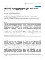
Within the observation time, local progression
occurred in 3 cases (12%) (Fig 1): they were treated
with RFA (2 cases) or PEI (1 case); 7 out of the 8 new
lesions were treated with RFA (6 cases) or surgical
resection (1 case), while no treatment was possible for
the remaining one due to multicentric disease con-
comitant with bone metastasis.
Fig 1 Local recurrence rates for the main tumor in HCC asso-
ciated with CLD without cirrhosis.
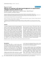
The new HCC lesions onset rates after 1, 3, and 5
years were 4%, 33%, and 41% of patients (Fig 2).
The 5 year probability to develop multicentric
disease was 16% (Fig 3). Actuarial survival rates after
1, 3, and 5 years were 92%, 72%, and 64% (Fig 4).
During the follow-up period, 7 out of 25 patients
died. Causes of death were neoplasia propagation (3
cases, one of which with bone metastasis), myocardial
infarction (1 case), acute leukemia (1 case), and were
unknown in the remaining 2 cases.
Only one major complication, subcutaneous tu-
moral seeding, was recorded: it occurs 24 months after
treatment and was surgically removed.
Minor complications were in 5 cases self-limited
Int. J. Med. Sci. 2008, 5
330
post-RFA syndrome (fatigue, low level fever and
flu-like syndrome), transient right shoulder pain in 2
cases for nodule sited in right sub-diaphragm liver’s
segment.
Fig 2 Overall actuarial probability curves for new hepatocellular
carcinoma associated with CLD without cirrhosis.
Fig 3 Overall actuarial probability curve for multicentric hepa-
tocellular carcinoma after RFA in patients without cirrhosis.
Fig 4 Overall actuarial survival rates in patients with hepato-
cellular carcinoma (HCC) associated to CLD without cirrhosis.
DISCUSSION
The prevalence of HCC association with CLD
without cirrhosis in our study group (12.5%) is com-
parable with that found in a previous local case-control
study (18).
Two recent Japanese reviews about HCC treat-
ment with RFA reported a prevalence of patients with
CLD without cirrhosis equal to 12% and 18% respec-
tively (8, 19). However, they did not describe the out-
come of this subgroup of patients. We can assume that
also surgical reviews on this topic found analogous
prevalence without reporting them.
In a recent meta-analysis evaluating RFA efficacy,
factors dependent on the tumor features, such as di-
ameter, pathology, proximity to large vessels, sub-
capsular location, as well as RFA electrodes and other
physician dependent factors, were considered, but not
pathology of the surrounding liver tissue (20,21).
It has been supposed that the cirrhotic tissue
could enable a better thermal ablation through a spe-
cific mechanism known as oven effect (21, 7). Due to
fibrosis and increased thickness, the cirrhotic tissue
around the nodule would work as a thermal insula-
tion, avoiding the dispersion of the heat generated
around the RFA needle electrode. In the current study
we found that the HCC nodules primary complete
ablation rate in patients affected by CLD without cir-
rhosis is very high (96%) and comparable with best
results reported in literature for cirrhotic patients.
Int. J. Med. Sci. 2008, 5
331
As recently reported for RFA in HCC sized less
than 3.5 cm, the technical efficacy (complete tumor
ablation) in 1 or 2 sessions range from 76 % to 96 % of
nodules, with a mean of 1.2 - 1.4 treatments (22,23,)
and up to 100 % with a mean of 2.2 treatments on
nodular basis (24).
The 5-years survival estimated from a cohort of
patients with cirrhosis and tumours below 3 cm or 3.5
cm treated by RF is 40 % and 33% respectively (25,26).
Recent evidence support percutaneous local ab-
lation therapy for small hepatocellular carcinoma con-
sidered as effective as surgical resection (27,28)
.
Data recently reported indicate that RFA can be
considered the treatment of choice for patients with
single HCC <or= 2.0 cm, even when surgical resection
is possible for patients with cirrhosis (28).
In our group survival rates after 1, 3, and 5 years
were 92%, 72%, and 64% (fig 4). The incidence of ad-
verse events of RFA shows mortality rates ranging
from 0.3% to 0.5%, and morbidity rates ranging from
2.2% to 8.9% (29,30). We had no deaths and few com-
plications without any impact on the outcome.
Local progression rates vary widely between 2%
to 60% (20). Shina et al. recorded the lower local pro-
gression rate of 2% at 3 years (24) while in our study
local progression occurred in 3 cases (12%) (Fig 1).
The new HCC lesions onset rates after 1, 3, and 5
years were 4%, 33%, and 41% of patients (Fig 2) less
than reported for patients with cirrhosis (81% at 5
years as reported by Lencioni et al (31)
and the 5 years
probability to develop multicentric disease was 16 %.
(Fig 3)
At the moment, no other studies of RFA therapy
of HCC associated with CLD without cirrhosis are
available and this is a limitation of actual guide lines.
Our results would need to be confirmed using larger
groups of patients and prospectively compared with
patients affected by hepatocellular carcinoma and cir-
rhosis.
The ideal treatment for HCC associated with
non-cirrhotic CLD is resection, even in case of large
tumors. However, in some cases it could not be feasi-
ble or safe, due to the nodule location or to the possible
comorbidity, and it could even be burdened with an
excessive mortality risk.
Our findings suggest that in cases of tumors less
than 3.5 cm in diameter RFA could be a treatment of
choice also for patients affected by CLD without cir-
rhosis not surgically resectable.
Fig 5. a: Ipoechoic nodule of the VII liver segment in a non
cirrhotic liver pattern, normal hepatic vein. b: The same nodule
after RF session.
Acknowledgments
Special thanks to Dr Anna Caroli for her help in
the English language revision of the manuscript.
Conflict of Interest
The authors have declared that no conflict of in-
terest exists.
References
1 Bruix J, Sherman M. Management of hepatocellular carcinoma.
Hepatology 2005;42:1208–1236.
2 Bolondi L, Sofia S, Siringo S et al. Surveillance programme of
cirrhotic patients for early diagnosis and treatment of hepato-
cellular carcinoma: a cost effectiveness analysis. Gut
2001;48:251–259.
3 Bolondi L. Screening for hepatocellular carcinoma in cirrhosis.
Journal of Hepatology 2003;39:1076–1084.
4 Bruix J, Llovet JM. Prognostic prediction and treatment strategy
in hepatocellular carcinoma. Hepatology 2002;35:519–524.
5 Martin H, Cosmi AB. Liver transplantation for malignancy. The
Oncologist 2005;10:269-281.
Int. J. Med. Sci. 2008, 5
332
6 Rossi S, Buscarini E, Garbagnati F, et al. Percutaneous treatment
of small hepatic tumors by an expandable RF needle electrode.
AJR 1998;170:1015-1022.
7 Gazelle GS, Goldberg SN, Solbiati L et al. State of the Art Tumor
Ablation with Radio-frequency Energy. Radiology
2000;217:633-646.
8 Shiina S, Teratani T, Obi S, et al. A randomized controlled trial of
radiofrequency ablation with ethanol injection for small hepa-
tocellular carcinoma. Gastroenterology 2005;129:122-130.
9 Lin SM, Lin C-J, Lin C-C, et al. Randomised controlled trial
comparing percutaneous radiofrequency thermal ablation, per-
cutaneous ethanol injection, and percutaneous acetic acid injec-
tion to treat hepatocellular carcinoma of 3 cm or less. Gut
2005;4:1151–1156
10 Ebara M, Okabe S, Kita K, et al. Percutaneous ethanol injection
for small hepatocellular carcinoma : therapeutic efficacy based
on 20 years observation. J Hepatol. 2005; 43 :377-80.
11 Sutherland LM, Williams J, Padbury R, et al. Radiofrequency
ablation of liver tumors A systematic review. Arch Surg
2006;141:181-190
12 Lu DSK,Yu NC, Raman SS, Lassman C, et al. Percutaneous ra-
diofrequency ablation of hepatocellular carcinoma as a bridge to
liver transplantation. Hepatology 2005;41:1130-37
13 Takano S,Yokosuka O, Imazeki F et al. Incidence of hepatocel-
lular carcinomain chronic hepatitis B and C : a prospective study
of 251patients. Hepatology 1995;21:650-655
14 Otto G. Heuschen U, Hofman WJ et al. Survival and recurrence
after liver transplantation versus liver resection for hepatocel-
lular carcinoma: a retrospective analysis. Ann Surg
1998;227:424-432
15 Desmet VJ, Gerber M, Hoofnagle JH et al. Classification of
chronic hepatitis:doagnosis,grading and staging. Hepatology
1994; 19:1513-1520.
16 Leevy CM, Sherlock S, et al. Diseases of the Liver and Biliary
Tract. Standardization of Nomenclature, Diagnostic Criteria and
Prognosis. New York: Raven Press, 1994.
17 Salmi A., Turrini R, Lanzani G, et al. Long term effectiveness of
radiofrequency ablation for hepatocellular carcinoma of 3.5 cm
or less. Hepatogastroenterology 2008; 55:191-196
18 Chiesa R, Donato F, Tagger A, et al. Etiology of hepatocellular
carcinoma in Italian patients with and without cirrhosis. Cancer
Epidemiol Biomarkers Prev 2000; 9(2):213-6.
19 Hong SN,Lee SY, Choi MS, et al. Comparing the outcomes of
radiofrequency ablation and surgery in patients with a single
small hepatocelular carcinoma and well preserved liver func-
tion. J Clin Gastroenterol 2005 ; 39:247-52.
20 Mulier S, Ni Y, Jamart J, Ruers T, et al. Local recurrence after
hepatic radiofrequency coagulation. Multivariate meta-analysis
and review of contributing factors. Ann Surg 2005;242:158-171.
21 Livraghi T, Goldberg SN, Lazzaroni S, et al. Small hepatocellular
carcinoma: treatment with radio-frequency ablation versus
ethanol injection. Radiology 1998; 210:655-661.
22 Lin SM, Lin C-J, Lin C-C, et al. Randomised controlled trial
comparing percutaneous radiofrequency thermal ablation, per-
cutaneous ethanol injection, and percutaneous acetic acid injec-
tion to treat hepatocellular carcinoma of 3 cm or less. Gut
2005;54:1151–1156.
23 Llovet JM, Vilana R, Bru´ C, et al. Increased risk of tumor seed-
ing after radiofrequency thermal ablation for single hepatocel-
lular carcinoma. Hepatology 2001;33:1124-1129.
24 Shiina S, Teratani T, Obi S, et al Sato S, Tateishi R, Fujishima T. A
randomized controlled trial of radiofrequency ablation with
ethanol injection for small hepatocellular carcinoma. Gastroen-
terology 2005;129:122-130.
25 Rossi S, Di Stasi M, Buscarini E, et al. Percutaneous RF interstitial
thermal ablation in the treatment of hepatic cancer. Am J
Roentgenol 1996;167: 759-68.
26 Buscarini L, Buscarini E, Di Stasi M,et al. Percutaneous radiof-
requency ablation of small hepatocellular carcinoma: long-term
results. Eur Radiol 2001;11:914–921.
27 Kaido T., Uemoto S. Recent evidence in the treatment of small
hepatocellular carcinoma. Hepatogastroenterology.
2008;55(85):1460-2.
28 Guglielmi A, Ruzzenante A, Valdegamberi A et al. Radiofre-
quency ablation versus surgical resection for the treatment of
hepatocellular carcinoma in cirrhosis. J Gastrointest Surg.
2008;12(1):192-8.
29 Livraghi T, Solbiati L, Meloni MF, et al. Treatment of focal liver
tumors with percutaneous radiofrequency ablation: complica-
tions encountered in a multicenter study. Radiology
2003;226:441–2008.
30 Mulier S, Mulier P, Ni Y, et al. Complications of radiofrequency
coagulation of liver tumors. Br J Surg 2002;89:1206 –1222.
31 Lencioni R, Cioni D, Crocetti L, et al. Early stage hepatocellular
carcinoma in patients with cirrhosis: long term results of percu-
taneous image-guided radiofrequency ablation. Radiology
2005;234:961-967.