Báo cáo y học: "Peripheral blood but not synovial fluid natural killer T cells are biased towards a Th1-like phenotype in rheumatoid arthritis" pptx
Bạn đang xem bản rút gọn của tài liệu. Xem và tải ngay bản đầy đủ của tài liệu tại đây (316.37 KB, 10 trang )
Open Access
Available online />R493
Vol 7 No 3
Research article
Peripheral blood but not synovial fluid natural killer T cells are
biased towards a Th1-like phenotype in rheumatoid arthritis
Loes Linsen, Marielle Thewissen, Kurt Baeten, Veerle Somers, Piet Geusens, Jef Raus and
Piet Stinissen
Biomedisch Onderzoeksinstituut, Limburgs Universitair Centrum and School of Life Sciences, Transnationale Universiteit Limburg, Universitaire
Campus, Diepenbeek, Belgium
Corresponding author: Piet Stinissen,
Received: 13 Oct 2004 Revisions requested: 17 Nov 2004 Revisions received: 14 Jan 2005 Accepted: 19 Jan 2005 Published: 18 Feb 2005
Arthritis Research & Therapy 2005, 7:R493-R502 (DOI 10.1186/ar1695)
This article is online at: />© 2005 Linsen et al.; licensee BioMed Central Ltd.
This is an Open Access article distributed under the terms of the Creative Commons Attribution License ( />2.0), which permits unrestricted use, distribution, and reproduction in any medium, provided the original work is properly cited.
Abstract
Natural killer T (NKT) cells have been implicated in the regulatory
immune mechanisms that control autoimmunity. However, their
precise role in the pathogenesis of rheumatoid arthritis (RA)
remains unclear. The frequency, cytokine profile and
heterogeneity of NKT cells were studied in peripheral blood
mononuclear cells (PBMCs) from 23 RA patients and 22 healthy
control individuals, including paired PBMC–synovial fluid
samples from seven and paired PBMC–synovial tissue samples
from four RA patients. Flow cytometry revealed a decreased
frequency of NKT cells in PBMCs from RA patients. NKT cells
were present in paired synovial fluid and synovial tissue
samples. Based on the reactivity of PBMC-derived NKT cells
toward α-galactosylceramide, RA patients could be divided into
responders (53.8%) and nonresponders (46.2%). However,
NKT cells isolated from synovial fluid from both responders and
nonresponders expanded upon stimulation with α-
galactosylceramide. Analysis of the cytokine profile of CD4
+
and
CD4
-
PBMC derived NKT cell lines from RA patients revealed a
significantly reduced number of IL-4 producing cells. In contrast,
synovial fluid derived NKT cell lines exhibited a Th0-like
phenotype, which was comparable to that in healthy control
individuals. This suggests that synovial fluid NKT cells are
functional, even in patients with nonresponding NKT cells in
their blood. We conclude that, because the number of
Vα24
+
Vβ11
+
CD3
+
NKT cells is decreased and the cytokine
profile of blood-derived NKT cells is biased toward a Th1-like
phenotype in RA patients, NKT cells might be functionally
related to resistance or progression of RA. Providing a local
boost to the regulatory potential of NKT cells might represent a
useful candidate therapy for RA.
Introduction
Natural killer T (NKT) cells are a distinct subset of lymphocytes
that share the characteristics of both T cells and natural killer
cells. They express a semi-invariant TCR (TCR Vα24Jα18 and
Vβ11 in human; Vα14Jα281 and Vβ8, Vβ7 or Vβ2 in mouse)
and recognize glycolipid antigens presented by the major his-
tocompatibility complex class I-like molecule CD1d [1]. Two
subsets can be distinguished [2,3]: CD4
+
NKT cells that pro-
duce T-helper (Th)1-type and Th2-type cytokines, and CD4
-
CD8
-
(double negative) NKT cells that primarily produce Th1-
type cytokines. The ability to secrete cytokines and chemok-
ines rapidly is thought to underlie their regulatory function in a
variety of diseases, including cancer and autoimmunity [4].
Although the natural ligand of NKT cells remains to be eluci-
dated, it has been reported that the sponge derived glycolipid
α-galactosylceramide (α-GalCer) is a potent activator of
mouse and human NKT cells, both in vitro and in vivo [5,6].
When α-GalCer is administered to mice it polarizes the adap-
tive immune response toward production of Th2 cytokines
[7,8], which therefore raises the possibility that α-GalCer can
temper or even prevent Th1-mediated autoimmune diseases.
Several studies have shown that NKT cells are decreased or
dysfunctional in autoimmune conditions such as insulin-
dependent diabetes mellitus, systemic sclerosis, systemic
lupus erythematosus, rheumatoid arthritis (RA) and multiple
α-GalCer = α-galactosylceramide; FITC = fluorescein isothiocyanate; IFN = interferon; IL = interleukin; NKT = natural killer T (cell); PBMC = periph-
eral blood mononuclear cell; PE = phycoerythrin; PCR = polymerase chain reaction; RA = rheumatoid arthritis; SFMC = synovial fluid mononuclear
cell; TCR = T-cell receptor; Th = T-helper (cell).
Arthritis Research & Therapy Vol 7 No 3 Linsen et al.
R494
sclerosis [9-12]. Significant therapeutic effects of α-GalCer
have been demonstrated in animal models of autoimmunity,
such as experimental allergic encephalomyelitis [13-15] and
nonobese diabetic mice [16,17].
Because the NKT/CD1d system is phylogenetically conserved
among mammals, findings in mice are expected to have a
direct parallel in humans. The NKT cell frequency in peripheral
blood mononuclear cells (PBMCs) is lower in humans than in
mice [1], which may be an obstacle in translating results from
animal studies to the clinic. However, results from a phase I
study conducted in advanced cancer patients revealed that
treating patients with α-GalCer can increase NKT cell num-
bers above pretreatment levels. This again indicates that α-
GalCer could be applied to the treatment of patients with
autoimmune disease [18].
RA is an autoimmune disease that is characterized by a
chronic inflammation of the joints, followed by progressive
destruction of cartilage and underlying bone [19]. Autoreac-
tive Th1 T cells are believed to play a major role in the disease
process [20-22]. In RA patients, the frequency of NKT cells is
decreased, but the functional characteristics of NKT cells have
not yet been fully elucidated. Chiba and coworkers [23] dem-
onstrated that administration of a truncated form of α-GalCer
to mice suffering from collagen-induced arthritis – a frequently
used animal model of RA – resulted in protection from disease,
indicating that this might represent a therapy that can enhance
NKT cell numbers in RA patients.
In the present study we analyzed the frequency, functional
characteristics and heterogeneity of NKT cells in peripheral
blood, synovial fluid and synovial tissue from RA patients. In
parallel, we assessed these parameters in α-GalCer-stimu-
lated short-term cell lines of both peripheral blood and synovial
Table 1
Patient characteristics
Patient Age (years)/sex Disease duration (years) Treatment
1 54/M 5 Azathioprine, methylprednisolone
2 38/F 6 Hydroxychloroquine, salazopyrine
364/F7NSAID
443/F5NSAID
546/M1Salazopyrine
646/M<1Salazopyrine
752/M11NSAID
853/F11NSAID
949/M10NSAID
10 52/F 4 NSAID
11 69/F 36 Salazopyrine
12 65/F <1 Untreated
13 35/M <1 Untreated
14
a
57/F 4 Methotrexate
15
a
46/M 2 Anti-TNF, salazopyrine
16
a
41/M 10 Salazopyrine
17
a
41/M 13 Methotrexate
18
a
60/M 2 Leflunomide
19
a
43/F 5 Methotrexate
20
b
63/M 17 Salazopyrine
21
a,b
65/F 4 Salazopyrine, hydroxychloroquine
22
b
62/F 12 Leflunomide
23
b
54/F 17 Methylprednisolone
a
Synovial fluid sample.
b
Synovial tissue sample. F, female; M, male; NSAID, nonsteroidal anti-inflammatory drug; TNF, tumour necrosis factor.
Available online />R495
fluid NKT cells. We found that NKT cells were decreased and
had altered functional properties in peripheral blood, but they
were not impaired in synovial fluid from RA patients. Our data
indicate that NKT cells may be involved in the disease process
of RA and that a strategy to boost the regulatory potential of
NKT cells might be useful in the treatment of RA.
Materials and methods
Patients and healthy control individuals
NKT cell characteristics were examined in 23 RA patients
(mean age 52.1 ± 2.0 years, 11 males and 12 females, mean
disease duration 8.0 ± 1.6 years), who were diagnosed in
accordance with the criteria of the American College of Rheu-
matology [24], and in 22 healthy individuals (mean age 48.6 ±
2.0 years, 10 males and 12 females). When RA patients pre-
sented with a swollen knee, paired peripheral blood and syno-
vial fluid samples were obtained. Synovial tissue samples were
obtained from four RA patients after total knee/hip arthro-
plasty. Patients were informed about the purpose of the study
and gave written consent. Approval for the study was granted
by our ethics committee. Patient characteristics are summa-
rized in Table 1.
Flow cytometric analysis of natural killer T cells
Expression of cell surface markers was analyzed by flow
cytometry. Fluorescein isothiocyanate (FITC)-labelled anti-
TCR Vα24 and phycoerythrin (PE)-labelled TCR Vβ11 were
purchased from Serotec Ltd (Oxford, UK). Anti-CD3-PE, anti-
CD3-PerCP, anti-CD4-FITC, anti-CD8-PE, anti-CD25-FITC,
anti-IFN-γ-FITC and anti-IL-4-PE were obtained from Becton
Dickinson (Erembodegem, Belgium). The frequency of invari-
ant NKT cells was estimated using three-colour anti-Vα24/
anti-Vβ11/anti-CD3 staining. For intracellular cytokine detec-
tion, α-GalCer expanded Vα24
+
Vβ11
+
NKT cells or Vα24
+
isolated NKT cell lines were stimulated with 25 ng/ml phorbol-
12-myristate-13-acetate and 1 µg/ml ionomycine in the pres-
ence of 10 µg/ml brefeldin A for 4 hours. Intracellular staining
was performed as previously described [25]. Cells were ana-
lyzed on a FACSCalibur flow cytometer using Cellquest soft-
ware (Becton Dickinson).
Direct ex vivo analysis of the cytokine profile of natural
killer T cells by ELISPOT
ELISPOT procedure was performed as previously described
[25]. Briefly, 2 × 10
5
PBMCs were stimulated with 100 ng/ml
α-GalCer in anti-IFN-γ or anti-IL-4 (Mabtech, Nacka, Sweden)
coated nitrocellulose bottomed plates (Millipore Corp, Bed-
ford, MA, USA). After 20 hours of culture, biotinylated anti-IFN-
γ or anti-IL-4 antibody (Mabtech) was added for 2 hours fol-
lowed by incubation with streptavidin-alkaline phosphatase
(Mabtech) and NBT/BCIP (Nitro Blue Tetrazolium/5-Bromo-4
Chloro-3-Indolyphosphate; Pierce, Rockford, IL, USA) as sub-
strate. The number of cytokine-secreting cells was calculated
by subtracting the number of spots in control wells (without
antigen) from the number of spots obtained in the presence of
α-GalCer.
Expansion and culture of Vα24
+
Vβ11
+
natural killer T
cells
PBMCs and synovial fluid mononuclear cells (SFMCs) were
isolated using Ficoll-Hypaque (Sigma Diagnostics, St Louis,
MO, USA) density gradient centrifugation. PBMCs and
SFMCs were cultured in the presence of 100 ng/ml α-GalCer
(Kirin Brewery Ltd, Gunma, Japan) at a density of 7.5 × 10
5
cells/ml RPMI supplemented with 10% heat-inactivated foetal
bovine serum, 1 mmol/l sodiumpyruvate and 1% nonessential
amino acids (Invitrogen, Merelbeke, Belgium). After 7 days,
cells were re-stimulated with irradiated autologous, α-GalCer
pulsed PBMCs and supplemented with 2 U/ml recombinant
human IL-2 (Roche Diagnostics, Brussels, Belgium). On day 7
after re-stimulation, NKT cells were isolated using Vα24
+
mag-
netic isolation (EasySep; Stemcell Technologies, Meylan,
France), in accordance with the manufacturer's instructions.
Reactivity of the isolated NKT cells toward α-GalCer was
tested in a standard [
3
H]thymidine incorporation assay. During
the last 16 hours of culture, cells were pulsed with 1 µCi
[
3
H]thymidine (Amersham, Buckinghamshire, UK) and subse-
quently harvested using an automated cell harvester (Pharma-
cia, Uppsala, Sweden). Incorporated radioactivity was
measured using a β-plate liquid scintillation counter (Wallac,
Turku, Finland). A NKT cell line was considered to be antigen
reactive when the mean counts per minute in the presence of
α-GalCer exceeded 1000 and the stimulation index (mean
counts with α-GalCer/mean counts without α-GalCer) was
greater than 3.
Analysis of clonal heterogeneity by T-cell receptor CDR3
region fragment length analysis
RNA was isolated from snap frozen synovial tissue samples
using the Absolutely RNA RT-PCR Miniprep Kit (Stratagene,
Amsterdam, The Netherlands). For isolation of total RNA from
PBMCs, SFMCs and isolated NKT cells, the High Pure total
RNA Isolation kit (Roche Diagnostics, Brussels, Belgium) was
used, in accordance with the manufacturer's instructions. RNA
was reverse transcribed into cDNA using AMV reverse tran-
scriptase and an oligo-dT primer (Promega, Madison, WI,
USA).
CDR3 spectratyping analysis was performed as described
previously [26]. Briefly, 2 µl cDNA was used for first-round
PCR analysis performed in 1 × PCR buffer, 0.9 U Taq
polymerase, 0.02 mmol/l dNTP mix (all from Roche Diagnos-
tics), 1 µmol/l forward primer specific for TCR Vα24 (5'-GAA
CGG AAG ATA TAC AGC AAC TC-3') or TCR Vβ11 (5'-TCC
ACA GAG AAG GGA GAT CTT TCC TCT GAG-3') region,
and 1 µmol/l reverse primer specific for TCR constant α (5'-
ATC ATA AAT TCG GGT AGG ATC C-3') or constant β (5'-
CTC TTG ACC ATG GCC ATC-3') region. PCR was per-
formed for 40 cycles (95°C for 20 s, 55°C for 20 s, and 72°C
Arthritis Research & Therapy Vol 7 No 3 Linsen et al.
R496
for 40 s) on a GeneAmp PCR system 9600 thermal cycler
(Perkin Elmer, Zaventem, Belgium). PCR amplicons were used
in a second amplification procedure of 25 cycles using the
TCR Vα24 or TCR Vβ11 specific primer as forward primer and
a FAM labelled TCR constant α (5'-FAM-CTG TTG CTC TTG
AAG TCC ATA G-3') or TCR constant β (5'-FAM-GTG GCA
AGG CAC ACC AGT GTG GGC C-3') as reverse primer
(Eurogentec, Liege, Belgium) under the same PCR conditions
as described above.
PCR amplicon lengths were analyzed on the 310 ABI DNA
sequencer (Applied Biosystems, Warrington, UK). Fragment
sizes of gene products were calculated using an internal
Genescan-500 ROX labelled standard and analysis was per-
formed with 672 Genescan Software (both from Applied Bio-
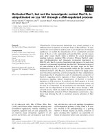
systems). The heterogeneity of the CDR3 spectratype profiles
provides an indication of the clonality of T-cell populations
(Fig. 1): monoclonal with one peak, oligoclonal with two to four
peaks, and polyclonal with more than four peaks. Identical
peak lengths strongly indicate the presence of identical T cell
clones in different samples. A 350 base pair fragment was
obtained for the invariant TCR.
Sequence analysis of the invariant T-cell receptor
Purified TCR Vα24 PCR amplicons obtained from first round
PCR (as described above) were sequenced with a TCR con-
stant α primer (5'-CTG TTG CTC TTG AAG TCC ATA G-3')
using the Big DyeTM Terminator Cycle Sequence Ready
Reaction Kit II (Applied Biosystems). Sequences were ana-
lyzed on a ABI Prism 310 Genetic Analyser (Applied
Biosystems).
Statistical analysis
Differences in the percentage of NKT cells between healthy
control individuals and RA patients and between peripheral
blood and synovial fluid from RA patients were analyzed using
the Mann–Whitney U-test. For comparisons between matched
peripheral blood and synovial fluid samples, the Wilcoxon
matched pairs signed rank test was used. P < 0.05 was con-
sidered statistically significant.
Results
Frequency of Vα24
+
Vβ11
+
CD3
+
natural killer T cells in
rheumatoid arthritis
The frequency of Vα24
+
Vβ11
+
CD3
+
NKT cells in PBMCs
from RA patients and healthy control individuals was analyzed
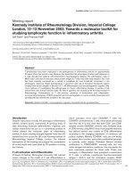
by flow cytometry (Fig. 2). Significantly fewer
Vα24
+
Vβ11
+
CD3
+
NKT cells were found in PBMCs from RA
patients (0.03 ± 0.01%) than in healthy control individuals
(0.11 ± 0.03%; P < 0.01). We simultaneously determined the
NKT cell frequency in paired blood–synovial fluid samples
from seven RA patients. Although a tendency toward a higher
frequency was observed in the synovial fluid (0.08 ± 0.03%)
as compared with the concordant PBMC samples (0.05 ±
0.02%), this finding could not be demonstrated for all patients.
These data indicate that the NKT cell frequency is decreased
in the blood of RA patients but not increased in synovial fluid
as compared with blood from these patients.
Cytokine profile of α-galactosylceramide stimulated
peripheral blood mononuclear cells
To assess the cytokine profile of NKT cells directly ex vivo, we
tested the reactivity of PBMCs to α-GalCer in 10 RA patients
and eight healthy control individuals using an ELISPOT tech-
nique with IFN-γ and IL-4 readout. Similar to the frequency
analysis by flow cytometry, a significantly decreased number
of α-GalCer reactive cells was found for IFN-γ as well as for IL-
4 in RA patients as compared with healthy control individuals
(2.3 ± 0.6 spots versus 24.3 ± 10.1 spots for IFN-γ and 0.2 ±
0.1 spots versus 3.9 ± 1.1 spots for IL-4 per 2 × 10
5
cells for
RA patients and healthy control individuals, respectively; P <
0.05). To determine whether this diminished frequency was
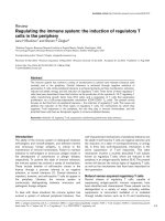
also associated with an altered cytokine profile, the IL-4/IFN-γ
ratio was calculated as the number of IL-4 producing cells to
Figure 1
Clonality of T-cell populationsClonality of T-cell populations. (a) Monoclonal: one peak. (b) Oligo-
clonal: two to four peaks. (c) Polyclonal: more than four peaks.
Available online />R497
the number of IFN-γ producing cells (Fig. 3). The IL-4/IFN-γ
ratio in RA patients was decreased as compared with that in
healthy control individuals (0.07 ± 0.03 in RA patients versus
0.30 ± 0.10 in healthy control individuals; P = 0.06). This was
mainly due to a reduced number of IL-4 producing cells,
because the frequency of IL-4 producing cells in RA patients
as compared with healthy control individuals was relatively
more reduced than that of IFN-γ producing cells. These data
indicate that NKT cells derived from RA patients are biased
toward a Th1-like phenotype.
Analysis of the invariant T-cell receptor in synovial tissue
NKT cells express the invariant Vα24Jα18 TCR-α chain com-
bined with a variable Vβ11 TCR-β chain. To compare the
Vα24 expression profile in PBMCs from RA patients and
healthy control individuals, PBMCs from five healthy control
individuals and paired PBMCs–SFMCs and PBMCs–synovial
tissue samples from four RA patients were subjected to TCR
CDR3 size analysis using primers for Vα24 and TCR-α con-
stant region. PBMCs from healthy control individuals exhibited
a polyclonal peak profile or a Gaussian-like distribution for
Vα24, containing a peak at 350 base pairs, which corre-
sponds to the invariant TCR-α chain that is characteristic for
NKT cells (not shown). Although PBMCs from RA patients
exhibited an oligoclonal or monoclonal distribution, indicating
a restricted usage for Vα24 (Table 2), the invariant TCR peak
was present in all patients. We determined whether the invar-
iant TCR could also be found in SFMCs and synovial tissue
samples. As in PBMCs, the TCR Vα24 usage in SFMCs and
synovial tissue tissue samples was skewed for some patients
but polyclonal for others. Again, the invariant TCR peak was
detected in SFMCs and synovial tissue samples for all RA
patients. Sequence analysis of the PCR products obtained
from the CDR3 fragment length analysis confirmed that the
peak size of the synovial tissue samples corresponded with
the invariant TCR sequence (not shown). These data show
that NKT cells are present in rheumatoid synovial fluid as well
as in synovial tissue.
Natural killer T-cell reactivity to α-galactosylceramide in
rheumatoid arthritis patients
To assess whether the reduced NKT cell frequency in periph-
eral blood from RA patients was due to an inadequate
response to the glycolipid antigen, we stimulated PBMCs from
nine healthy control individuals and 13 RA patients and
SFMCs from five RA patients with α-GalCer. At day 7, cells
were re-stimulated with autologous α-GalCer pulsed, irradi-
ated PBMCs. The NKT cell frequency was determined by flow
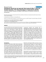
cytometry at day 14 (Fig. 4). NKT cells from healthy control
individuals expanded in response to α-GalCer to 15.8 ± 2.7%,
whereas the number of peripheral blood and synovial fluid NKT
cells from RA patients was significantly lower after α-GalCer
stimulation (8.4 ± 2.9% and 4.4 ± 1.6%, respectively; P <
0.01). A more detailed analysis revealed that this decrease
was due to the existence of two subpopulations of RA patients
based on the NKT cell numbers reached after 14 days of α-
GalCer stimulation. As shown in Fig. 5, NKT cells from six out
13 RA patients did not respond to α-GalCer stimulation (mean
frequency after 14 days: 1.0 ± 0.2%, P < 0.01; nonrespond-
ers), whereas NKT cells from the remaining seven patients
reached frequencies comparable with those in healthy control
individuals (14.7 ± 4.0%; responders). Moreover, NKT cells of
responder patients appeared to have increased ability to
respond to α-GalCer because the expansion was greater than
that in healthy control individuals (294-fold versus 149-fold,
respectively). No relation between disease parameters (dis-
ease duration, disease status) or treatment and responsive-
ness/nonresponsiveness of NKT cells could be demonstrated.
Remarkably, synovial fluid NKT cells, even from nonrespond-
Figure 2
Frequency of natural killer T (NKT) cells in rheumatoid arthritis (RA) patients and healthy control individualsFrequency of natural killer T (NKT) cells in rheumatoid arthritis (RA)
patients and healthy control individuals. NKT cell frequency in freshly
isolated peripheral blood (PB) mononuclear cells from 22 healthy con-
trol individuals and 23 RA patients, and in synovial fluid (SF) mononu-
clear cells from seven RA patients was determined by flow cytometry.
Cells were stained with anti-Vα24, anti-Vβ11 and anti-CD3 monoclonal
antibody. Error bars indicate the standard error of the mean. *P < 0.01.
Figure 3
IL-4/IFN-γ ratio in α-galactosylceramide (α-GalCer) stimulated periph-eral blood mononuclear cells (PBMCs) evaluated by ELISPOTIL-4/IFN-γ ratio in α-galactosylceramide (α-GalCer) stimulated periph-
eral blood mononuclear cells (PBMCs) evaluated by ELISPOT. PBMCs
(2 × 10
5
cells/well) from 10 rheumatoid arthritis patients and eight
healthy control individuals were stimulated with α-GalCer or no antigen
for 20 hours. The number of cytokine secreting cells was calculated by
subtracting the number of spots in control wells (without antigen) from
the number of spots obtained in the presence of each stimulating
agent. The IL-4/IFN-γ ratio is the number of IL-4 producing cells divided
by the number of IFN-γ producing cells. Error bars indicate standard
error of the mean.
Arthritis Research & Therapy Vol 7 No 3 Linsen et al.
R498
ing RA patients, did expand after α-GalCer stimulation (4.94 ±
1.90%). These findings indicate that the reactivity of peripheral
blood NKT cells to α-GalCer is impaired in some RA patients,
whereas it is intact and even increased in others.
Cytokine profile of peripheral blood and synovial fluid
natural killer T cell lines
Next, we analyzed the cytokine profile of peripheral blood
derived NKT cells from five healthy control individual and five
RA patients, and synovial fluid derived NKT cells from five RA
patients by intracellular staining of 14-day-old, α-GalCer stim-
ulated cultures gated on Vα24
+
cells. Figure 6 shows that the
Vα24
+
NKT cell fraction of healthy control individuals con-
tained 64.5 ± 13.1% IFN-γ producing cells, 15.7 ± 6.9% IL-4
producing cells, and 19.7 ± 6.4% cells producing both IFN-γ
and IL-4. In contrast, peripheral blood NKT cells from RA
patients consisted of significantly more IFN-γ producing cells
and significantly fewer cells producing both IFN-γ and IL-4
(92.5 ± 2.7% and 6.1 ± 2.3%, respectively; P < 0.05).
Remarkably, synovial fluid derived NKT cells exhibited a
cytokine profile similar to that of healthy control individuals,
although the number of IL-4 producing cells tended to be
lower and the number of cells producing both IFN-γ and IL-4
was somewhat higher (5.3 ± 5.3% and 28.7 ± 6.7%, respec-
tively; P > 0.05). No differences were found between the
cytokine profiles of NKT cells of α-GalCer responding and
nonresponding patients. Furthermore, no relation with treat-
ment or any disease parameter was found. These observations
show that, although NKT cells in PBMCs from RA patients are
biased toward a Th1-like cytokine profile, NKT cells in the syn-
ovial fluid exhibit a Th0-like cytokine profile that is comparable
with that in healthy control individuals.
Cytokine profile of CD4
+
and CD4
-
natural killer T cell
subsets in patients with rheumatoid arthritis and healthy
control individuals
The observed Th1-like bias in NKT cells from RA patients
might be due to an increased number of double-negative NKT
cells or a decreased number of CD4
+
NKT cells. To analyze
the frequency of these NKT cell subtypes, we isolated the
Vα24
+
cells of α-GalCer stimulated, 14-day-old cultures
derived from PBMCs from nine healthy control individuals and
seven RA patients by immunomagnetic selection. Positively
selected cells were tested for α-GalCer reactivity to ensure
the NKT cell nature of the cells. The presence of CD4 was
assessed by flow cytometry. NKT cells of healthy control indi-
viduals consisted of 33.3 ± 6.7% CD4
+
NKT cells and 66.7 ±
Table 2
T cell receptor Vα24 usage in peripheral blood mononuclear cells, synovial fluid mononuclear cells and synovial tissue from
rheumatoid arthritis patients
Vα24 Vβ11
PBMCs SFMCs ST PBMCs SFMCs ST
RA 1 mono mono NA oligo (2) mono NA
RA 2 mono poly oligo (2) mono poly oligo (2)
RA 3 mono oligo (2) NA Poly oligo (2) NA
RA 4 poly poly NA Poly poly NA
RA 5 oligo (3) NA mono mono NA mono
RA 6 poly NA poly poly NA poly
RA 7 oligo (3) NA mono poly NA oligo (3)
The clonality of the T-cell receptor (TCR) Vα24 family was assessed by CDR3 spectratyping of peripheral blood mononuclear cells (PBMCs),
synovial fluid mononuclear cells (SFMCs) and synovial tissue (ST) from rheumatoid arthritis (RA) patients. (See Fig. 1 for representative
monoclonal [panel a], oligoclonal [panel b] and polyclonal [panel c] profiles.) mono, monoclonal profile; NA, not available; oligo, oligoclonal profile;
poly, polyclonal profile.
Figure 4
Reactivity of peripheral blood (PB) and synovial fluid (SF) derived natu-ral killer T (NKT) cells to α-galactosylceramide (α-GalCer)Reactivity of peripheral blood (PB) and synovial fluid (SF) derived natu-
ral killer T (NKT) cells to α-galactosylceramide (α-GalCer). PB mononu-
clear cells (1.5 × 10
6
cells/well) of nine healthy control individuals and
13 rheumatoid arthritis (RA) patients as well as SF mononuclear cells of
five RA patients were stimulated with α-GalCer and re-stimulated on
day 7 with autologous, α-GalCer pulsed, irradiated PB mononuclear
cells in the presence of 2 U/ml IL-2. NKT cell numbers were determined
by flow cytometry at day 14. Error bars indicate standard error of the
mean. *P < 0.01.
Available online />R499
6.7% CD4
-
(double-negative) NKT cells. The frequency of
CD4
+
and CD4
-
NKT cells in RA patients did not differ signifi-
cantly from that in healthy control individuals (49.8 ± 6.3% and
50.2 ± 6.3%, respectively; data not shown).
Figure 7 shows the cytokine profile of each NKT cell subset,
as determined by intracellular staining. Peripheral blood
derived CD4
-
NKT cells from healthy control individuals pre-
dominantly consisted of IFN-γ producing cells (IFN-γ
+
57.6 ±
8.8%; IL-4
+
19.4 ± 6.6%; IFN-γ
+
IL-4
+
23.0 ± 6.0%), whereas
CD4
+
NKT cells contained almost as many IL-4 producing
cells as IFN-γ producing cells (IFN-γ
+
40.1 ± 7.4%; IL-4
+
25.1
± 7.5%; IFN-γ
+
IL-4
+
34.8 ± 6.4%). However, the CD4
+
as well
as the CD4
-
NKT cell fractions in RA patients contained signif-
icantly fewer IL-4 producing cells as compared with their
counterparts in healthy control individuals (for CD4
+
NKT
cells: IFN-γ
+
57.2 ± 12.9%; IL-4
+
5.8 ± 1.5%; IFN-γ
+
IL-4
+
37.0
± 13.2%; and for CD4
-
NKT cells: IFN-γ
+
72.1 ± 12.4%; IL-4
+
3.3 ± 1.9%; IFN-γ
+
IL-4
+
24.6 ± 11.9%), indicating that both
CD4
+
and CD4
-
NKT cells in the peripheral blood of RA
patients are biased toward a Th1-like cytokine profile.
To exclude the possibility that the observations in NKT cell
lines of RA patients were caused by the clonal expansion of
one or a few NKT cells, we analyzed the heterogeneity of the
Vα24 and Vβ11 TCR by means of CDR3 fragment length
analysis. We found that the NKT cell lines of both RA patients
and healthy control individuals exhibited a monoclonal Vα24
and polyclonal Vβ11 profile (data not shown), which shows
that the differences between NKT cells from RA patients and
healthy control individuals found in response to α-GalCer are
not due to a skewed outgrowth of only one or a few NKT cells.
Discussion
Several studies have provided evidence that NKT cells are
involved in autoimmune conditions [27]. Attempts to increase
the number of NKT cells in animal models of autoimmunity by
transgenic expression of the invariant TCR or by passive trans-
fer of NKT cells resulted in a protective effect against disease
induction [28,29]. Additionally, administration of α-GalCer
resulted in prevention or suppression of disease. These stud-
ies indicate that NKT cells can play a role in the regulation of
autoimmunity and that they are therefore an interesting subject
for further investigation in human autoimmune diseases.
In the present study we demonstrated a decreased frequency
of NKT cells in PBMCs from RA patients. Because we used
anti-Vα24 and anti-Vβ11 monoclonal antibodies to identify
invariant NKT cells, it is possible that conventional T cells were
also stained by this combination. However, Araki and cowork-
ers [12] showed that the frequency of Vα24
+
Vβ11
+
CD3
+
T
cells, even at low numbers, corresponded well with the NKT
cell frequency determined by CD1d tetramers, which supports
the specificity of anti-Vα24 and anti-Vβ11 staining for NKT
cells.
Several mechanisms may account for NKT cell reduction in the
peripheral blood of RA patients. First, NKT cells might prefer-
entially migrate into the joint to fulfill their regulatory function.
We therefore studied the frequency of NKT cells in synovial
fluid and synovial tissue of RA patients. We found that the NKT
cell frequency is not elevated in synovial fluid, but that the
invariant TCR can be detected in both synovial tissue and syn-
ovial fluid samples from RA patients. Preferential migration of
NKT cells into the synovium may have resulted in a monoclonal
or oligoclonal Vα24 profile in synovial samples. However, we
Figure 5
Rheumatoid arthritis (RA) patients can be divided into responder and nonresponder patients, based on peripheral blood derived natural killer T (NKT) cell reactivity to α-galactosylceramide (α-GalCer)Rheumatoid arthritis (RA) patients can be divided into responder and nonresponder patients, based on peripheral blood derived natural killer T (NKT)
cell reactivity to α-galactosylceramide (α-GalCer). Peripheral blood (PB) mononuclear cells (1.5 × 10
6
cells/well) from nine healthy control individu-
als and 13 RA patients, as well as synovial fluid (SF) mononuclear cells from five RA patients, were stimulated with α-GalCer and re-stimulated on
day 7 with autologous, α-GalCer pulsed, irradiated PB mononuclear cells in the presence of 2 U/ml IL-2. NKT cell numbers were determined by flow
cytometry on day 14. Patients were considered nonresponders when the frequency of Vα24
+
Vβ11
+
CD3
+
NKT cells derived from PB mononuclear
cells was lower than 2% after 14 days of culture. Error bars indicate standard error of the mean. *P < 0.01.
Arthritis Research & Therapy Vol 7 No 3 Linsen et al.
R500
did not find such a profile in the synovial fluid or synovial tissue
of all patients, indicating that the decrease cannot be
accounted for by a selective migration of NKT cells toward the
joint. A similar conclusion was reached by others for RA [30]
and multiple sclerosis [31].
A second possibility might be that the reduced NKT cell fre-
quency is caused by a selective loss of a limited number of
NKT cell clones. It was shown in mice that NKT cells exhibit a
highly diverse TCR-β repertoire and a small clone size [32],
and hence a loss of NKT cells should result in a reduced diver-
sity of TCR Vβ11. However, the Vβ11 profile of α-GalCer
expanded peripheral blood NKT cells from RA patients was
polyclonal, which suggests that RA patients do not suffer from
a specific loss of NKT cells.
A third possible cause is a decreased reactivity toward the nat-
ural NKT cell ligand. To examine this possibility, we stimulated
PBMCs of RA patients with α-GalCer and found that, in
53.8% of the patients ('responders'), NKT cells expanded
upon α-GalCer stimulation and reached levels comparable to
those in healthy control individuals. This suggests that an
inadequate expression of CD1d [33] or an aberrant presenta-
tion of the natural NKT cell antigen, but not decreased reactiv-
ity, might account for the NKT cell reduction in these
responder patients. In contrast, in 46.2% of the patients ('non-
responders') NKT cells did not react to α-GalCer. This
impaired NKT cell function was also reported previously by
Kojo and coworkers [11], who proposed that this decreased
reactivity might result from an inherent NKT cell defect or a
dysfunctional antigen presentation. However, those authors
could exclude the possibility that antigen-presenting cells
were dysfunctional in nonresponder patients. Remarkably,
synovial fluid NKT cells of both responders and nonrespond-
ers expanded upon stimulation, indicating that the impaired
NKT cell function in nonresponders is restricted to the blood
compartment.
Additional mechanisms may account for the reduced fre-
quency, including a decreased thymic output, as was
described previously for conventional T cells in RA [34], and a
chronic over-stimulation of NKT cells resulting in a decreased
frequency due to TCR downregulation after activation [35].
Moreover, it is possible that a chronic activation might also
lead to nonresponsiveness because it was shown that NKT
cells in α-GalCer injected mice are anergic for an extended
period of time [36].
When we analyzed the cytokine profiles of in vitro expanded
NKT cells, we found that CD4
-
NKT cells from healthy control
Figure 6
Cytokine profile of α-galactosylceramide (α-GalCer) expanded natural killer T (NKT) cellsCytokine profile of α-galactosylceramide (α-GalCer) expanded natural
killer T (NKT) cells. Peripheral blood (PB) mononuclear cells (1.5 × 10
6
cells/well) from five healthy control individuals and five RA patients as
well as synovial fluid (SF) mononuclear cells from five RA patients were
stimulated with α-GalCer and re-stimulated on day 7 with autologous,
α-GalCer pulsed, irradiated PB mononuclear cells in the presence of 2
U/ml IL-2. The cytokine profile was analyzed by intracellular staining and
gating on the Vα24
+
subset. Error bars indicate standard error of the
mean. *P < 0.05.
Figure 7
Cytokine profile of CD4
+
and CD4
-
natural killer T (NKT) cell lines derived from peripheral blood mononuclear cells from rheumatoid arthri-tis (RA) patients and healthy control individualsCytokine profile of CD4
+
and CD4
-
natural killer T (NKT) cell lines
derived from peripheral blood mononuclear cells from rheumatoid arthri-
tis (RA) patients and healthy control individuals. Vα24
+
cells of α-galac-
tosylceramide (α-GalCer) stimulated, 14-day-old cultures from nine
healthy control individuals and nine RA patients were isolated using
biomagnetic selection. The cytokine profile of (a) CD4
+
and (b) CD4
-
NKT cells was assessed by intracellular staining. Error bars indicate
standard error of the mean. *P < 0.05.
Available online />R501
individuals mainly consisted of IFN-γ producing cells, whereas
CD4
+
NKT cells can produce both Th1-like and Th2-like
cytokines. This reflects the direct ex vivo situation reported by
others [2,3]. We observed that peripheral blood derived NKT
cells from RA patients exhibited a Th1-like phenotype, which
was due to a decreased number of IL-4 producing cells in both
the CD4
+
and CD4
-
NKT cell subsets compared with healthy
control individuals. Although these data were obtained from in
vitro cultured cells, our data obtained from direct ex vivo stim-
ulation of PBMCs with α-GalCer confirm a Th1-like bias of
NKT cells in RA patients. Strikingly, NKT cells in the synovial
fluid do not show this Th1-like bias, but have a Th0-like profile
that is similar to that of peripheral blood NKT cells from healthy
control individuals. A Th1-like bias of peripheral blood derived
NKT cells was also found in diabetes [9] and multiple sclerosis
[12], indicating that NKT cell dysfunction is not specific for RA
but might play a major role in the aetiology of autoimmune
diseases.
Although no relation between reactivity to α-GalCer or NKT
cell cytokine profiles and drug treatment was found, a possible
effect of the medication cannot be excluded.
In summary, the presence, even in nonresponder patients, of
functional NKT cells that exhibit a Th0-like cytokine profile in
the synovial fluid may indicate that unimpaired NKT cells
migrate from the peripheral blood toward the synovium in
order to exert their regulatory function. NKT cells express a
chemokine receptor profile similar to Th1-type inflammatory
homing cells, which suggests that these cells perform their
function mainly in the tissue [37]. However, their number and/
or function are probably insufficient to resolve the ongoing
autoimmune reaction. Hence, a strategy to enhance locally the
number of NKT cells by α-GalCer represents a potential treat-
ment for RA.
Conclusion
Because the number of Vα24
+
Vβ11
+
CD3
+
NKT cells is
decreased and the cytokine profile of blood derived NKT cells
is biased toward a Th1-like phenotype in RA patients, NKT
cells might be functionally related to resistance or progression
of RA and are therefore an interesting target for the treatment
of RA.
Competing interests
The author(s) declare that they have no competing interests.
Authors' contributions
LL carried out all experiments and drafted the manuscript. MT
participated in frequency analysis of NKT cells. KB partici-
pated in reactivity assays. PG provided clinical material. VS
and JR critically revised the manuscript. PS coordinated the
study. All authors read and approved the final manuscript.
Acknowledgements
The authors wish to thank Kirin Brewery Ltd for kindly providing α-Gal-
Cer, Dr J Vanhoof and H Leroi for collecting patient material, and J Bleus
for expert technical help. This study was supported by a grant of the 'Bij-
zonder onderzoeksfonds, LUC'.
References
1. Godfrey DI, Hammond KJ, Poulton LD, Smyth MJ, Baxter AG: NKT
cells: facts, functions and fallacies. Immunol Today 2000,
21:573-583.
2. Lee PT, Benlagha K, Teyton L, Bendelac A: Distinct functional lin-
eages of human V(alpha)24 natural killer T cells. J Exp Med
2002, 195:637-641.
3. Gumperz JE, Miyake S, Yamamura T, Brenner MB: Functionally
distinct subsets of CD1d-restricted natural killer T cells
revealed by CD1d tetramer staining. J Exp Med 2002,
195:625-636.
4. Wilson MT, Singh AK, Van Kaer L: Immunotherapy with ligands
of natural killer T cells. Trends Mol Med 2002, 8:225-231.
5. Brossay L, Chioda M, Burdin N, Koezuka Y, Casorati G, Dellabona
P, Kronenberg M: CD1d-mediated recognition of an alpha-
galactosylceramide by natural killer T cells is highly conserved
through mammalian evolution. J Exp Med 1998,
188:1521-1528.
6. Nieda M, Nicol A, Koezuka Y, Kikuchi A, Takahashi T, Nakamura H,
Furukawa H, Yabe T, Ishikawa Y, Tadokoro K, et al.: Activation of
human Valpha24NKT cells by alpha-glycosylceramide in a
CD1d-restricted and Valpha24TCR-mediated manner. Hum
Immunol 1999, 60:10-19.
7. Singh N, Hong S, Scherer DC, Serizawa I, Burdin N, Kronenberg
M, Koezuka Y, Van Kaer L: Cutting edge: activation of NK T cells
by CD1d and alpha-galactosylceramide directs conventional T
cells to the acquisition of a Th2 phenotype. J Immunol 1999,
163:2373-2377.
8. Burdin N, Brossay L, Kronenberg M: Immunization with alpha-
galactosylceramide polarizes CD1-reactive NK T cells towards
Th2 cytokine synthesis. Eur J Immunol 1999, 29:2014-2025.
9. Wilson SB, Kent SC, Patton KT, Orban T, Jackson RA, Exley M,
Porcelli S, Schatz DA, Atkinson MA, Balk SP, et al.: Extreme Th1
bias of invariant Valpha24JalphaQ T cells in type 1 diabetes.
Nature 1998, 391:177-181.
10. van der Vliet HJ, von Blomberg BM, Nishi N, Reijm M, Voskuyl AE,
van Bodegraven AA, Polman CH, Rustemeyer T, Lips P, van den
Eertwegh AJ, et al.: Circulating V(alpha24+) Vbeta11+ NKT cell
numbers are decreased in a wide variety of diseases that are
characterized by autoreactive tissue damage. Clin Immunol
2001, 100:144-148.
11. Kojo S, Adachi Y, Keino H, Taniguchi M, Sumida T: Dysfunction
of T cell receptor AV24AJ18+, BV11+ double-negative regula-
tory natural killer T cells in autoimmune diseases. Arthritis
Rheum 2001, 44:1127-1138.
12. Araki M, Kondo T, Gumperz JE, Brenner MB, Miyake S, Yamamura
T: T(h)2 bias of CD4(+) NKT cells derived from multiple scle-
rosis in remission. Int Immunol 2003, 15:279-288.
13. Singh AK, Wilson MT, Hong S, Olivares-Villagomez D, Du C, Sta-
nic AK, Joyce S, Sriram S, Koezuka Y, Van Kaer L: Natural killer T
cell activation protects mice against experimental autoim-
mune encephalomyelitis. J Exp Med 2001, 194:1801-1811.
14. Jahng AW, Maricic I, Pedersen B, Burdin N, Naidenko O, Kronen-
berg M, Koezuka Y, Kumar V: Activation of natural killer T cells
potentiates or prevents experimental autoimmune
encephalomyelitis. J Exp Med 2001, 194:1789-1799.
15. Furlan R, Bergami A, Cantarella D, Brambilla E, Taniguchi M, Del-
labona P, Casorati G, Martino G: Activation of invariant NKT
cells by alphaGalCer administration protects mice from
MOG35-55-induced EAE: critical roles for administration route
and IFN-gamma. Eur J Immunol 2003, 33:1830-1838.
16. Hong S, Wilson MT, Serizawa I, Wu L, Singh N, Naidenko OV,
Miura T, Haba T, Scherer DC, Wei J, et al.: The natural killer T-
cell ligand alpha-galactosylceramide prevents autoimmune
diabetes in non-obese diabetic mice. Nat Med 2001,
7:1052-1056.
17. Sharif S, Arreaza GA, Zucker P, Mi QS, Sondhi J, Naidenko OV,
Kronenberg M, Koezuka Y, Delovitch TL, Gombert JM, et al.: Acti-
vation of natural killer T cells by alpha-galactosylceramide
Arthritis Research & Therapy Vol 7 No 3 Linsen et al.
R502
treatment prevents the onset and recurrence of autoimmune
Type 1 diabetes. Nat Med 2001, 7:1057-1062.
18. Okai M, Nieda M, Tazbirkova A, Horley D, Kikuchi A, Durrant S,
Takahashi T, Boyd A, Abraham R, Yagita H, et al.: Human periph-
eral blood Valpha24+ Vbeta11+ NKT cells expand following
administration of alpha-galactosylceramide-pulsed dendritic
cells. Vox Sang 2002, 83:250-253.
19. VanderBorght A, Geusens P, Raus J, Stinissen P: The autoim-
mune pathogenesis of rheumatoid arthritis: role of autoreac-
tive T cells and new immunotherapies. Semin Arthritis Rheum
2001, 31:160-175.
20. Moots RJ: A fistful of T cells. Br J Rheumatol 1998, 37:602-611.
21. Weyand CM, Goronzy JJ: T-cell responses in rheumatoid arthri-
tis: systemic abnormalities-local disease. Curr Opin Rheumatol
1999, 11:210-217.
22. Kusaba M, Honda J, Fukuda T, Oizumi K: Analysis of type 1 and
type 2 T cells in synovial fluid and peripheral blood of patients
with rheumatoid arthritis. J Rheumatol 1998, 25:1466-1471.
23. Chiba A, Oki S, Miyamoto K, Hashimoto H, Yamamura T, Miyake S:
Suppression of collagen-induced arthritis by natural killer T
cell activation with OCH, a sphingosine-truncated analog of
alpha-galactosylceramide. Arthritis Rheum 2004, 50:305-313.
24. Arnett FC, Edworthy SM, Bloch DA, McShane DJ, Fries JF, Cooper
NS, Healey LA, Kaplan SR, Liang MH, Luthra HS, et al.: The Amer-
ican Rheumatism Association 1987 revised criteria for the
classification of rheumatoid arthritis. Arthritis Rheum 1988,
31:315-324.
25. van der AA, Hellings N, Bernard CC, Raus J, Stinissen P: Func-
tional properties of myelin oligodendrocyte glycoprotein-reac-
tive T cells in multiple sclerosis patients and controls. J
Neuroimmunol 2003, 137:164-176.
26. VanderBorght A, van der AA, Geusens P, Vandevyver C, Raus J,
Stinissen P: Identification of overrepresented T cell receptor
genes in blood and tissue biopsies by PCR-ELISA. J Immunol
Methods 1999, 223:47-61.
27. Van Kaer L: Natural killer T cells as targets for immunotherapy
of autoimmune diseases. Immunol Cell Biol 2004, 82:315-322.
28. Hammond KJ, Godfrey DI: NKT cells: potential targets for
autoimmune disease therapy? Tissue Antigens 2002,
59:353-363.
29. Wilson MT, Van Kaer L: Natural killer T cells as targets for ther-
apeutic intervention in autoimmune diseases. Curr Pharm Des
2003, 9:210-220.
30. Maeda T, Keino H, Asahara H, Taniguchi M, Nishioka K, Sumida T:
Decreased TCR AV24AJ18+ double-negative T cells in rheu-
matoid synovium. Rheumatology (Oxford) 1999, 38:186-188.
31. Illes Z, Kondo T, Newcombe J, Oka N, Tabira T, Yamamura T: Dif-
ferential expression of NK T cell V alpha 24J alpha Q invariant
TCR chain in the lesions of multiple sclerosis and chronic
inflammatory demyelinating polyneuropathy. J Immunol 2000,
164:4375-4381.
32. Matsuda JL, Gapin L, Fazilleau N, Warren K, Naidenko OV, Kronen-
berg M: Natural killer T cells reactive to a single glycolipid
exhibit a highly diverse T cell receptor beta repertoire and
small clone size. Proc Natl Acad Sci USA 2001,
98:12636-12641.
33. Kojo S, Tsutsumi A, Goto D, Sumida T: Low expression levels of
soluble CD1d gene in patients with rheumatoid arthritis. J
Rheumatol 2003, 30:2524-2528.
34. Koetz K, Bryl E, Spickschen K, O'Fallon WM, Goronzy JJ, Weyand
CM: T cell homeostasis in patients with rheumatoid arthritis.
Proc Natl Acad Sci USA 2000, 97:9203-9208.
35. Wilson MT, Johansson C, Olivares-Villagomez D, Singh AK, Stanic
AK, Wang CR, Joyce S, Wick MJ, Van Kaer L: The response of
natural killer T cells to glycolipid antigens is characterized by
surface receptor down-modulation and expansion. Proc Natl
Acad Sci USA 2003, 100:10913-10918.
36. Fujii S, Shimizu K, Kronenberg M, Steinman RM: Prolonged IFN-
gamma-producing NKT response induced with alpha-galacto-
sylceramide-loaded DCs. Nat Immunol 2002, 3:867-874.
37. Thomas SY, Hou R, Boyson JE, Means TK, Hess C, Olson DP,
Strominger JL, Brenner MB, Gumperz JE, Wilson SB, et al.: CD1d-
restricted NKT cells express a chemokine receptor profile
indicative of Th1-type inflammatory homing cells. J Immunol
2003, 171:2571-2580.