Báo cáo y học: "ANKH variants associated with ankylosing spondylitis: gender difference" ppsx
Bạn đang xem bản rút gọn của tài liệu. Xem và tải ngay bản đầy đủ của tài liệu tại đây (439.37 KB, 13 trang )
Open Access
Available online />R513
Vol 7 No 3
Research article
ANKH variants associated with ankylosing spondylitis: gender
differences
Hing Wo Tsui
1
, Robert D Inman
1,2
, Andrew D Paterson
2,3
, John D Reveille
4
and
Florence WL Tsui
1,2
1
Toronto Western Research Institute, Toronto, Ontario, Canada
2
University of Toronto, Toronto, Ontario, Canada
3
The Hospital for Sick Children, Toronto, Ontario, Canada
4
The University of Texas-Houston Health Science Center, Houston, Texas, USA
Corresponding author: Florence WL Tsui,
Received: 27 Oct 2004 Revisions requested: 16 Dec 2004 Revisions received: 21 Jan 2005 Accepted: 24 Jan 2005 Published: 25 Feb 2005
Arthritis Research & Therapy 2005, 7:R513-R525 (DOI 10.1186/ar1701)
This article is online at: />© 2005 Tsui et al.; licensee BioMed Central Ltd.
This is an Open Access article distributed under the terms of the Creative Commons Attribution License ( />2.0), which permits unrestricted use, distribution, and reproduction in any medium, provided the original work is properly cited.
Abstract
The ank (progressive ankylosis) mutant mouse, which has a
nonsense mutation in exon 12 of the inorganic pyrophosphate
regulator gene (ank), exhibits aberrant joint ankylosis similar to
human ankylosing spondylitis (AS). We previously performed
family-based association analyses of 124 Caucasian AS families
and showed that novel genetic markers in the 5' flanking region
of ANKH (the human homolog of the murine ank gene) are
modestly associated with AS. The objective of the present study
was to conduct a more extensive evaluation of ANKH variants
that are significantly associated with AS and to determine
whether the association is gender specific. We genotyped 201
multiplex AS families with nine ANKH intragenetic and two
flanking microsatellite markers, and performed family-based
association analyses. We showed that ANKH variants located in
two different regions of the ANKH gene were associated with
AS. Results of haplotype analyses indicated that, after
Bonferroni correction, the haplotype combination of rs26307
[C] and rs27356 [C] is significantly associated with AS in men
(recessive/dominant model; P = 0.004), and the haplotype
combination of rs28006 [C] and rs25957 [C] is significantly
associated with AS in women (recessive/dominant model; P =
0.004). A test of interaction identified rs26307 (i.e. the region
that was associated in men with AS) as showing a difference in
the strength of the association by gender. The region associated
with AS in women only showed significance in the test of
interaction among the subset of families with affected individuals
of both genders. These findings support the concept that ANKH
plays a role in genetic susceptibility to AS and reveals a gender–
genotype specificity in this interaction.
Introduction
Ankylosing spondylitis (AS) is a disorder that results in chronic
joint and entheseal inflammation, and ankylosis of axial and
peripheral joints. It affects approximately 0.1–0.8% of Cauca-
sians [1]. The disease usually begins in young adulthood and
can be associated with chronic pain and significant disability.
AS is strongly associated with HLA-B27 [2], but analyses of
recurrence risk among family members [3] suggest that at
least three other genetic loci in addition to HLA-B27 are
required to confer full susceptibility to AS. However, genome-
wide linkage studies have detected very few strongly linked
non-major histocompatibility complex (MHC) loci [4-6], imply-
ing that non-MHC susceptibility loci have small effects and/or
that heterogeneous sets of loci combine with HLA-B27 to
confer susceptibility to AS. This complexity highlights the stra-
tegic advantage of testing predetermined candidate genes. In
addition, although several chromosomal regions showed
potential linkage in several genome-wide linkage studies con-
ducted in AS families [5,6], the identities of the predisposing
genes in these regions remain largely unknown.
Normal osteogenesis depends critically on maintaining the
physiological level of inorganic pyrophosphate (PPi). Abnor-
mal PPi levels can be associated with aberrant bone formation.
AIMS = Arthritis Impact Measurement Scales; ARE = androgen response element; AS = ankylosing spondylitis; bp = base pairs; FBAT = family-
based association testing; HBAT = haplotype-based association testing; LD = linkage disequilibrium; MHC = major histocompatibility complex; PPi
= inorganic pyrophosphate; SNP = single nucleotide polymorphism; TDT = transmission disequilibrium test.
Arthritis Research & Therapy Vol 7 No 3 Tsui et al.
R514
PPi export from the cell is regulated by the ANK protein [7],
and mutant mice (ank/ank), which have a premature stop
codon in the 3' end of the ank gene, develop severe ankylosis.
As a first step in testing the hypothesis that specific polymor-
phisms in the ANKH gene might contribute to AS susceptibil-
ity, we previously reported the identification of two novel
polymorphic sites, one in the 5' noncoding region (ANKH-OR)
and the other in the promoter region (ANKH-TR), of ANKH [8].
These two marker alleles are in complete linkage disequilib-
rium (LD). Our results from a linkage analysis of 124 North
American AS families [8] indicated that AS is genetically linked
to ANKH, and the locus-specific sibling recurrence risk of
ANKH to AS susceptibility (λ
S
) is 1.9 (λ
S
for HLA-B27 is 5.2).
Our family-based association analysis on the same families [8]
showed that AS is modestly associated with ANKH-OR allele
1 (additive model: P = 0.03). Because of insufficient numbers
of informative families, our results did not allow us to distin-
guish between different modes of inheritance. In addition, our
analyses were focused on the 5' end of the gene, using only
two markers. For these reasons, we have now carried out fine
mapping of the complete ANKH region, including not only the
AS families used in the previous study but also an additional
77 multiplex AS families (a total of 201 multiplex AS families).
The prevalence of AS is 2.5 times higher in men than in women
[9]. Extensive fusion of the spine is a phenotype of the mouse
model ank. There has been a clinical impression that radio-
graphic severity (e.g. the bamboo spine) may be relatively less
common in affected women than in men [10-13]. It has also
been observed that long-term outcome in AS is worse in men
than in women [14,15], but the basis for this difference in
severity of clinical expression remains unclear. It is unlikely that
the major genetic factors that account for these differences
are X-linked because there is no linkage of AS susceptibility
with X-chromosome markers [16]. Gender also has a signifi-
cant impact on heritability in AS. AS has a higher prevalence
in the offspring of women than men with AS, and sons of men
with AS are 2.5 times more likely than daughters to inherit the
disease [17,18]. It remains unclear whether there is gender
heterogeneity in non-MHC loci that confer susceptibility to AS.
In the present study, we asked whether there is any gender dif-
ference in the association of ANKH with AS in multiplex
families.
Materials and methods
Ankylosing spondylitis families
The study group comprised 201 Caucasian AS families (a
total of 226 nuclear families; Tables 1 and 2). This group was
recruited from the Toronto Western Spondylitis Clinic (23
families) and from other sites in the North American Spondyli-
tis Consortium (178 families). All patients met modified New
York criteria for the diagnosis of AS [19], which include radio-
graphic evidence of sacroiliitis. Of the affected and unaffected
individuals, 60% and 47% were men, respectively. The ages
of the individuals ranged from 8 to 75 years. The study was
approved by the University Health Network Research Ethics
Board and the Committee for the Protection of Human Sub-
jects at the University of Texas Health Science Center-Hou-
ston.
Genotyping
DNA from the affected and unaffected family members was
prepared from peripheral blood lymphocytes using standard
techniques.
Microsatellite markers
Genotyping was performed using three microsatellite markers
flanking ANKH on chromosome 5p: D5S1953, D5S1991 and
D5S1954. Polymerase chain reaction fragments were run on
native polyacrylamide gel, stained with ethidium bromide and
visualized using an imager (Bio-Rad, Hercules, CA, USA).
Single nucleotide polymorphisms
Genotyping was performed using seven intronic single nucle-
otide polymorphisms (SNPs; rs26307 [C/T], rs27356 [C/T],
3088132 [G/C], rs153929 [A/G], rs258215 [A/T], rs28006
[C/T] and rs25957 [C/G]). Optimized allelic discrimination
assays for SNPs were purchased from Applied Biosystems
(Foster City, CA, USA). The plates were read on an ABI
PRISM 7900 sequence detection system (Applied
Biosystems).
Statistical analysis
Error checking
To minimize data errors, extensive error checking procedures
were used. For microsatellite markers, allele assignment was
checked manually for all genotypes by two independent indi-
viduals. Size data were converted into discrete allele numbers;
samples not following Mendelian patterns of inheritance were
identified using Pedmanager (available online at ftp://ftp-
genome.wi.mit.edu/distribution/software/pedmanager), and
these samples were subjected to repeat genotyping.
Family-based association analyses
The transmission disequilibrium test (TDT) was used to test for
transmission of specific alleles from heterozygous parents to
affected offspring [20]. We computed the test statistics using
the empirical variance option of family-based association test-
ing (FBAT) software, version 1.5.5 (available online at http://
www.biostat.harvard.edu/~fbat/default.html) [21]. This option
is used when testing for associations in an area of known link-
age (the null hypothesis assumes no association but linkage)
with multiple affected siblings in a family or when multiple
nuclear families in a pedigree are considered. This program
uses data from nuclear families, sibships, pedigrees or any
combination, and provides unbiased tests with or without
founder genotypes. Biallelic tests were performed using addi-
tive, dominant/recessive genetic models. Haplotype analyses
were carried out using the haplotype-based association test-
ing (HBAT) empirical variance (-e) option in the FBAT pro-
Available online />R515
gram. For Bonferroni correction, because eight tests (four
haplotypes and two models) were carried out in the HBAT-e
analyses, P < 0.00625 (0.05/8) was considered statistically
significant.
For analysis of affected men/women, the FBAT command
'setafftrait' was used. The unaffected siblings and parents from
the families were coded as unknown (0) phenotype, the
affected men were coded as 2, and the affected women as 1.
Table 1
Characteristics of 226 nuclear families included in the family-based association studies
Number of affected siblings Number of unaffected siblings Number of unaffected parents Number of affected parents Number of nuclear families
200071
202033
201014
20115
21004
22003
21013
21202
22201
30009
30206
30104
30112
31001
31101
40001
40202
40101
41201
102025
10118
10102
10017
11003
11013
11201
11111
12001
12011
12201
13001
13201
02001
02112
01013
Arthritis Research & Therapy Vol 7 No 3 Tsui et al.
R516
FBAT-e analyses using the setafftrait 1 0 0 command were
used to test specifically for affected men, and analyses using
the setafftrait 0 -1 0 command were used to test specifically
for affected women. To test for differences between family-
based association for affected men and women, the setafftrait
1 -1 0 command was used.
TDT was used to estimate the frequency of transmission to the
affected men or women of the haplotypes of interest. Findings
in one affected individual, randomly selected from each of the
multiplex families, were used in the calculations.
Table 2
Gender information for affected individuals in the 201 ankylosing spondylitis families
Number of affected men/women in a family Number of families
Families with both affected men and women 94
1/1 60
2/1 17
3/1 1
1/2 8
2/2 3
3/2 1
1/3 2
2/3 2
Families with only affected men 74
1/0 5
2/0 54
3/0 12
4/0 2
5/0 1
Families with only affected women 33
0/1 2
0/2 26
0/3 4
0/4 1
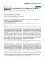
Figure 1
Locations and spacings of genetic markers used for genotypingLocations and spacings of genetic markers used for genotyping. D5S1991 and ANKH-OR are located at the 5' flanking region of ANKH. All seven
single nucleotide polymorphisms used are located in the introns of ANKH.
Available online />R517
Results
Association between specific ANKH variants and
ankylosing spondilitis
The ANKH gene encodes for ANKH transcripts with different
lengths at the 3' untranslated region. The longer transcript
(3928 bp; AB046801) is derived from 12 exons, whereas the
shorter transcript (2426 bp; AK001799, which contains the
last 1721 bp of this transcript) is derived from 13 exons. We
fine-mapped the ANKH gene using 11 markers (Fig. 1): three
microsatellite markers (D5S1954, D5S1991 and D5S1953),
one 5' untranslated region variant (ANKH-OR), and seven
intronic SNPs (rs25957 and rs28006 in intron 1, rs258215 in
intron 2, rs153929 in intron 7, 3088132 and rs27356 in
intron 8, and rs26307 in intron 12).
As an extension to our previous study [8], we included a total
of 201 multiplex AS families in a family-based association anal-
ysis (77 additional multiplex AS families were included, in
addition to the 124 AS families considered in the first study).
All of the families were genotyped with 11 markers in the
ANKH region (D5S1953, rs26307, rs27356, 3088132,
rs153929, rs258215, rs28006, rs25957, ANKH-OR,
D5S1991 and D5S1954). FBAT analyses showed two
regions in the ANKH gene where associations between
ANKH variants and AS were detected. Using both additive
and recessive models, rs27356 [C] was significantly associ-
ated with AS (additive model: Z score = 2.54, P = 0.011;
recessive model: Z score = 2.32, P = 0.020). However,
depending on the model used for the analysis, two different
ANKH markers were also associated with AS. Using an addi-
tive model, an intron 1 SNP, namely rs25957 [C], was associ-
ated with AS (Z score = 2.02, P = 0.043). Using a dominant
model, ANKH-OR allele 1 was associated with AS (Z score =
2.20, P = 0.027). The results are summarized in Table 3. How-
Table 3
FBAT-e analyses conducted in 226 ankylosing spondylitis nuclear families (201 pedigrees, 894 persons)
Marker Allele Allele frequency Number of informative
families
Z score P
Additive model: biallelic test
D5S1953 2 0.45 54 0.57 0.569
rs26307 C 0.81 35 1.73 0.084
rs27356 C 0.80 39 2.54 0.011*
3088132 G 0.79 28 1.28 0.198
rs153929 A 0.76 48 1.61 0.107
rs258215 A 0.59 43 1.58 0.113
rs28006 C 0.74 34 1.62 0.104
rs25957 C 0.76 36 2.02 0.043*
ANKH-OR 1 0.47 59 1.62 0.105
D5S1991 2 0.48 58 1.33 0.181
D5S1954 1 0.64 54 0.65 0.515
Recessive model: biallelic test
D5S1953 2 0.43 48 0.45 0.683
rs26307 C 0.80 40 1.49 0.137
rs27356 C 0.79 44 2.32 0.020*
3088132 G 0.79 31 1.19 0.233
rs153929 A 0.76 50 1.86 0.062
rs258215 A 0.58 32 1.55 0.120
rs28006 C 0.74 37 1.46 0.142
rs25957 C 0.76 38 1.65 0.098
ANKH-OR 2 0.52 37 -2.20 0.027*
D5S1991 1 0.52 45 -1.96 0.050*
D5S1954 1 0.64 54 0.65 0.517
*Statistically significant findings. FBAT, family-based association testing.
Arthritis Research & Therapy Vol 7 No 3 Tsui et al.
R518
ever, these markers are located in different haplotype or LD
blocks (see below), implying that there is more than one sus-
ceptibility locus in the ANKH gene.
Thus, our analyses of 201 multiplex AS families showed that
ANKH variants found in two different regions of the ANKH
gene are modestly associated with AS. Our working hypothe-
sis was that there are two subsets of AS patients, each with a
different predisposing polymorphism in the ANKH locus.
Because ANKH has been shown to be an androgen respon-
sive gene [22-24], we considered whether there are gender
differences between family-based associations of ANKH vari-
ants to AS.
Men with ankylosing spondilitis differ from affected
women for association with different ANKH variants
Radiographic features of AS vary between men and women,
with extensive spinal ankylosis being relatively infrequent in
women with AS [10]. Table 2 summarizes gender information
for the affected individuals in the 201 North American multi-
plex AS families. There were 94 families with both affected
men and women in each family, 74 families with affected men
only, and 33 families with affected women only. In this cohort
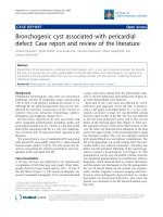
of North American multiplex AS families, men have a signifi-
cantly earlier age at diagnosis than that for women (mean [±
standard deviation] age of diagnosis for affected men = 28 ±
11 years [n = 213]; mean age of diagnosis for affected women
= 30 ± 11 years [n = 149]; including family as an independent
variable [using SAS PROC GLM; SAS Institute Inc., Cary, NC,
USA]: F = 5.10, P = 0.025; Fig. 2). In addition, analysis of age
Table 4
FBAT-e analyses using setafftrait 0 -1 0, testing specifically for affected women
Marker Allele Allele frequency Number of informative families Z score P
Additive model: biallelic test
D5S1953 1 0.56 47 0.47 0.635
rs26307 T 0.19 22 0.32 0.751
rs27356 T 0.20 24 1.03 0.302
3088132 C 0.21 20 0.37 0.712
rs153929 G 0.24 46 1.31 0.191
rs258215 T 0.41 30 1.54 0.123
rs28006 T 0.26 31 2.82 0.004*
rs25957 G 0.23 23 2.82 0.004*
ANKH-OR 2 0.52 59 0.94 0.347
D5S1991 1 0.52 53 1.10 0.270
D5S1954 2 0.36 45 1.38 0.168
Recessive model: biallelic test
D5S1953 2 0.44 39 -1.21 0.227
rs26307 C 0.81 26 0.52 0.606
rs27356 T 0.20 10 1.81 0.069
3088132 G 0.79 22 0.08 0.930
rs153929 A 0.76 44 -1.39 0.162
rs258215 A 0.58 21 -1.76 0.077
rs28006 C 0.74 28 -2.49 0.012*
rs25957 C 0.76 23 -2.25 0.024*
ANKH-OR 2 0.52 34 1.14 0.254
D5S1991 1 0.51 33 1.52 0.127
D5S1954 1 0.64 45 -1.54 0.122
*Statistically significant findings. FBAT, family-based association testing.
Available online />R519
at AS diagnosis in affected men did not reveal a normal distri-
bution; rather the distribution was skewed toward an earlier
onset.
In view of these gender differences, we re-analyzed our geno-
typing results along gender lines in two separate FBAT analy-
ses using the setafftrait command. FBAT analysis of
transmission of alleles to affected women showed that both
rs25957 [G] and rs28006 [T] were associated with AS
(additive model and biallelic test: rs25957 [G], Z score = 2.82,
P = 0.004; rs28006 [T], Z score = 2.82, P = 0.004; Table 4).
These results indicate that only ANKH variants at the 5' end,
and not those at the 3' end, of ANKH are associated with AS
in affected women. This also suggested that ANKH variants at
the 3' end of the gene might be associated with AS only in
affected men.
To test this hypothesis, we analyzed transmission of alleles to
affected men. FBAT analysis of transmission of alleles to
affected men using the setafftrait command showed that two
neighbouring ANKH variants at the 3' end of the gene, namely
rs26307 [C] and rs27356 [C] (16 kb apart), were associated
with AS in affected men as was predicted (additive model:
rs26307 [C], Z score = 2.06, P = 0.039; rs27356 [C], Z
score = 2.63, P = 0.008; recessive model: rs26307 [C], Z
score = 2.51, P = 0.012; rs27356 [C], Z score = 2.99, P =
0.002; Table 5).
Identification of ANKH haplotypes that are associated
with ankylosinig spondylitis
Where the aetiological variant is not typed, haplotype-based
analysis is more powerful for association studies in which
there is significant LD in the region of interest. We took advan-
tage of the data from the HapMap project (12 October 2004
Table 5
FBAT-e analyses using setafftrait 1 0 0, testing specifically for affected men
Marker Allele Allele frequency Number of informative families Z score P
Additive model: biallelic test
D5S1953 2 0.44 47 0.25 0.805
rs26307 C 0.80 22 2.06 0.039*
rs27356 C 0.79 25 2.63 0.008*
3088132 G 0.79 18 1.23 0.216
rs153929 A 0.76 36 0.49 0.619
rs258215 A 0.58 32 0.81 0.419
rs28006 T 0.26 37 0.15 0.877
rs25957 C 0.77 33 0.76 0.446
ANKH-OR 1 0.48 64 1.04 0.297
D5S1991 2 0.49 64 0.39 0.696
D5S1954 2 0.36 49 0.33 0.736
Recessive model: biallelic test
D5S1953 1 0.56 40 -0.83 0.406
rs26307 C 0.81 25 2.51 0.012*
rs27356 C 0.80 28 2.99 0.002*
3088132 G 0.79 22 1.44 0.149
rs153929 A 0.76 39 0.71 0.477
rs258215 A 0.59 23 0.60 0.543
rs28006 T 0.26 16 0.33 0.739
rs25957 C 0.76 29 0.64 0.518
ANKH-OR 2 0.52 44 -1.26 0.205
D5S1991 1 0.51 46 -0.71 0.472
D5S1954 1 0.64 43 -0.65 0.510
*Statistically significant findings. FBAT, family-based association testing.
Arthritis Research & Therapy Vol 7 No 3 Tsui et al.
R520
release 12; [25]). The markers we
used for genotyping are located in four different haplotype
blocks (block 1: rs26307, rs27356; block 2: 3088132 and
rs153929; block 3: rs28006 and rs25957; block 4: ANKH-
OR and D5S1991).
We carried out haplotype analyses based on this information,
using the HBAT empirical variance option in the FBAT pro-
gram, and the results are summarized in Table 6. For HBAT
analyses considering all 226 AS nuclear families, in each of
three different haplotype blocks (blocks 1, 2 and 4) there was
one haplotype with a significant P value, suggesting that there
is heterogeneity in this locus. When HBAT analyses were car-
ried out specifically for affected women, a haplotype with a sig-
nificant P value was found in haplotype block 3 located at the
5' end of the gene. When HBAT analyses were conducted
specifically for affected men, one haplotype with a significant
P value was present in block 1, which is located at the 3' end
of the gene. These results are consistent with those from sin-
gle-marker tests in the FBAT analyses. Furthermore, after Bon-
ferroni correction for the number of haplotypes and models (n
= 8), the haplotype combination of rs26307 [C] and rs27356
[C] remained significantly associated with AS in men (reces-
sive/dominant model: P = 0.004), and the haplotype combina-
tion of rs28006 [C] and rs25957 [C] was significantly
associated with AS in women (recessive/dominant model: P =
0.004).
A direct test for differences between family-based
association with affected men and women
In order to conclude that there are gender differences in
ANKH variants associated with AS, one must show significant
heterogeneity between affected men and women. For this pur-
pose, we used the setafftrait 1 -1 0 command to conduct the
FBAT-e analyses. We coded unaffected siblings and parents
from the families as unknown phenotype (0), affected men as
phenotype 2, and affected women as phenotype 1. The
setafftrait 1 -1 0 command converted affect status to trait 1
(affected men), -1 (affected women) and 0 (unaffected sib-
lings and parents), and the results are summarized in Table 7.
The only marker with a significant P value was rs26307 [C]
(dominant/recessive model: P = 0.03), suggesting that this
marker was significantly associated with AS only in affected
men.
Table 6
HBAT-e analyses using ANKH markers in four haplotype blocks defined in HapMap
Markers in the haplotype block AS nuclear families (n = 226) Testing specifically for affected women Testing specifically for affected men
Additive model
rs26307, rs27356 [C,C]; 0.78; 39; 0.04 NS [C,C]; 0.79; 24; 0.014
3088132, rs153929 NS NS NS
rs28006, rs25957 NS [T,G]; 0.25; 19; 0.007 NS
ANKH-OR, D5S1991 [1,2]; 0.43; 57; 0.013 NS NS
Recessive/dominant model
rs26307, rs27356 [C,C]; 0.78; 40; 0.02 NS [C,C]; 0.79; 25; 0.004*
3088132, rs153929 [G,A]; 0.70; 32; 0.02 NS NS
rs28006, rs25957 NS [C,C]; 0.71; 18; 0.004* NS
ANKH-OR, D5S1991 NS NS NS
Data are expressed as [allele]; allele frequency; number of informative families; P value.
*Significant P value after Bonferroni correction. (Because eight tests [four haplotypes and two models] were carried out in the haplotype-based
association testing [HBAT]-e analyses, P < 0.00625 [0.05/8] is considered statistically significant.) AS, ankylosing spondylitis; NS, not
significant.
Figure 2
Age of diagnosis for (a) men and (b) women in the North American multiplex ankylosing spondilitis familiesAge of diagnosis for (a) men and (b) women in the North American
multiplex ankylosing spondilitis families.
Available online />R521
In view of this finding, we considered whether there is a subset
of AS multiplex families in which ANKH variants were signifi-
cantly associated with AS only in affected women. As summa-
rized in Table 2, there were two types of families in our cohort
of multiplex AS families: families with affected individuals of
both genders; and families with only one gender of affected
individuals (either affected men or affected women).
To assess whether there was significant heterogeneity
between affected men and women in the families of the first
family type (with affected men and women in each family), we
used the setafftrait 1 -1 0 command to conduct the FBAT-e
analyses. The results are summarized in Table 8. Two markers
(rs28006 [T] and rs25957 [G]) exhibited significant P values
(additive model: P = 0.004 for rs28006 and P = 0.017 for
rs25957), suggesting that these two markers were associated
with AS only in affected women in the subset of AS families
with affected individuals of both genders.
We also conducted FBAT-e analysis using setafftrait com-
mand 1 -1 0 in families with only one gender of affected indi-
viduals (data not shown). However, there were few informative
families (<20 families from which we could track the transmis-
sion of alleles), and so the results might not be reliable.
Selective transmission of haplotypes of interest to the
affected men/women
In order to estimate the magnitude of the effect, we calculated
the frequency at which the haplotypes of interest were
transmitted to the affected men or women using TDT. For the
haplotype rs28006 [C] rs25957 [C], the frequency of trans-
mission was 74% (17/23) to affected women and 40% (12/
Table 7
FBAT-e analyses considering 226 ankylosing spondylitis nuclear families (201 pedigrees, 894 persons): summary of results using
setafftrait 1 -1 0
Marker Allele Allele frequency Number of informative
families
Z score P
Additive model: biallelic test
D5S1953 1 0.56 63 0.15 0.879
rs26307 C 0.81 35 1.29 0.195
rs27356 C 0.80 39 1.12 0.261
3088132 G 0.79 29 0.69 0.489
rs153929 G 0.24 52 0.61 0.541
rs258215 T 0.41 41 0.46 0.638
rs28006 T 0.26 44 1.69 0.090
rs25957 G 0.23 39 0.90 0.366
ANKH-OR 1 0.48 80 0.20 0.841
D5S1991 1 0.52 80 0.29 0.769
D5S1954 2 0.36 63 1.21 0.227
Recessive model: biallelic test
D5S1953 1 0.56 46 -0.89 0.370
rs26307 C 0.81 37 2.17 0.030*
rs27356 C 0.80 41 1.91 0.055
3088132 G 0.79 29 1.17 0.240
rs153929 G 0.24 18 0.55 0.582
rs258215 A 0.59 29 -0.75 0.453
rs28006 C 0.74 40 -1.13 0.186
rs25957 C 0.77 36 -0.66 0.507
ANKH-OR 2 0.52 50 -0.34 0.733
D5S1991 2 0.49 52 -0.28 0.779
D5S1954 1 0.64 61 -1.53 0.126
*Statistically significant findings. FBAT, family-based association testing.
Arthritis Research & Therapy Vol 7 No 3 Tsui et al.
R522
30) to affected men. Thus, the 'odds ratio' for increased risk is
1.85 (0.74/0.4). More dramatic proportions were seen in the
subset of families with affected individuals of both genders. In
these families, this haplotype was transmitted to affected
women 79% of the time (15/19) but to affected men only 27%
of the time (3/11). In this case, the 'odds ratio' for increased
risk approaches 3.0 (0.79/0.27 = 2.92).
For the haplotype rs26307 [C] rs27356 [C], the frequency of
transmission was 70% (21/30) to affected men and 43% (13/
30) to affected women (an 'odds ratio' for increased risk of
1.75). In the subset of families with only affected men, 94%
(16/17) of the time this haplotype was transmitted to affected
men. There were too few informative families with only affected
women with this variant (n = 6), and so we do not have a reli-
able assessment of the frequency at which this haplotype was
transmitted to affected women in this subset for comparison.
Discussion
In this study of the association of ANKH genetic markers with
AS, including 201 AS multiplex families, we found that ANKH
variants located in two different regions of the ANKH gene
were associated with AS. A more striking finding was that the
genetic association for men and women with AS differed. In
men, AS was associated with genetic markers at the 3' end of
the ANKH gene, whereas in women AS appeared to be asso-
ciated with genetic markers at the 5' end of the ANKH gene.
As expected, when the genders of AS patients were analyzed
separately, we observed more than one SNP in each region
(within the same haplotype block) showing significant associ-
Table 8
FBAT-e analyses considering 108 ankylosing spondylitis nuclear families (94 pedigrees, 425 persons) in which both affected men
and women are present in each family: summary of the results using setafftrait 1 -1 0
Marker Allele Allele frequency Number of informative
families
Z score P
Additive model: biallelic test
D5S1953 1 0.56 39 0.77 0.437
rs26307 C 0.90 10 0.50 0.617
rs27356 C 0.77 17 0.15 0.875
3088132 C 0.21 13 0.63 0.527
rs153929 G 0.23 32 0.81 0.418
rs258215 T 0.49 21 1.62 0.104
rs28006 T 0.29 27 2.81 0.004*
rs25957 G 0.32 22 2.37 0.017*
ANKH-OR 1 0.48 51 0.25 0.801
D5S1991 2 0.50 48 0.13 0.891
D5S1954 2 0.33 31 1.13 0.254
Recessive model: biallelic test
D5S1953 2 0.44 31 -1.34 0.177
rs26307 C 0.79 19 1.00 0.314
rs27356 C 0.80 20 1.17 0.239
3088132 G 0.79 14 -0.26 0.788
rs153929 G 0.23 10 1.06 0.288
rs258215 A 0.51 12 -2.49 0.012*
rs28006 C 0.71 20 -2.344 0.019*
rs25957 C 0.67 15 -1.97 0.048*
ANKH-OR 1 0.48 27 0.83 0.406
D5S1991 2 0.49 29 0.98 0.322
D5S1954 1 0.66 31 -1.67 0.093
*Statistically significant findings. FBAT, family-based association testing.
Available online />R523
ation with AS. Haplotype analyses appeared to confirm the
results of the single-marker tests (FBAT analyses), indicating
that the predisposing polymorphism(s) for men with AS prob-
ably lies at the 3' end of the ANKH gene, whereas those for
affected women are probably at the 5' end of the gene. After
Bonferroni correction for the number of haplotypes and mod-
els, the haplotype combination of rs26307 [C] and rs27356
[C] was significantly associated with men with AS; and the
haplotype combination of rs28006 [C] and rs25957 [C] was
significantly associated with women with AS. However, in both
cases, the significance level was modest. We attribute this to
the fact that we have not identified the aetiological variants in
the men/women with AS. Despite the modest P values (which
are a function of sample size), the calculated 'odds ratios' for
increased risk (which provide estimates of the magnitude of
the effect) were close to 2 for the transmission of rs26307 [C]
rs27356 [C] to affected men, and close to 3 for the transmis-
sion of rs28006 [C] rs25957 [C] to affected women in the
subset of families with affected individuals of both genders.
A test of interaction identified the region that was associated
in men with AS (rs26307) as showing a difference in the
strength of the association by gender. The region associated
with AS in women only showed significance of the test of inter-
action among the subset of families with affected individuals of
both genders. Our current efforts are to identify and analyze
more common SNPs in these two regions, ultimately finding
the predisposing polymorphisms in men and women.
The rationale for studying multiplex AS families is to enhance
the chances of identifying the genes involved. There are very
few studies that directly compare familial versus sporadic AS.
In one study [26], familial versus sporadic Dutch AS patients
exhibited no difference in age at disease onset, age at diagno-
sis, or prevalence of peripheral arthritis and acute anterior uvei-
tis. In another study, familial AS disease was significantly
milder than sporadic disease, as assessed by spinal mobility
score, Arthritis Impact Measurement Scales (AIMS) overall
impact score, AIMS physical activity score, AIMS social func-
tion score and AIMS pain score [27]. Thus, findings from mul-
tiplex families might not be directly applicable to individuals
affected with sporadic AS. Most studies assessing the impact
gender has on age at AS onset or diagnosis have been con-
ducted without addressing whether the individuals had familial
or sporadic disease [28-30]; these studies showed that the
age at disease onset is similar between genders. However, in
our cohort of AS multiplex families, men had a significantly
earlier age at diagnosis compared with that for women (for
men 28 ± 11 years [n = 213]; and for women 30 ± 11 years
[n = 149]). Because these are AS multiplex families, it is
unlikely that there is a bias leading physicians to delay diagno-
sis in affected women. The misconception that AS is exclu-
sively a male disease may yet be a confounding factor. In the
subset of families with affected individuals of both genders,
men have an even earlier age at diagnosis (27.8 ± 11 years [n
= 94]) compared with that for women (32.6 ± 11 years [n =
101]), whereas both men and women have similar ages of
diagnosis in the subsets of families with affected individuals of
only one gender (for men 28.6 ± 12 years [n = 130]; for
women 29.9 ± 12 years [n = 53]). This finding suggests that
there is heterogeneity even in multiplex AS families.
The ANKH variants that were significantly associated with AS
are located in introns 1, 8 and 12. It is likely that the predispos-
ing polymorphisms affect gender-specific regulation of ANKH
expression. Very little is known regarding the molecular mech-
anisms that underlie the regulation of ANKH expression. One
study [31] reported that ANKH is a growth factor responsive
gene. Three recent reports [22-24] showed that ANKH is an
androgen responsive gene. In androgen-treated prostate can-
cer cell lines, the abundance of ANKH transcripts was sixfold
higher than in the untreated cells. In the ANKH promoter, there
is a sequence at position -1015 (AGAACAcacTtTcCT) with
83% match to an androgen response element (ARE) consen-
sus sequence [22]. It remains unclear whether this ARE-like
motif is functional. In view of the locations of the ANKH vari-
ants associated with AS, it remains unclear whether this ARE-
like motif at the promoter region can directly contribute to the
regulation of ANKH expression by the predisposing polymor-
phisms. It is also unknown whether there is a different mode of
ANKH regulation in women.
A report recently concluded that ANKH did not significantly
contribute to susceptibility or specific disease expression in
AS patients from the UK [32]. In that report, a case–control
study was conducted using five ANKH SNPs within the cod-
ing region and flanking splice sites and three known promoter
variants. There was no association between these polymor-
phisms and AS or the clinical pattern of the disease. In addi-
tion, using 185 affected sib-pair AS families, no linkage
between ANKH and AS was observed. However, the exact
linkage results were not shown. Using multipoint exclusion
mapping of the ANKH region, the presence of a gene contrib-
uting more than 10% of the recurrence risk to AS (λ
S
= 1.4)
was excluded. Using λ
S
of 1.4 as the cutoff may exclude genes
with modest effects. In that report, the LD between markers
was not shown. In situations where the aetiological variant is
not typed, haplotype-based analysis may be a more powerful
analytical method when there is significant LD.
The basis for the discrepancy between the UK results [32] and
ours is not entirely clear, but there are several possible expla-
nations. First, the UK group focused on analyzing exonic vari-
ants, variants near splice junctions and in the promoter region.
Their analysis did not include any ANKH variants in the 3'
region, where we detected association with AS in men. Sec-
ond, although the UK group included a gender breakdown of
their patients (63.5% men and 36.5% women), the analysis
did not include a breakdown of AS patients by gender, and
variants with modest gender-specific effects might have been
Arthritis Research & Therapy Vol 7 No 3 Tsui et al.
R524
missed. Third, it is possible that there are some intrinsic differ-
ences between the two populations (UK versus North Ameri-
can). Genome-wide linkage scans performed in the two
groups revealed some similar susceptibility regions, such as
on chromosomes 6p (the MHC), 5q and 10q [5,6]. However,
the linkage identified on chromosome 11q23 in the North
American Spondylitis Consortium families was not seen in the
UK study. In addition, the linkage identified on chromosome 2q
in the UK study was not seen in the North American Spondyli-
tis Consortium study. The intrinsic differences could reflect
clinical differences in the patient population recruited, or they
could be due to population-specific mechanisms of genetic
susceptibility. Finally, because both groups analyzed about
200 AS families, there might not be sufficient power to detect
genes with 'small effects' consistently, leading to discrepan-
cies between results.
In our cohort of North American multiplex AS families, the age
at diagnosis was significantly younger in men than in women.
However, FBAT analyses using the offset option (-o; an option
which works for both quantitative and qualitative traits) did not
show any significant association of age at diagnosis in the men
or women with AS, even in subsets of families, using the
ANKH markers (data not shown), suggesting that ANKH vari-
ants are responsible for disease susceptibility. Our finding of
gender-specific polymorphisms in the ANKH gene conferring
differential susceptibility to AS might shed light on the biolog-
ical basis of these clinical observations.
In view of the difficulty in locating susceptibility loci with mod-
est effects in recent genome-wide linkage studies conducted
in AS families, it will be of interest to assess whether gender
subsetting in the analyses of genome-wide linkage studies
might yield further insights into the genetic basis of rheumatic
diseases, many of which have a strong gender predilection.
Conclusion
Taken together, our findings showed that, after Bonferroni cor-
rection, two intronic markers at the 3' end of the ANKH gene
were significantly associated with AS only in affected men,
and two intronic markers at the 5' end of the ANKH gene were
significantly associated with AS only in affected women. This
may partly account for the gender difference in the prevalence
of AS.
Competing interests
The author(s) declare that they have no competing interests.
Authors' contributions
HWT conducted all of the genotyping and analyzed the data.
RDI conceived the study, provided some of the patients'
blood/cells for extracting DNA and reviewed the manuscript.
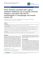
Figure 3
A summary of family-based association analyses (FBAT; additive model and biallelic tests)A summary of family-based association analyses (FBAT; additive model and biallelic tests). Each column in the charts represents the -log FBAT P
value for each marker located at a distance relative to one another. D5S1953 is positioned at 0 distance. Chart ALL shows analysis of 226 ankylos-
ing spondylitis (AS) nuclear families with 894 persons. Chart Women shows analysis of 127 AS families with 184 affected women. Chart Men
shows analysis of 168 AS families with 282 affected men.
Available online />R525
ADP designed the study, supervised the statistical analyses
and revised the manuscript. JDR coordinated the recruitment
of individuals from AS families, provided most of the DNA sam-
ples and reviewed the manuscript. FWLT conceived, designed
and coordinated the study, analyzed and interpreted the data,
performed statistical analyses, and drafted and revised the
manuscript. All authors read and approved the final
manuscript.
Acknowledgements
We thank Dr Cathy Barr for making the ABI sequence detection system
available, Karen Wigg for reading the plates, and Dr Celia Greenwood
for review of the manuscript and helpful suggestions on the statistical
analyses. We also thank the Ontario Spondylitis Association and the
Spondylitis Association of America for their assistance in recruiting the
families included in this study.
This work was supported by grants from the Canadian Institutes of
Health Research, the Arthritis Center of Excellence, Genome Canada,
and by the NIH (National Institute of Arthritis and Musculoskeletal and
Skin Diseases grant R01-AR-46208 to Dr Reveille). Dr Paterson is a
Canada Research Chair.
References
1. Calin A: Ankylosing spondylitis. In Oxford Textbook of Rheuma-
tology Volume 2. Edited by: Maddison PJ, Isenberg DA, Woo P,
Glass DN. Oxford: Oxford University Press; 1998:1058-1070.
2. Braun J, Bollow M, Remlinger G, Eggens U, Rudwaleit M, Distler
A, Sieper J: Prevalence of spondylarthropathies in HLA-B27
positive and negative blood donors. Arthritis Rheum 1998,
41:58-67.
3. Brown MA, Laval SH, Brophy S, Calin A: Recurrence risk mode-
ling of the genetic susceptibility to ankylosing spondylitis. Ann
Rheum Dis 2000, 59:883-886.
4. Brown MA, Pile KD, Kennedy LG, Campbell D, Andrew L, March
R, Shatford JL, Weeks DE, Calin A, Wordsworth BP, et al.: A
genome-wide screen for susceptibility loci in ankylosing
spondylitis. Arthritis Rheum 1998, 41:588-595.
5. Laval SH, Timms A, Edwards L, Bradbury L, Brophy S, Milicic A,
Rubin L, Siminovitch KA, Weeks DE, Calin A, et al.: Whole-
genome screening in ankylosing spondylitis: evidence of non-
MHC genetic-susceptibility loci. Am J Hum Genet 2001,
68:918-926.
6. Zhang G, Luo J, Bruckel J, Weisman MA, Schumacher HR, Kahn
MA, Inman RD, Mahowald M, Maksymowych WP, Martin TM, et al.:
Genetic studies in familial ankylosing spondylitis
susceptibility. Arthritis Rheum 2004, 50:2246-2254.
7. Ho AM, Johnson MD, Kingsley DM: Role of the mouse ank gene
in control of tissue calcification and arthritis. Science 2000,
289:265-270.
8. Tsui FWL, Tsui HW, Cheng EY, Stone M, Payne U, Reveille JD,
Paterson AD, Inman RD: Novel genetic markers in the 5'-flank-
ing region of ANKH are associated with ankylosing spondylitis.
Arthritis Rheum 2003, 48:791-797.
9. Calin A: Ankylosing spondylitis. Clin Rheum Dis 1985,
11:41-60.
10. Eustace S, Coughlan RJ, McCarthy C: Ankylosing spondylitis. A
comparison of clinical and radiographic features in men and
women. Ir Med J 1993, 86:120-122.
11. Gran JT, Husby G, Hordvik M, Stormer J, Romberg-Andersen O:
Radiological changes in men and women with ankylosing
spondylitis. Ann Rheum Dis 1984, 43:570-575.
12. Will R, Edmunds L, Elswood J, Calin A: Is there sexual inequality
in ankylosing spondylitis? A study of 498 women and 1202
men. J Rheumatol 1990, 17:1649-1652.
13. Kidd B, Mullee M, Frank A, Cawley M: Disease expression of
ankylosing spondylitis in males and females. J Rheumatol
1988, 15:1407-1409.
14. Guillemin F, Briancon S, Pourel J, Gaucher A: Longterm disability
and prolonged sick leave as outcome measures in ankylosing
spondylitis. Arthritis Rheum 1990, 33:1001-1006.
15. Doran MF, Brophy S, MacKay K, Taylor G, Calin A: Predictors of
longterm outcome in ankylosing spondylitis. J Rheum 2003,
30:316-320.
16. Hoyle E, Laval SH, Calin A, Wordsworth BP, Brown MA: The X-
chromosome and susceptibility to ankylosing spondylitis.
Arthritis Rheum 2000, 43:1353-1355.
17. Calin A, Brophy S, Blake D: Impact of sex on inheritance of
ankylosing spondylitis: a cohort study. Lancet 1999,
354:1687-1690.
18. Brophy S, Taylor G, Blake D, Calin A: The interrelationship
between sex, susceptibility factors, and outcome in ankylosing
spondylitis and its associated disorders including inflamma-
tory bowel disease, psoriasis and iritis. J Rheumatol 2003,
30:2054-2058.
19. Van der Linden S, Valkenburg H, Cats A: Evaluation of diagnostic
criteria for ankylosing spondylitis: a proposal for modification
of the New York criteria. Arthritis Rheum 1984, 27:361-368.
20. Spielman RS, McGinnis RE, Ewens WJ: Transmission test for
linkage disequilibrium: the insulin gene region and insulin-
dependent diabetes mellitus (IDDM). Am J Hum Genet 1993,
52:506-516.
21. Lake SL, Blacker D, Laird NM: Family-based tests of association
in the presence of linkage. Am J Hum Genet 1993, 52:506-516.
22. Nelson PS, Clegg N, Arnold H, Ferguson C, Bonham M, White J,
Hood L, Lin B: The program of androgen-responsive genes in
neoplastic prostate epithelium. Proc Natl Acad Sci USA 2002,
99:11890-11895.
23. DePrimo SE, Diehn M, Belson JB, Reiter RE, Matese J, Fero M, Tib-
shirani R, Brown PO, Brooks JD: Transcriptional programs acti-
vated by exposure of human prostate cancer cells to
androgen. Genome Biol 2002, 3:32.1-32.12.
24. Segawa T, Nau ME, Xu LL, Chilukuri RN, Makaren M, Zhang W,
Petrovics G, Sesterhenn IA, McLeod DG, Moul JW, et al.: Andro-
gen-induced expression of endoplasmic reticulum (ER) stress
response genes in prostate cancer cells. Oncogene 2002,
21:8749-8758.
25. The international HapMap Consortium: The international Hap-
Map project. Nature 2003, 426:789-795.
26. Paardt M, Dijkmans B, Giltay E, vander Horst-Bruinsma I: Dutch
patients with familial and sporadic ankylosing spondylitis do
not differ in disease phenotype. J Rheumatol 2002,
29:2583-2584.
27. Calin A, Kennedy LG, Edmunds L, Will R: Familial versus spo-
radic ankylosing spondylitis. Two different diseases? Arthritis
Rheum 1993, 36:676-681.
28. Van der Linden SM, Valkenburg HA, de Jongh BM, Cats A: The
risk of developing ankylosing spondylitis in HLA-B27 positive
individuals. Arthritis Rheum 1984, 27:241-249.
29. Gran JT, Ostensen M, Husby G: A clinical comparison between
males and females with ankylosing spondylitis. J Rheumatol
1985, 12:126-129.
30. Feldtkeller E, Khan MA, van der Heijde Désirée, van der Linden S,
Braun J: Age at disease onset and diagnosis delay in HLA-B27
negative vs. positive patients with ankylosing spondylitis.
Rheumatol Int 2003, 23:61-66.
31. Guo Y, Hsu DKW, Feng SY, Richards CM, Winkles JA: Polypep-
tide growth factors and phorbol ester induce progressive
ankylosis (ank) gene expression in murine and human
fibroblasts. J Cell Biochem 2001, 84:27-38.
32. Timms AE, Zhang Y, Bradbury L, Wordsworth BP, Brown MA:
Investigation of the role of ANKH in ankylosing spondylitis.
Arthritis Rheum 2003, 48:2898-2902.