Báo cáo y học: "Signalling pathway involved in nitric oxide synthase type II activation in chondrocytes: synergistic effect of leptin with interleukin-1" pdf
Bạn đang xem bản rút gọn của tài liệu. Xem và tải ngay bản đầy đủ của tài liệu tại đây (1000.58 KB, 11 trang )
Open Access
Available online />R581
Vol 7 No 3
Research article
Signalling pathway involved in nitric oxide synthase type II
activation in chondrocytes: synergistic effect of leptin with
interleukin-1
Miguel Otero
1
, Rocío Lago
1
, Francisca Lago
2
, Juan Jesús Gomez Reino
3,4
and Oreste Gualillo
1
1
NEIRID (NeuroEndocrine Interactions in Rheumatology and Inflammatory Diseases) Laboratory, Santiago University Clinical Hospital, Research
Laboratory 4, Santiago de Compostela, Spain
2
Laboratory of Molecular and Cellular Cardiology, Santiago University Clinical Hospital, Research Laboratory 1, Santiago de Compostela, Spain
3
Rheumatology Division, Santiago University Clinical Hospital, Santiago de Compostela, Spain
4
Department of Medicine, School of Medicine, University of Santiago de Compostela, Santiago de Compostela, Spain
Corresponding author: Oreste Gualillo,
Received: 11 Aug 2004 Revisions requested: 16 Sep 2004 Revisions received: 14 Jan 2005 Accepted: 3 Feb 2005 Published: 4 Mar 2005
Arthritis Research & Therapy 2005, 7:R581-R591 (DOI 10.1186/ar1708)
This article is online at: />© 2005 Otero et al.; licensee BioMed Central Ltd.
This is an Open Access article distributed under the terms of the Creative Commons Attribution License ( />2.0), which permits unrestricted use, distribution, and reproduction in any medium, provided the original work is properly cited.
Abstract
The objective of the present study was to investigate the effect
of leptin, alone or in combination with IL-1, on nitric oxide
synthase (NOS) type II activity in vitro in human primary
chondrocytes, in the mouse chondrogenic ATDC5 cell line, and
in mature and hypertrophic ATDC5 differentiated chondrocytes.
For completeness, we also investigated the signalling pathway
of the putative synergism between leptin and IL-1. For this
purpose, nitric oxide production was evaluated using the Griess
colorimetric reaction in culture medium of cells stimulated over
48 hours with leptin (800 nmol/l) and IL-1 (0.025 ng/ml), alone
or combined. Specific pharmacological inhibitors of NOS type II
(aminoguanidine [1 mmol/l]), janus kinase (JAK)2 (tyrphostin
AG490 and Tkip), phosphatidylinositol 3-kinase (PI3K;
wortmannin [1, 2.5, 5 and 10 µmol/l] and LY294002 [1, 2.5, 5
and 10 µmol/l]), mitogen-activated protein kinase kinase
(MEK)1 (PD098059 [1, 5, 10, 20 and 30 µmol/l]) and p38
kinase (SB203580 [1, 5, 10, 20 and 30 µmol/l]) were added 1
hour before stimulation. Nitric oxide synthase type II mRNA
expression in ATDC5 chondrocytes was investigated by real-
time PCR and NOS II protein expression was analyzed by
western blot. Our results indicate that stimulation of
chondrocytes with IL-1 results in dose-dependent nitric oxide
production. In contrast, leptin alone was unable to induce nitric
oxide production or expression of NOS type II mRNA or its
protein. However, co-stimulation with leptin and IL-1 resulted in
a net increase in nitric oxide concentration over IL-1 challenge
that was eliminated by pretreatment with the NOS II specific
inhibitor aminoguanidine. Pretreatment with tyrphostin AG490
and Tkip (a SOCS-1 mimetic peptide that inhibits JAK2)
blocked nitric oxide production induced by leptin/IL-1. Finally,
wortmannin, LY294002, PD098059 and SB203580
significantly decreased nitric oxide production. These findings
were confirmed in mature and hypertrophic ATDC5
chondrocytes, and in human primary chondrocytes. This study
indicates that leptin plays a proinflammatory role, in synergy with
IL-1, by inducing NOS type II through a signalling pathway that
involves JAK2, PI3K, MEK-1 and p38 kinase.
Introduction
Chondrocytes are the predominant cells in mature cartilage
that synthesize and maintain the integrity of cartilage-specific
extracellular matrix. In rheumatoid arthritis and osteoarthritis
the phenotype of chondrocytes changes, and apoptosis and
extracellular matrix degradation occur [1-3]. These severe per-
turbations in cartilage homeostasis may be mediated in part by
nitric oxide (NO). This gaseous mediator is induced by several
proinflammatory cytokines, including IL-1.
Leptin, the OB gene product, is a 16 kDa hormone that is syn-
thesized by adipocytes. Leptin regulates food intake and
ERK = extracellular signal-regulated kinase; GAPDH = glyceraldehyde-3-phosphate dehydrogenase; IFN = interferon; IL = interleukin; JAK = janus
kinase; MAPK = mitogen-activated protein kinase; MEK = mitogen-activated protein kinase kinase; MMP = matrix metalloproteinase; NF-κB = nuclear
factor-κB; NO = nitric oxide; NOS = nitric oxide synthase; PBS = phosphate-buffered saline; PI3K = phosphatidylinositol 3-kinase; RT-PCR = reverse
transcription polymerase chain reaction; SOCS = suppressor of cytokine signalling.
Arthritis Research & Therapy Vol 7 No 3 Otero et al.
R582
energy expenditure, but it also modulates neuroendrocrine
function [4]. It is involved in immune modulation in that it influ-
ences the innate immune response by promoting activation of
monocyte/macrophages, chemotaxis and activation of neu-
trophils, and activation of natural killer cells [5]. Furthermore,
leptin influences adaptive immunity by increasing the expres-
sion of adhesion molecules by CD4
+
T cells, and promoting
proliferation and secretion of IL-2 by naïve CD4
+
T cells [5-7].
Leptin has also been found to influence bone growth [8] and
inflammation [9].
High leptin levels are associated with obesity, which is a risk
factor for osteoarthritis [10-12]. Interestingly, in patients with
osteoarthritis leptin is present in synovial fluid and is expressed
by articular chondrocytes [13], and normal human chondro-
cytes express the functional Ob-Rb leptin receptor isoform
[14]. It is unlikely that leptin alone acts on cartilage to trigger
an inflammatory response; rather, it may associate with other
proinflammatory cytokines to amplify inflammation and
enhance damage to cartilage. We recently demonstrated a
synergistic effect of leptin with IFN-γ on nitric oxide synthase
(NOS) type II activity in cultured chondrocytes that was medi-
ated by the janus kinase (JAK)2 [15]. In the present study we
investigated whether leptin synergizes with IL-1, an abundant
mediator of inflammation and cartilage destruction [16,17], to
activate NOS type II in chondrocytes. To gain further insights
into the mechanism of action of this putative synergism, we
also analyzed the role played by several intracellular kinases by
using specific pharmacological inhibitors.
Materials and methods
Reagents
Foetal bovine serum, tissue culture media, media supple-
ments, mouse and human recombinant leptin, mouse recom-
binant IL-1, tyrphostin AG490, wortmannin, LY294002,
PD098059 and SB203580 were purchased from Sigma (St
Louis, MO, USA) unless otherwise specified. RT-PCR rea-
gents were purchased from Invitrogen (Carlsbad, CA, USA)
and Stratagene (La Jolla, CA, USA). Tkip (WLVFFVIFYFFR), a
suppressor of cytokine signalling (SOCS)-1 mimetic peptide
that inhibits JAK2 autophosphorylation, was generously pro-
vided by Dr Howard M Johnson (Institute of Food and Agricul-
tural Science, Department of Microbiology and Cell Science,
University of Florida, Gainesville, FL, USA).
Cell culture
The clonal chondrogenic cell line ATDC5 was chosen for
these studies because it has been shown to be a useful in vitro
model for examining the multistep differentiation of chondro-
cytes. Undifferentiated ATDC5 cells proliferate rapidly until
they reach confluence, at which point they undergo growth
arrest. When treated with insulin, transferrin and sodium
selenite, confluent ATDC5 cells re-enter a proliferative phase
and form cartilaginous matrix nodules (mature chondrocytes).
As differentiation progresses, these cells undergo a late differ-
entiation phase, becoming hypertrophic, calcifying chondro-
cytes that synthesize type X collagen and osteopontin – a
marker of terminal chondrocyte differentiation [18]. ATDC5
cells were a kind gift from Dr Agamemnon E Grigoriadis
(Department of Craniofacial Development, King's College,
London Guy's Hospital, London, UK). Unless otherwise spec-
ified, cells were cultured in Dulbecco's modified Eagle's
medium/Hams' F12 medium supplemented with 5% foetal
bovine serum, 10 µg/ml human transferrin, 3 × 10
-8
mol/l
sodium selenite and antibiotics (50 U/ml penicillin and 50 µg/
ml streptomycin).
In some experiments, conducted to demonstrate that leptin/IL-
1 synergism does not appear to depend on the differentiation
state of the chondrocytes, chondrogenic ATDC5 cells were
differentiated into mature and hypertrophic chondrocytes, as
described by Thomas and coworkers [19]. Briefly, cells were
plated at an initial density of 2 × 10
4
cells/well in 24-well
plates. Cells were cultured in the above-mentioned medium
supplemented with 10 µg/ml of human recombinant insulin
(Novo Nordisk A/S, Bagsvaerd, Denmark). Culture was contin-
ued for a further 15 or 21 days, with replacement of medium
every other day. As expected, ATDC5 cultures treated with
insulin underwent progressive differentiation from 0 to 21 days
as compared with untreated cultures. This differentiation was
qualitatively characterized by increased formation of cartilage
nodules and enhanced staining with alcian blue dye, which is
indicative of cartilage proteoglycan accumulation.
In other experiments (data not shown), the differentiation from
days 0 to 21 was further evidenced by sequential increases in
type II collagen, aggrecan and type X collagen mRNAs. The
early and mature chondrocyte marker type II collagen was
expressed in undifferentiated ATDC5 cells; the level began to
increase at day 3, peaked at days 7–10 and gradually declined
after day 15. The expression profile of aggrecan mimicked that
of type II collagen but with a slight delay of a couple of days.
The decline in expression of both chondrocyte markers coin-
cided with the onset of late-stage chondrocyte differentiation.
The expression of the hypertrophic chondrocyte marker type X
collagen began at days 12 and 13. The expression patterns of
these early and late chondrocyte markers were consistent with
previous findings in ATDC5 cells regarding in vivo chondro-
cyte differentiation. We do not illustrate findings regarding the
differentiation of ATDC5 cells because they are extensively
reported in literature [19].
Cartilage harvest and human chondrocyte isolation
Human normal articular cartilage samples were obtained from
knee joints of patients undergoing leg amputations from above
the knee because of peripheral vascular disease. (Permission
from the local ethical committee was granted.) None of the
patients had a clinical history of arthritis or any other pathology
affecting the cartilage, and the specimens appeared normal on
morphological examination (no change in colour and no
Available online />R583
fibrillation). For chondrocyte isolation, aseptically dissected
cartilage was subjected to sequential digestion with pronase
(catalogue number 165921; Roche Molecular Biochemicals,
Indianapolis, IN, USA) and collagenase P (catalogue number
1213873; Roche Molecular Biochemicals) at a final concen-
tration of 1 mg/ml in Dulbecco's modified Eagle's medium/F12
plus 10% foetal calf serum and sterilized by filtration, in
accordance with the manufacturer's instructions. In our hands,
this procedure was superior to enzymatic isolation with colla-
genase alone in terms of chondrocyte yields and capacity for
attachment. Cartilage specimens were finely diced in phos-
phate-buffered saline (PBS), and after removing PBS diced
tissue was incubated for 30 min with pronase in a shaking
water bath at 37°C. Pronase was subsequently removed from
the digestion flask and the cartilage pieces were washed with
PBS. After removal of PBS, digestion was continued with
addition of collagenase P; this was done over 6–8 hours in a
shaking water bath at 37°C. The resulting cell suspension was
filtered through a 40 µm nylon cell strainer (BD Biosciences
Europe, Erembodegem, Belgium) in order to remove debris.
Cells were centrifuged and washed twice with PBS, counted
and plated in 24-well tissue culture plates for chondrocyte cul-
ture. Cells were serially passaged to obtain a sufficient number
of cells and used between the first and second passages.
Cell treatments and nitrite assay
ATDC5 cells and human primary chondrocytes, with a viability
greater than 95% as evaluated using the trypan blue exclusion
method, were cultured (as described above) in 24-well plates.
After 12 hours of starvation in serum-free medium, cells were
stimulated for 48 hours with leptin (800 nmol/l), alone or in
combination with IL-1 (0.025 ng/ml). We wished to determine
whether increased NO production was due to NOS type II
activation and to the involvement of JAK2, phosphatidylinositol
3-kinase (PI3K), mitogen-activated protein kinase kinase
(MEK)1 and p38 kinase. For this purpose, the following spe-
cific pharmacological inhibitors were added 1 hour before
cytokine stimulation: aminoguanidine (1 mmol/l) for NOS type
II; tyrphostin AG490 (5 and 10 µmol/l) and Tkip (20 and 50
µmol/l) for JAK2; wortmannin (1, 2.5, 5 and 10 µmol/l) and
LY294002 (1, 2.5, 5 and 10 µmol/l) for PI3K; PD098059 (1,
5, 10, 20 and 30 µmol/l) for MEK-1; and SB203580 (1, 5, 10,
20 and 30 µmol/l) for p38 kinase. Cytokines and pharmaco-
logical inhibitor doses were selected on the basis of prior
dose–response experiments (data not shown) or previously
published literature [15].
Nitrite accumulation was measured in culture medium using
the Griess reaction. Briefly, 100 µl cell culture medium was
mixed with 100 µl Griess reagent (equal volumes of 1%
[weight/vol] sulfanilamide in 5% [vol/vol] phosphoric acid and
0.1% [weight/vol] naphtylethylenediamine-HCl), incubated at
room temperature for 10 min, and then the absorbance at 550
nm was measured using a microplate reader (Titertek-Multi-
scan, Labsystem, Helsinki, Finland). Fresh culture medium was
used as blank in all of the experiments. The amount of nitrite in
the samples (in micromolar units) was calculated from a
sodium nitrite standard curve freshly prepared in culture
medium.
RNA isolation and real-time RT-PCR
ATDC5 chondrogenic cells were seeded in P6 well plates to
reach 85–90% confluence. After 8 hours of starvation in
serum-free medium, cells were treated with leptin alone or in
combination with IL-1. In order to test the involvement of JAK2,
PI3K, MEK-1 and p38 kinase on NOS type II mRNA expres-
sion, specific inhibitors (tyrphostin AG490 10 µmol/l, wort-
mannin and LY294002 10 µmol/l, PD098059 30 µmol/l and
SB203580 30 µmol/l) were added 1 hour before cytokine
stimulation. After 48 hours of treatment, RNA was isolated
from cell culture using the Trizol-LS
®
TM method (Gibco-BRL,
Life Technologies, Grand Island, NY USA), in accordance with
the manufacturer's instructions. Briefly, 5 × 10
5
cells were
lysed in 1000 µl Trizol-LS
®
reagent, and recovery of total RNA
after isopropanol precipitation was measured using a spectro-
photometer (Beckman DU62, Amersham Biosciences, Chal-
font St. Giles, UK) at 260 nm.
Analysis of nitric oxide synthase type II gene expression
using real-time RT-PCR
Real-time RT-PCR analyses were performed in a fluorescent
temperature cycler (MX3000P Real Time PCR System; Strat-
agene), in accordance with the manufacturer's instructions.
Total RNA 1 µg was used for each RT reaction. cDNAs were
synthesized using 200 units of Moloney murine leukaemia
reverse transcriptase (Gibco-BRL) and 6 µl dNTPs mix (10
mmol/l of each dNTP), 6 µl of first strand buffer (250 mmol/l
Tris-HCl [pH 8.3], 375 mmol/l KCl, 15 mmol/l MgCl
2
; Gibco-
BRL), 1.5 µl of 50 mmol/l MgCl
2
, 0.17 µl random hexamer
solution (3 µg/µl; Gibco-BRL) and 0.25 µl of RNAse OutTM
(recombinant ribonuclease inhibitor 40 µg/µl; Gibco-BRL), in
a total volume of 30 µl. Reaction mixtures were incubated at
37°C for 50 min and at 42°C for 15 min. The RT reaction was
terminated by heating at 95°C for 5 min and subsequently
quick chilled on ice. The 50 µl amplification mixture (Brilliant
SYBR Green QPC Master Mix; Stratagene) contained 2 µl of
RT reaction products plus 0.75 µl (30 nmol/l) diluted refer-
ence dye, 150 nmol/l of each primer and nuclease-free, PCR
grade water to adjust the final volume to 50 µl.
After a first enzyme activation step (95°C for 10 min), reac-
tions were cycled 33 times using the following parameters for
NOS type II detection: denaturation at 95°C for 40 s, anneal-
ing at 60°C for 1 min and extension at 72°C for 1 min. Mouse
glyceraldehyde-3-phosphate dehydrogenase (GAPDH) cDNA
(5'-TCCATGACAACTTTGGCATCGTGG-3' for upstream
primer and 5'-GTTGCTGTTGAAGTCACAGGAGAC-3' for
downstream primer; Genebank M32599) was amplified under
the same conditions and was used as a normalizer gene. The
amount of PCR products formed in each cycle was evaluated
Arthritis Research & Therapy Vol 7 No 3 Otero et al.
R584
on the basis of SYBR Green I fluorescence. A final extension
at 72°C over 10 min was followed by melting curve profiles as
follows: 95°C for 1 min, ramping down to 45°C at a rate of
0.2°C/s, and heating slowly (0.5°C/cycle) to 95°C for a total
of 81 cycles (30 s/cycle). Fluorescence was measured contin-
uously to confirm amplification of specific transcripts (data not
shown).
The oligonucleotide primers specific for mouse NOS type II
were as follows: upstream primer 5'-CTCACTGGGACAG-
CACAGAA-3' and downstream primer 5'-TGGT-
CAAACTCTTGGGGTTC-3' (from Genbank U43428).
Cycle-to-cycle fluorescence emission readings were moni-
tored and quantified using the second derivative maximum
method from the MX3000P Real Time software package
(Stratagene). This method determines the crossing points of
individual samples using an algorithm that identifies the first
turning point of the fluorescence curve. This turning point cor-
responds to the first maximum of the second derivative curve
and correlates inversely with the log of the initial template con-
centration. NOS type II mRNA levels were normalized with
respect to mouse GAPDH level in each sample.
Nitric oxide synthase type II western blot analysis
ATDC-5 chondrogenic cells were seeded in P100 plates until
they reached 85–90% confluence. After overnight starvation
in serum-free medium, cells were stimulated for 24 hours with
leptin (800 nmol/l), alone or in combination with IL-1 (0.025
ng/ml). In order to demonstrate the involvement of JAK2, PI3K,
MEK-1 and p38 kinase, the following specific pharmacological
inhibitors were added 1 hour before cytokine stimulation: tyr-
phostin AG490 (5 and 10 µmol/l) and Tkip (20 and 50 µmol/
l) for JAK2; LY294002 (1, 5 and 10 µmol/l) for PI3K;
PD098059 (1, 10 and 30 µmol/l) for MEK-1; and SB203580
(1, 10 and 30 µmol/l) for p38 kinase. After stimulation, cells
were rapidly washed with ice cold PBS and scraped in lysis
buffer: 10 mmol/l Tris-HCl (pH 7.5), 5 mmol/l EDTA, 150
mmol/l NaCl, 30 mmol/l sodium pyrophosphate, 50 mmol/l
sodium fluoride, 1 mmol/l sodium orthovanadate (Na
3
VO
4
),
10% glycerol, 0.5% Triton X-100, 1 mmol/l phenylmethylsul-
fonilfluoride, aprotinin, leupeptin and pepstatin A (10 mg/ml).
Lysed cells were centrifuged at 13000 g for 15 min. Lysates
from control or stimulated cells were collected and separated
by SDS-PAGE on a 10% polyacrylamide gel. Proteins were
subsequently transferred to a polyvinylidene difluoride transfer
membrane (Hybond TM-P; Amersham International, Little
Chalfont, UK) using a transfer semidry blot cell (BioRad Labo-
ratories, Hercules, CA, USA). Blots were incubated with the
appropriate antibody (mouse anti-NOS II antibody; purchased
from Upstate Biotech, Lake Placid, NY, USA). Immunoblots
were visualized using ECLPlus detection Kit (Amersham-Phar-
macia Biotech, Barcelona, Spain) using horseradish peroxi-
dase labelled secondary antibody. To confirm equal load in
each sample, after stripping in glycine buffer at pH 3, mem-
branes were reblotted with anti-actin antibody (Santa Cruz
Biotechnology Inc., Santa Cruz, CA, USA). The images of
autoradiograms were captured and analyzed using a Typhoon
9410 digital variable mode imager (Amersham Biotech, Little
Chalfont, UK).
Data analysis
Data are expressed as mean ± standard error of the mean of
at least three independent experiments, each with at least
three or more independent observations. Statistical analysis
was performed using analysis of variance followed by the Stu-
dent–Newman–Keuls or Bonferroni multiple comparison test
with the Instat computerized package (GraphPad Software
Inc., San Diego, CA, USA). i < 0.05 was considered statisti-
cally significant.
Results
Leptin synergistic effect over IL-1 induced nitrite
production in chondrocytes
A leptin concentration of 800 nmol/l was found to be optimal
for co-stimulatory experiments. This concentration was
selected based on a braod set of previous dose–response
experiments (data not shown). Because NOS type II stimula-
tion with IL-1 at 0.05 ng/ml was maximal, a dose of 0.025 ng/
ml was selected in order to avoid masking leptin synergism. As
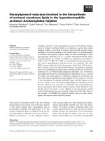
shown in Fig. 1, ATDC5 cells and human primary chondro-
cytes did not accumulate nitrites when stimulated with leptin
alone; however, leptin was able to increase significantly nitrite
accumulation induced by IL-1 when cells were co-stimulated
with both cytokines (Fig 1a,c). This result was confirmed in
terms of protein expression. Indeed, a clear-cut increase in lev-
els of NOS type II protein was observed when cells were co-
stimulated with leptin and IL-1 (Fig. 1b).
To confirm whether NO formation was produced via NOS type
II, ATDC5 cells and human chondrocytes were incubated for
48 hours with both cytokines in the presence of the NOS type
II inhibitor aminoguanidine (1 mmol/l), added 1 hour before
cytokine administration. Aminoguanidine completely inhibited
nitrite accumulation in the culture supernatant of human pri-
mary chondrocytes (Fig. 1c) and ATDC5 cells (Fig. 1d).
Janus kinase-2 inhibition blocks leptin/IL-1 induced
nitric oxide production and nitric oxide synthase type II
protein expression
We also investigated the role played by JAK2 in nitrite produc-
tion evoked by co-stimulation with leptin and IL-1 by using tyr-
phostin AG490. This JAK2 inhibitor, added 1 hour before
cytokine co-stimulation, completely blocked nitrite production
(Fig. 2a). This result was confirmed in terms of protein expres-
sion, because cell pretreatment with tyrphostin AG490 signif-
icantly decreased NOS II protein expression in leptin/IL-1 co-
stimulated cells (Fig. 2d). Intriguingly, tyrphostin AG490 was
also able to inhibit nitrite accumulation induced by IL-1 alone,
suggesting that leptin synergizes with fundamental pathways
Available online />R585
in IL-1 responses. To gain further insights into the involvement
of JAK2, Tkip (a 12-mer SOCS-1 mimetic peptide that binds
to the autophosphorylation site of JAK2) was added to ATDC5
cells 1 hour before they were stimulated with leptin or IL-1, or
both cytokines. Tkip at 50 µmol/l was able to blunt completely
leptin/IL-1 induced nitrite accumulation and NOS II protein
expression (Fig. 2b,e). A lipophilic irrelevant peptide, MuIFN-
γ
95–125
(AKFEVNNPQVQRQAFNELIRVVHQLLPESSL), was
used as control. Intriguingly, Tkip was also able to inhibit, in a
dose–response manner, nitrite accumulation and NOS II pro-
tein expression in ATDC5 cells stimulated with IL-1 alone (Fig.
2c,e).
Effect of the specific signalling pathways inhibitors
LY294002, PD098059 and SB203580 on leptin/IL-1 co-
stimulation
In order to define the signalling pathway involved in the syner-
gistic induction of NOS type II mediated by co-stimulation with
leptin and IL-1 in cultured ATDC5 cells, we evaluated the
effects of specific pharmacological inhibitors on other kinases,
specifically PI3K, MEK-1 and p38 kinase.
We first investigated the effect of a specific inhibitor of PI3K,
namely LY294002 (1, 2.5, 5 and 10 µmol/l) on leptin/IL-1
induced NO production. The addition of LY294002 1 hour
before cytokine co-stimulation resulted in significant and dose-
dependent decreases in NO production and NOS type II pro-
tein expression (Fig. 3a,a1).
In order to test whether MEK-1 (the mitogen-activated protein
kinase [MAPK] kinase involved in extracellular signal-regulated
kinase [ERK]-1 and ERK-2 phosphorylation/activation) partici-
pates in NOS type II induction via leptin/IL-1 co-stimulation,
we used the specific MEK-1 inhibitor PD98059. When this
inhibitor was added 1 hour before cytokine co-stimulation, sig-
nificant dose-dependent decreases in NO production and
NOS II protein expression were observed (Fig. 3b,b1).
Figure 1
Leptin synergizes with IL-1 in inducing nitric oxide synthase (NOS) type IILeptin synergizes with IL-1 in inducing nitric oxide synthase (NOS) type II. Synergistic effect of leptin (OB) on nitrite (NO
2
-
) accumulation and NOS
type II protein expression induced by IL-1. Stimulations were conducted in serum-free conditions (a,b) in ATDC5 chondrogenic cells and (c) in
human primary chondrocytes. NO
2
-
accumulation is selectively inhibited by aminoguanidine (AG) both in (d) ATDC5 cells and in (panel c) human pri-
mary chondrocytes. Values are expressed as mean ± standard error of the mean. WB, western blot.
Arthritis Research & Therapy Vol 7 No 3 Otero et al.
R586
Finally, because it has been shown that p38 kinase is involved
in apoptotic processes induced by NO in chondrocytes, we
tested whether this MAPK is also involved in NOS type II syn-
ergistic activation stimulated by leptin/IL-1. For this purpose,
we used the specific p38 kinase inhibitor SB203580. Addition
of this inhibitor 1 hour before leptin/IL-1 co-stimulation caused
significant and dose-dependent decreases in NO production
and NOS II protein expression (Fig. 3c,c1 [lower panel]).
Leptin synergism does not depend on chondrocyte
differentiation state
In order to determine whether leptin/IL-1 synergism and its sig-
nalling pathway depend on the differentiation state of chondro-
cytes, we conducted similar experiments in mature and
hypertrophic chondrocytes. We differentiated ATDC5 cells
(see Materials and methods, above) into mature and hyper-
trophic chondrocytes, and tested co-stimulation and treat-
ments with all specific inhibitors. Nitrite accumulation,
evaluated in 15-day (mature) and in 21-day (hypertrophic) dif-
ferentiated ATDC5 cells at 24 and 48 hours after treatment,
was similar to that observed in the ATDC5 chondrogenic
undifferentiated cell line (Fig. 4a–d). Note that in order to eval-
uate the involvement of PI3K, in some experiments we also
used wortmannin at 10 µmol/l (a classical but not very specific
PI3K inhibitor), yielding results similar to those obtained with
LY294002.
Finally, a similar pattern was observed in human cultured pri-
mary chondrocytes. In these cells, leptin induced a strong
increase in nitrite accumulation over that induced by IL-1, and
Figure 2
Janus kinase (JAK)2 inhibition blocks leptin/IL-1-induced nitric oxide (NO) production and nitric oxide synthase (NOS) type II protein expressionJanus kinase (JAK)2 inhibition blocks leptin/IL-1-induced nitric oxide (NO) production and nitric oxide synthase (NOS) type II protein expression.
Effect of tyrphostin AG490 and Tkip on NO production and NOS II protein expression. The effect of tyrphostin AG490 was evaluated in terms of (a)
nitrite accumulation in ATDC5 cells stimulated with leptin and IL-1, and in terms of (d) NOS II protein expression. The effect of Tkip was evaluated by
nitrite accumulation in (b) leptin/IL-1 ATDC5 co-stimulated cells and in (c) IL-1 stimulated cells (panel c). (e) Effect of Tkip on NOS type II protein
expression in leptin/IL-1 co-stimulated cells.
Available online />R587
the synergistic response was significantly inhibited by
tyrphostin AG490, wortmannin, LY294002, PD98059 and
SB203580 (Fig. 5).
Effect of leptin/IL-1 co-stimulation on nitric oxide
synthase type II RNA expression
We finally studied NOS II mRNA expression in order to deter-
mine whether NO increase/inhibition was due to modulation of
NOS type II mRNA expression. As shown in Fig. 6, NOS type
II mRNA, evaluated using real-time PCR, was strongly
expressed when cells were co-stimulated with leptin plus IL-1,
and this expression was significantly reduced by tyrphostin
AG490, wortmannin, LY294002, PD098059 and SB203580.
Discussion
In the present study we investigated the effect of leptin on NO
production stimulated by IL-1. We found that leptin had a syn-
ergistic effect in the ATDC5 murine chondrogenic cell line, in
differentiated mature and hypertrophic ATDC5 chondrocytes,
and in human primary chondrocytes.
Leptin has been classified as a cytokine-like hormone,
because of its structure and the homology of its receptors with
members of the class I cytokine receptor superfamily. A proin-
flammatory role for leptin has previously been proposed. Sev-
eral data show that leptin levels are increased by
proinflammatory cytokine administration and in animal models
of acute inflammation [9]. In addition, leptin regulates not only
humoral but also cellular immune responses in antigen-
induced arthritis models [20]. Nevertheless, there are only few
Figure 3
Involvement of phosphatidylinositol 3-kinase (PI3K), mitogen-activated protein kinase kinase (MEK)-1 and p38-kinase in leptin/IL-1-induced nitric oxide synthase (NOS)Involvement of phosphatidylinositol 3-kinase (PI3K), mitogen-activated protein kinase kinase (MEK)-1 and p38-kinase in leptin/IL-1-induced nitric
oxide synthase (NOS). Dose-dependent effect of (a,a1) LY294002, (b,b1) PD098059 and (c,c1) SB203580 on nitrite (NO
2
-
) production and NOS
type II protein expression in stimulated and unstimulated ATDC5 cells. Stimulations were conducted in serum-free conditions. Each inhibitor was
added 1 hour before cytokine co-stimulation. Values are expressed as mean ± standard error of the mean. OB, leptin; WB, western blot.
Arthritis Research & Therapy Vol 7 No 3 Otero et al.
R588
reports of a direct action of leptin at the cellular level in carti-
lage [14,15].
NO controls a variety of cartilage functions, including loss of
chondrocyte phenotype, chondrocyte apoptosis, and extracel-
lular matrix degradation [2,3]. NOS type II is mainly expressed
by immune cells in response to a wide range of proinflamma-
tory cytokines [21,22]. In vitro, human articular cartilage is able
to produce large amounts of NO [23], which can be enhanced
by proinflammatory cytokines. In addition, NO production can
be significantly increased by the presence of leptin, as shown
in our previous work [15] and in the present study.
Here, we show that the IL-1 induced production of NO by
ATDC5 murine chondrocytes and by human chondrocytes is
significantly enhanced by leptin. It is noteworthy that, apart
from blood, several sources of leptin and IL-1 have been iden-
tified in or around the joints in pathological conditions. IL-1 is
produced by inflamed synovium and periarticular fat pad [24].
Interestingly, multipotent stromal cells from the infrapatellar fat
produce leptin [25]. In addition, osteoarthritic human chondro-
cytes produce leptin, and leptin administration in rats induces
over-expression of this hormone by articular chondrocytes
[13]. Thus, in patients with inflammatory synovitis or osteoar-
thritis, there is a unique microenvironment in the cartilage char-
acterized by elevated levels of both leptin and IL-1, due not
only to local production but also to systemic increase
[10,13,26]. It is conceivable that in this scenario leptin plays a
significant proinflammatory role, as suggested by the findings
presented here. Of further interest is our previous report [15]
of the co-stimulatory effect of leptin and IFN-γ at the chondro-
cyte level.
Figure 4
Leptin synergism does not depend upon chondrocyte differation stateLeptin synergism does not depend upon chondrocyte differation state. Effect of different inhibitors on nitrite (NO
2
-
) accumulation in 15-day differen-
tiated ATDC5 cells stimulated or not with leptin, alone or in combination with IL-1, during (a) 24 and (b) 48 hours. The effect of inhibitors was also
evaluated in 21-day differentiated ATDC5 cells, after (c) 24 or (d) 48 hours of stimulation with leptin and IL-1 (alone or in combination). Values are
expressed as mean ± standard error of the mean. OB, leptin.
Available online />R589
We previously established that the early event in leptin/IFN-γ
synergistic NOS type II activation was the involvement of JAK2
[15]; the present results confirm that JAK2 activation is also an
early step in leptin/IL-1 induced NOS type II co-stimulation.
The fact that tyrphostin AG490 blocks the leptin/IL-1
response implies that leptin synergizes with critical pathways
in IL-1 response. It was surprising that tyrphostin AG490 also
blocked the response to IL-1 alone, because JAK2 is not
known to be required for IL-1 receptor transduction, and so
one would expect the effect of tyrphostin AG490 to be partial.
However, our results are in agreement with those reported by
other investigators [27,28].
We also used Tkip in our experiments; Tkip is a 12-mer
SOCS-1 mimetic lipophilic peptide (WLVFFVIFYFFR) that
inhibits JAK2 autophosphorylation [29]. Interestingly, the
behaviour of this peptide was similar to that of tyrphostin
AG490 in terms of NOS II inhibition. It is conceivable that this
peptide, because of its SOCS-1 mimetic properties, could
inhibit IL-1/Toll-like receptor function in chondrocytes. SOCS-
1 is a negative regulator of lipopolysaccharide-induced macro-
phage activation [30,31] and has been shown to bind to IL-1
receptor associated kinase [32]. This disrupts the cascade
that leads to nuclear factor-κB (NF-κB) signalling and causes
NOS inhibition. Of note, it has been demonstrated that tyr-
phostin AG490 inhibits IL-1 induced NF-κB activation in con-
centrations that also inhibit NOS II mRNA and protein
synthesis. These findings suggest that JAK2 is required for
NF-κB activation, which in turn mediates IL-1 induced NOS II
expression in chondrocytes [28].
To gain further insights into the mechanism by which leptin,
together with IL-1, promotes NO production, we evaluated the
roles played by downstream signalling cascades using spe-
cific pharmacological inhibitors. First, we analyzed the involve-
ment of PI3K. The role played by this kinase in the activation of
NOS type II is quite controversial and remains the subject of
debate. A number of studies support the view that PI3K activ-
ity down-regulates NOS type II, but there are several caveats
Figure 5
Leptin acts synergistically with IL-1 in human primary chondrocytesLeptin acts synergistically with IL-1 in human primary chondrocytes.
Nitrite (NO
2
-
) accumulation in leptin (OB)/IL-1 co-stimulated human pri-
mary chondrocytes. Stimulations were conducted in serum-free condi-
tions in the presence or absence of tyrphostin AG490, wortmannin,
LY294002, PD98059 and SB203580 inhibitors. Values are expressed
as mean ± standard error of the mean.
Figure 6
Effect of leptin/IL-1 co-stimulation on nitric oxide synthase (NOS) type II mRNA expressionEffect of leptin/IL-1 co-stimulation on nitric oxide synthase (NOS) type II
mRNA expression. Real-time RT-PCR analysis of the expression of the
inducible NOS type II mRNA in leptin (OB)/IL-1 co-stimulated ATDC5
cells. Stimulations (24 hours) were conducted in serum-free conditions.
Specific inhibitors were added 1 hour before cytokine co-stimulation.
Values are expressed as mean ± standard error of the mean.
Arthritis Research & Therapy Vol 7 No 3 Otero et al.
R590
to this view. For instance, insulin-like growth factor-II
stimulates NOS type II expression and activity in myoblasts via
a PI3K-dependent mechanism involving IκBα degradation and
increased p65 NF-κB DNA binding activity [33], which is in
agreement with recent evidence indicating that PI3K/protein
kinase B is involved in NF-κB activation [34,35]. In addition,
PI3K homologues (mammalian target of rapamycin/FKBP12–
rapamycin associated protein) have been implicated in the
phosphorylation and activation of NOS type II [36]. It should
therefore be stressed that the interaction between NOS type
II and PI3K may vary depending on the cell model, and so this
interaction may be subject to tissue-specific regulation.
Our results also suggest that ERK-1/2 and p38 kinase play
pivotal roles in the activation of NOS type II mediated by leptin/
IL-1 co-stimulation. We found that ERK-1/2-specific pharma-
cological inhibition significantly decreased NO production
induced by leptin/IL-1 co-stimulation in cultured chondrocytes.
This result is in agreement with previous studies that associ-
ated ERK-1/2 activation with NOS type II induction by a com-
bination of proinflammatory stimuli [37-40].
Finally, we found that the blockade of p38 kinase caused a sig-
nificant decrease in NO production, NOS II mRNA expression
and NOS II protein level. These data are concordant with pre-
vious reports that implicate p38 kinase in NOS type II upregu-
lation in macrophages [41], neural cells [42,43] and
chondrocytes [44].
Synergistic interactions of IL-1 with other soluble factors are
not novel and have been reported in chondrocytes and other
cell types. For instance, IL-1 synergizes with oncostatin M to
induce markedly the expression of matrix metalloproteinase
(MMP)-1, MMP-3, MMP-8 and MMP-13, as well as aggreca-
nase ADAM-TS4 [45]. In addition, a synergistic induction of
MMP-1 by IL-1 and oncostatin M has been observed in human
chondrocytes via a novel mechanism that involves STAT (sig-
nal transducer and activator of transcription) and activator pro-
tein-1 [46].
As far as we are aware, this is the first report that demon-
strates the cooperative interaction between leptin and IL-1 in
the induction of NO production in activated chondrocytes.
Conclusion
The present study shows that in human and ATDC5 murine
cultured chondrocytes, leptin, together with IL-1, significantly
increases the production of NO by a mechanism that implies
upregulation of NOS type II mRNA and protein. In this modu-
lation, an intracellular signalling pathway that involves JAK2
tyrosine kinase, PI3K and two members or the MAPK pathway
(ERK and p38) is at play. The functional interplay of these
pathways may be important for the onset as well as the main-
tenance of inflammatory responses in cartilage.
Competing interests
The author(s) declare that they have no competing interests.
Acknowledgements
This work was supported by grants from Spanish Ministry of Health (FIS
01/3137 and PI-020431). Oreste Gualillo and Francisca Lago are
recipients of a research contract from Spanish Ministry of Health, Insti-
tuto de Salud Carlos III (EXP 00/3051 and 99/3040). Miguel Otero is a
recipient of a predoctoral fellowship funded by Xunta de Galicia. Rocío
Lago is a recipient of a fellowship funded by Instituto de Salud Carlos III
(Red Temática G03/152). We would like to thank Prof. Carlos Dieguez
for his helpful advice and for his continued support during the realization
of this work. The authors are very grateful to Dr Antonio Mera from Rheu-
matology Division and to Dr Jorge Fernadez Noya from Vascular Surgery
Division of Santiago Univeristy Clinical Hospital for helping us in harvest-
ing human tissues.
References
1. Goldring MB: The role of the chondrocyte in osteoathritis.
Arthritis Rheum 2000, 43:1916-1926.
2. Kim SJ, Ju JW, Oh CD, Yoon YM, Song WK, Kim JH, Yoo YJ, Bang
OS, Kang SS, Chun JS: ERK-1/2 and p38 kinase oppositely
regulate nitric oxide-induced apoptosis of chondrocytes in
association with p53, caspase-3 and differentiation status. J
Biol Chem 2002, 277:1332-1339.
3. Sasaki K, Hattori T, Fujisawa T, Takahashi K, Inoue H, Takigawa M:
Nitric oxide mediates interleukin-1-induced gene expression
of matrix metalloproteinases and basic fibroblast growth fac-
tor in cultured rabbit articular chondrocytes. J Biochem (Tokyo)
1998, 123:431-439.
4. Sandoval DA, Davis SN: Leptin: metabolic control and
regulation. J Diabetes Complications 2003, 17:108-113.
5. La Cava A, Matarese G: The weight of leptin in immunity. Nature
Rev Immunol 2004, 4:371-379.
6. Fantuzzi G, Faggioni R: Leptin in the regulation of immunity,
inflammation and hematopoiesis. J Leukoc Biol 2000,
68:437-446.
7. Matarese G, La Cava A: The intricate interface between
immune system and metabolism. Trends Immunol 2004,
25:193-200.
8. Steppan CM, Crawford DT, Chidsey-Frink KL, Ke H, Swick AG:
Leptin is a potent stimulator of bone growth in ob/ob mice.
Regul Pept 2000, 92:73-78.
9. Gualillo O, Eiras S, Lago F, Dieguez C, Casanueva FF: Elevated
serum leptin concentrations induced by experimental acute
inflammation. Life Sci 2000, 67:2433-2441.
10. Felson DT, Anderson JJ, Naimark A, Walker AM, Meenan RF:
Obesity and knee osteoarthritis: the Framingham Study. Ann
Intern Med 1988, 109:18-24.
11. Karlson EW, Mandl LA, Aweh GN, Sangha O, Liang MH, Grod-
stein F: Total hip replacement due to osteoarthritis: the impor-
tance of age, obesity an other modifiable risk factors. Am J
Med 2003, 114:93-98.
12. Oliveria SA, Felson DT, Cirillo PA, Reed JI, Walker AM: Body
weight, body mass index and incident symptomatic osteoar-
thritis of the hand, hip and knee. Epidemiology 1999,
10:161-166.
13. Dumond H, Presle N, Terlain B, Mainard D, Loeuille D, Netter P,
Pottie P: Evidence for a key role of leptin in osteoarthritis.
Arthritis Rheum 2003, 48:3118-3129.
14. Figenschau Y, Knutsen G, Shahazeydi S, Johansen O, Svein-
bjornsson B: Human articular chondrocytes express functional
leptin receptors. Biochem Biophys Res Commun 2001,
287:190-197.
15. Otero M, Gómez-Reino JJ, Gualillo O: Synergistic induction of
nitric synthase type II: in vitro effect of leptin and interferon-γ
in human chondrocytes and ATDC5 chondrogenic cells. Arthri-
tis Rheum 2003, 48:404-409.
16. Dayer JM: The pivotal role of interleukin-1 in the clinical mani-
festations of rheumatoid arthritis. Rheumatology 2003,
42(Suppl):ii3-ii10.
Available online />R591
17. Goldring SR: Pathogenesis of bone and cartilage destruction
in rheumatoid arthritis. Rheumatology 2003,
42(Suppl):ii11-ii16.
18. Akiyama H, Shukunami C, Nakamura T, Hiraki Y: Differential
expressions of BMP family genes during chondrogenic differ-
entiation of mouse ATDC5 cells. Cell Struct Funct 2000,
25:195-204.
19. Thomas DP, Sunters A, Gentry A, Grigoriadis AE: Inhibition of
chondrocyte differentiation in vitro by constitutive and induci-
ble overexpression of the c-fos proto-oncogene. J Cell Sci
2000, 113:439-450.
20. Busso N, So A, Chobaz-Peclat V, Morard C, Martinez-Soria E, Tal-
abot-Ayer D, Gabay C: Leptin signaling deficiency impairs
humoral and cellular immune responses and attenuates
experimental arthritis. J Immunol 2002, 168:875-882.
21. Brune B, von Knethen A, Sandau KB: Nitric oxide and its role in
apoptosis. Eur J Pharmacol 1998, 351:261-272.
22. Del Carlo M, Loeser RF: Nitric oxide-mediated chondrocyte cell
death requires the generation of additional reactive oxygen
species. Arthritis Rheum 2002, 46:394-403.
23. Vuolteenaho K, Moilanen T, Al-Saffar N, Knowles RG, Moilanen E:
Regulation of the nitric oxide production resulting from the
glucocorticoid-insensitive expression of NOS type II in human
osteoarthritic cartilage. Osteoarthritis Cartilage 2001,
9:597-605.
24. Ushiyama T, Chano T, Inoue K, Matsusue Y: Cytokine production
in the infrapatellar fat pad: another source of cytokines in knee
synovial fluids. Ann Rheum Dis 2003, 62:108-112.
25. Wickham MQ, Erickson GR, Gimble JM, Vail TP, Guilak F:
Multipotent stromal cells derived from the infrapatellar fat pad
of the knee. Clin Orthop 2003, 412:196-212.
26. Bokarewa M, Bokarewa D, Hultgren O, Tarkowski A: Leptin con-
sumption in the inflamed joints of patients with rheumatoid
arthritis. Ann Rheum Dis 2003, 62:952-956.
27. Doi M, Shichiri M, Katsuyama K, Ishimaru S, Hirata Y: Cytokine-
activated Jak-2 is involved in inducible nitric oxide synthase
expression independent from NF-kappaB activation in vascu-
lar smooth muscle cells. Atherosclerosis 2002, 160:123-132.
28. Mendes AF, Caramona MM, Carvalho AP, Lopes MC: Role of
mitogen-activated protein kinases and tyrosine kinases on IL-
1-Induced NF-kappaB activation and iNOS expression in
bovine articular chondrocytes. Nitric Oxide 2002, 6:35-44.
29. Flowers LO, Johnson HM, Mujtaba MG, Ellis MR, Haider SM, Sub-
ramaniam PS: Characterization of a peptide inhibitor of Janus
kinase 2 that mimics suppressor of cytokine signaling 1
function. J Immunol 2004, 172:7510-7518.
30. Kinjyo I, Hanada T, Inagaki-Ohara K, Mori H, Aki D, Ohishi M, Yosh-
ida H, Kubo M, Yoshimura A: SOCS1/JAB is a negative regulator
of LPS-induced macrophage activation. Immunity 2002,
17:583-5891.
31. Nakagawa R, Naka T, Tsutsui H, Fujimoto M, Kimura A, Abe T, Seki
E, Sato S, Takeuchi O, Takeda K, et al.: SOCS-1 participates in
negative regulation of LPS responses. Immunity 2002,
17:677-687.
32. Akira S, Takeda K: Toll-like receptor signalling. Nat Rev Immunol
2004, 4:499-511.
33. Kaliman P, Canicio J, Testar X, Palacin M, Zorzano A: Insulin-like
growth factor-II, phosphatidylinositol 3-kinase, nuclear factor-
kappaB and inducible nitric-oxide synthase define a common
myogenic signaling pathway. J Biol Chem 1999,
274:17437-17444.
34. Sizemore N, Leung S, Stark GR: Activation of phosphatidylinosi-
tol 3-kinase in response to interleukin-1 leads to phosphoryla-
tion and activation of the NF-kappaB p65/RelA subunit. Mol
Cell Biol 1999, 19:4798-4805.
35. Kane LP, Shapiro VS, Stokoe D, Weiss A: Induction of NF-kap-
paB by the Akt/PKB kinase. Curr Biol 1999, 9:601-604.
36. Salh B, Wagey R, Marotta A, Tao JS, Pelech S: Activation of
phosphatidylinositol 3-kinase, protein kinase B, and p70 S6
kinases in lipopolysaccharide-stimulated Raw 264.7 cells: dif-
ferential effects of rapamycin, Ly294002, and wortmannin on
nitric oxide production. J Immunol 1998, 161:6947-6954.
37. Blanchette J, Jaramillo M, Olivier M: Signalling events involved in
interferon-gamma-inducible macrophage nitric oxide
generation. Immunology 2003, 108:513-522.
38. Kan H, Xie Z, Finkel MS: TNF-alpha enhances cardiac myocyte
NO production through MAP kinase-mediated NF-kappaB
activation. Am J Physiol 1999, 277:H1641-H1646.
39. Kristof AS, Marks-Konczalik J, Moss J: Mitogen-activated protein
kinases mediate activator protein-1-dependent human induc-
ible nitric-oxide synthase promoter activation. J Biol Chem
2001, 276:8445-8452.
40. Han IO, Kim HS, Kim HC, Joe EH, Kim WK: Synergistic expres-
sion of inducible nitric oxide synthase by phorbol ester and
interferon-gamma is mediated through NF-kappaB and ERK in
microglial cells. J Neurosci Res 2003, 73:659-669.
41. Ajizian SJ, English BK, Meals EA: Specific inhibitors of p38 and
extracellular signal-regulated kinase mitogen-activated pro-
tein kinase pathways block inducible nitric oxide synthase and
tumor necrosis factor accumulation in murine macrophages
stimulated with lipopolysaccharide and interferon-gamma. J
Infect Dis 1999, 179:939-944.
42. Bhat NR, Zhang P, Lee JC, Hogan EL: Extracellular signal-regu-
lated kinase and p38 subgroups of mitogen-activated protein
kinases regulate inducible nitric oxide synthase and tumor
necrosis factor-alpha gene expression in endotoxin-stimu-
lated primary glial cultures. J Neurosci 1998, 18:1633-1641.
43. Da Silva J, Pierrat B, Mary JL, Lesslauer W: Blockade of p38
mitogen-activated protein kinase pathway inhibits inducible
nitric-oxide synthase expression in mouse astrocytes. J Biol
Chem 1997, 272:28373-28380.
44. Badger AM, Cook MN, Lark MW, Newman-Tarr TM, Swift BA, Nel-
son AH, Barone FC, Kumar S: SB 203580 inhibits p38 mitogen-
activated protein kinase, nitric oxide production, and inducible
nitric oxide synthase in bovine cartilage-derived chondrocytes.
J Immunol 1998, 161:467-473.
45. Koshy PJ, Lundy CJ, Rowan AD, Porter S, Edwards DR, Hogan A,
Clark IM, Cawston TE: The modulation of matrix metalloprotei-
nase and ADAM gene expression in human chondrocytes by
interleukin-1 and oncostatin M: a time-course study using
real-time quantitative reverse transcription-polymerase chain
reaction. Arthritis Rheum 2002, 46:961-967.
46. Catterall JB, Carrere S, Koshy PJ, Degnan BA, Shingleton WD,
Brinckerhoff CE, Rutter J, Cawston TE, Rowan AD: Synergistic
induction of matrix metalloproteinase 1 by interleukin-1alpha
and oncostatin M in human chondrocytes involves signal
transducer and activator of transcription and activator protein
1 transcription factors via a novel mechanism. Arthritis Rheum
2001, 44:2296-2310.