Báo cáo y học: "Dynamic magnetic resonance of the wrist in psoriatic arthritis reveals imaging patterns similar to those of rheumatoid arthritis" pps
Bạn đang xem bản rút gọn của tài liệu. Xem và tải ngay bản đầy đủ của tài liệu tại đây (242.88 KB, 7 trang )
Open Access
Available online />R725
Vol 7 No 4
Research article
Dynamic magnetic resonance of the wrist in psoriatic arthritis
reveals imaging patterns similar to those of rheumatoid arthritis
Marco A Cimmino
1
, Massimiliano Parodi
1
, Stefania Innocenti
2
, Giulia Succio
3
, Simone Banderali
3
,
Enzo Silvestri
3
and Giacomo Garlaschi
3
1
Clinica Reumatologica, Dipartimento di Medicina Interna e Specialità Mediche, Università di Genova, Italy
2
ESAOTE Biomedica, Genova, Italy
3
Sezione di Diagnostica Radiologica, Dipartimento di Medicina Sperimentale, Università di Genova, Italy
Corresponding author: Marco A Cimmino,
Received: 14 Dec 2004 Revisions requested: 17 Jan 2005 Revisions received: 2 Mar 2005 Accepted: 7 Mar 2005 Published: 1 Apr 2005
Arthritis Research & Therapy 2005, 7:R725-R731 (DOI 10.1186/ar1734)
This article is online at: />© 2005 Cimmino et al.; licensee BioMed Central Ltd.
This is an Open Access article distributed under the terms of the Creative Commons Attribution License ( />2.0), which permits unrestricted use, distribution, and reproduction in any medium, provided the original work is properly cited.
Abstract
This dynamic magnetic resonance imaging (MRI) study is
concerned with a prospective evaluation of wrist synovitis in
patients with psoriatic arthritis (PsA) in comparison with patients
with rheumatoid arthritis (RA) and healthy controls. Fifteen
consecutive patients with PsA, 49 consecutive patients with RA,
30 RA patients matched for disease severity with those with
PsA, and 8 healthy controls were studied. MRI was performed
with a low-field (0.2T), extremity-dedicated machine. After an
intravenous bolus injection of gadolinium-
diethylenetriaminepentaacetic acid, 20 consecutive fast spin-
echo axial images of the wrist were obtained every 18 s. The
enhancement ratio was calculated both as rate of early
enhancement (REE), which shows the slope of the curve of
contrast uptake per second during the first 55 s, and as relative
enhancement (RE), which indicates the steady state of
enhancement. The REE was 1.0 ± 0.6 in patients with PsA, 1.6
± 0.7 in consecutive patients with RA, and 0.1 ± 0.1 in controls
(p <0.001). The RE was 87.1 ± 39.2 in patients with PsA, 125.8
± 48.0 in consecutive RA patients, and 15.5 ± 19.2 in controls
(p <0.001). However, the same figures in matched RA patients
were 1.3 ± 0.7 and 107.3 ± 48.2, respectively (not significant in
comparison with PsA). Rheumatoid-like PsA and oligoarticular
PsA did not differ from each other in terms of synovial
enhancement. Dynamic MRI shows the same pattern of synovitis
in patients with PsA and RA when the two groups are matched
for disease severity. This technique cannot be used to
differentiate PsA from RA. However, REE and RE were
significantly higher in PsA than in normal controls, with only one
instance of overlap between values found for the two groups.
Introduction
Psoriatic arthritis (PsA), defined as the occurrence of seroneg-
ative arthritis and psoriasis, is a debated entity. The reasons for
this uncertainty include disparity in the subgroups identified in
the original description [1], lack of a validated case definition
of PsA, possible inclusion of patients with enthesitis but with-
out clear arthritis, the elusive link between skin and joint dis-
eases, and similarities with rheumatoid arthritis (RA). It is not
known if PsA and RA are completely distinct diseases, or if
PsA is a form of RA modified by the coexisting psoriasis. This
second hypothesis is brought into doubt by the original obser-
vation that articular involvement is chiefly symmetric and pol-
yarticular in RA, but oligoarticular and asymmetric in PsA.
However, subsequent studies have shown that the most com-
mon subset of PsA is a symmetric polyarthritis resembling RA
[2]. In particular, oligoarticular disease may be a characteristic
of PsA mainly at presentation [3], and symmetry is a function
of the number of joints involved but not of the type of arthritis
[4]. On average, however, PsA is characterized by a milder
degree of synovitis than RA, with only 8% of patients develop-
ing erosions of the hand. Therefore, therapy with disease-mod-
ifying anti-rheumatic drugs is rarely needed [5].
Magnetic resonance imaging (MRI) has helped in the evalua-
tion of PsA by suggesting that the primary site of inflammation
is extrasynovial and that synovial inflammation may be a
ANOVA = analysis of variance; Gd-DTPA = gadolinium-diethylenetriaminepentaacetic acid; MRI = magnetic resonance imaging; ns = not significant;
PsA = psoriatic arthritis; RA = rheumatoid arthritis; RE = relative enhancement; REE = rate of early enhancement; ROI = region of interest.
Arthritis Research & Therapy Vol 7 No 4 Cimmino et al.
R726
secondary phenomenon [6]. In addition to morphologic stud-
ies of the joint, MRI may be used also to evaluate synovial
membrane inflammation through a dynamic, contrast-
enhanced technique. By this method, we evaluated gadolin-
ium perfusion of the synovial membrane to differentiate active
RA from RA in remission [7]. In the present paper, the same
technique has been used to study patients with PsA. The main
goals of the study were to investigate whether synovitis differs
between PsA and RA, and, if so, whether the difference is
intrinsic to the type of arthritis or is due to the severity and
duration of the disease.
Materials and methods
Patients
Fifteen consecutive patients with PsA, defined as the simulta-
neous occurrence of active arthritis and psoriasis, were pro-
spectively studied. Eight patients had a polyarticular
rheumatoid-like pattern of arthritis and seven had monoarthritis
or oligoarthritis, associated in one patient with axial involve-
ment. Dynamic MRI data for these patients were compared
with those of 49 consecutive patients with active RA (group I),
diagnosed according to the criteria of the American Rheuma-
tism Association [8]. An additional group of 30 patients with
RA, matched with those with PsA in terms of age, disease
duration, and number of involved joints, was considered
(group II). These patients were identified in ongoing follow-up
studies. Because of difficulties in matching, nine of these
patients were recruited from groupI RA patients. All the
patients considered in this study were seen at the Rheumato-
logical Clinic of the University of Genoa, Italy, either in the clin-
ical ward or as outpatients, and had clinical inflammatory
involvement of at least one wrist. Clinical parameters, such as
duration of arthritis, early-morning stiffness, fatigue, number of
tender and swollen joints, and type of treatment, were evalu-
ated before MRI. At the same time, blood was drawn for the
determination of the erythrocyte sedimentation rate (Wester-
gren method), C-reactive protein, and IgM rheumatoid factor
by standard laboratory methods. In addition, a control group of
eight healthy volunteers, who did not report any history of joint
disease and were negative for signs of arthritis on clinical
examination, was also studied. All subjects gave their informed
consent to the protocol, which had been approved by the eth-
ics committee of the Department of Internal Medicine of the
University of Genoa. Demographic and clinical characteristics
of the three groups of patients and the group of controls are
reported in Table 1.
Methods
MRI of the wrist was performed with a low-field (0.2T), extrem-
ity-dedicated machine (Artoscan™, Esaote, Genoa, Italy)
equipped with a permanent magnet and with a dedicated
hand-and-wrist coil 13 cm in diameter, as previously described
[6]. The hand was fixed in neutral position and the fingers in
extended position with the thumb up, by the application of
several cushions. The field of view was 120 mm and allowed
the evaluation of the carpal bones, the metacarpal bases, and
the distal radius and ulna. Slice thickness was 5 mm and the
interslice gap was 0.3 mm. The sequence used was a spin
echo (TR/TE = 100/16 ms, matrix = 160 × 128, FOV = 150
× 150), which was acquired in the axial plane. In patients with
arthritis, the more severely affected wrist was examined. In
patients with equal involvement of the wrists, and in normal
controls, the right wrist was examined. Patients and controls
were instructed to avoid intense activity involving the wrists in
the 24 hours preceding the examination.
After the wrist was positioned in the gantry, the first image was
acquired. Then an intravenous bolus injection of 0.2 ml/kg of
Gd-DTPA (gadolinium-diethylenetriaminepentaacetic acid)
(Omniscan, Schering, Germany) was given manually in 30 s
through a 21-mm butterfly needle into a cubital vein. Twenty
consecutive fast images of three slices of the wrist, the first of
which was positioned tangential to the radius, were repeated
every 18 s thereafter. The rate of enhancement was evaluated
by a radiologist on the slice that showed the highest visual
enhancement. It was calculated as ∆ on a small, elliptical
region of interest (ROI) of synovial membrane of approximately
25 mm
2
positioned in the area of highest visual enhancement
(Fig. 1). Entheses and synovial sheaths were not included in
the ROI. In healthy controls, the synovial membrane was more
difficult to identify. In these cases, the area where the syn-
ovium was thought to be located was chosen on the basis of
anatomic landmarks and comparison of pre- and post-
enhancement images. For this reason, the elliptical ROI was
usually smaller in controls than in patients. This corresponded
usually to the median and dorsal area of the wrist.
The images were processed blind to the clinical and laboratory
findings. The enhancement ratio was calculated both as rate
of early enhancement (REE) per second during the first 55 s
according to the formula
REE
55
= (S
55
- S
0
)/(S
0
× 55) × 100%
and as relative enhancement (RE) at t seconds according to
the formula
RE
t
= (S
t
- S
0
)/S
0
× 100%
where S
0
and S
t
are the signal-to-noise ratios, before and t
seconds after contrast injection, calculated as ratio between
the signal measured in the ROI and the standard deviation of
the background noise. The study of enhancement after 55 s
was chosen because it showed maximal enhancement differ-
ence between knees with clinically inactive or active disease
in a previous study [9]. In addition, the signal was normalized
to the bone to reduce noise. The REE shows the slope of the
curve of contrast uptake tangential to the α angle and is
steeper if inflammation is higher. The RE indicates the steady
state of enhancement. The intra- and inter-observer mean per-
Available online />R727
centage variations for REE were 3.9% (range 0.5% to 14.3%)
and 2.8% (range 0% to 5.1%), respectively, in 18 wrists. Intra-
and inter observer mean percentage variation for RE were
1.9% (range 0% to 9.3%) and 1.9% (range 0.05% to 6.4%)
(manuscript in preparation).
Statistical evaluation
Means were compared by the Student's t-test or by one-way
analysis of variance (ANOVA) if their distribution was normal
and by the Wilcoxon test with Mann–Whitney correction or
Kruskal–Wallis ANOVA when the distribution was nonpara-
metric. Frequencies were compared using the Fisher exact
test. Correlations were calculated by the Pearson or Spear-
man rank tests. P values less than 0.05 were considered
significant.
Results
Comparison between PsA and consecutive RA patients
The demographic and clinical data for patients and controls
are reported in Table 1 in comparison with RA patients of
group I. PsA patients had a less skewed female-to-male ratio,
had a lower tender joint count, and were less frequently posi-
tive for IgM rheumatoid factor. All the other clinical character-
istics were similar in the two groups. Controls were younger
than the patients of the other two groups. Dynamic MRI was
performed in all subjects without causing any discomfort or
adverse events. The mean duration of the complete examina-
tion was 15 min. REE and RE were significantly different in the
three groups (p <0.001) (Table 1; Fig. 2). The values were
highest for the group with RA, followed by that with PsA and
controls. In PsA and RA patients, the REE ranged from 0.14%
to 2.13% per second, and from 0.08% to 3.49% per second,
respectively. In controls, it ranged from -0.02% to 0.37% per
second.
Nonsteroidal anti-inflammatory drugs were being used by 14
(93.3%) of 15 PsA patients, and by 40 (81.6%) of 49 RA
patients (ns). Sulphasalazine was used in 7 (47.7%) of 15 PsA
patients and in 17 (34.7%) of 49 RA patients (ns). Conversely,
patients with PsA were treated less frequently with prednisone
(4 of 15, or 26.7%, vs 36 of 49, or 73.5%; P = 0.002) or meth-
otrexate (2 of 15, or 13.3%, vs 21 of 49, or 42.9%, P = 0.06)
than patients with RA. Dosages were similar in the two groups
of patients (data not shown).
Comparison between PsA and matched RA patients
To exclude the possibility that a lower disease activity in PsA
patients could account for the observed dynamic MRI differ-
ence, 2 RA patients were matched for age and number of ten-
der and swollen joints to each of the 15 patients with PsA. The
clinical and laboratory characteristics of the PsA patients and
of the new RA control group (group II) were similar, with no
statistically significant difference. Only positivity for IgM rheu-
matoid factor was higher in RA patients (P < 0.05) (Table 1).
Table 1
Demographic, clinical, and laboratory characteristics of patients with psoriatic arthritis, rheumatoid arthritis, and controls
Characteristic Psoriatic arthritis Rheumatoid arthritis
(group I)
Matched rheumatoid arthritis
(group II)
Controls
Number of patients 15 49 30 8
Age (years) 55.7 ± 10.7 57.6 ± 14.6 56.3 ± 16.3 38.1 ± 21.9
Sex (women/men)* 8/7 42/7 20/10 4/4
Disease duration (months) 63.5 ± 62.7 93.5 ± 98.7 78.2 ± 86.2 NA
Morning stiffness (minutes) 70.4 ± 55.7 96.2 ± 96.3 76.8 ± 92.1 NA
Number of tender joints* 8.0 ± 5.5 13.7 ± 9.4 9.1 ± 7.3 0
Number of swollen joints 5.3 ± 4.4 8.0 ± 5.8 5.0 ± 3.9 0
Ritchie index 8.3 ± 5.7 11.6 ± 6.9 7.0 ± 5.1 0
ESR (mm/h) 34.7 ± 21.8 49.1 ± 31.6 45.5 ± 35.7 ND
CRP (mg/l) 30.4 ± 39.4 30.3 ± 34.4 26.6 ± 36.1 ND
Number of patients with IgM
rheumatoid factor**
1 (6.7%) 33 (67.3%) 18 (60%) ND
Rate of early enhancement
§
1.0 ± 0.6 1.6 ± 0.7 1.3 ± 0.7 0.1 ± 0.1
Relative enhancement
§
87.1 ± 39.2 125.8 ± 48.0 107.3 ± 48.2 15.5 ± 19.2
Values are expressed as means ± standard deviations except for sex and IgM rheumatoid factor, for which the percentage of positive patients is
reported. *P < 0.05 for the comparison between PsA and groupI RA; **P < 0.001 for the comparison between PsA and groupI RA and P < 0.05
for the comparison between PsA and groupII RA;
§
P < 0.001 by one-way analysis of variance between PsA patients, groupI RA patients, and
controls.
Arthritis Research & Therapy Vol 7 No 4 Cimmino et al.
R728
Treatment in the matched RA group (II) was similar to that
seen in the groupII RA patients. The use of nonsteroidal anti-
inflammatory drugs and sulphasalazine was not different in
PsA patients from that in matched RA patients. Patients with
PsA were treated less frequently with prednisone (26.7% vs
70%; P = 0.01) and with methotrexate (13.3% vs 46.6%, P =
0.046) than patients with RA, but dosages were similar.
REE and RE were not different between PsA and RA of similar
severity (Table 1; Fig. 2). Figure 3 compares the mean values
of the curves in the three subgroups, that is, patients with PsA,
groupII RA patients, and healthy controls. Before Gd-DTPA
infusion and for the first 36 s after infusion, the three curves
were almost identical. However, a highly significant difference
in enhancement was seen by ANOVA at all the following time
points (P = 0.003 at t = 156 s, p <0.001 at t = 174 s, and P
< 0.001 thereafter). The curves identified two groups of
patients, one being patients with PsA or groupII RA and the
other being controls.
Correlations between dynamic MRI and clinical and
laboratory findings
REE was 0.8 ± 0.5 in patients with rheumatoid-like PsA and
1.2 ± 0.6 in those with monoarthritis or oligoarthritis (ns). Val-
ues for RE were 78 ± 28.2 and 97.5 ± 49.2, respectively. This
difference was not significant. REE and RE were not
correlated with clinical and laboratory findings in PsA. There
was a tendency to an association between REE and number
of swollen joints in the RA patients of group II (P = 0.07).
Discussion
Dynamic MRI is a promising method for the investigation of
patients with arthritis. In our previous experience, it could dif-
ferentiate patients with active RA from those in remission and
from controls [7]. In another study of arthritic knees, dynamic
gadolinium-enhanced MRI showed increased contrast diffu-
sion in comparison with controls [9]. These results are in
keeping with the well-known ability of MRI to detect synovitis
in diseased joints [10]. We therefore decided to evaluate
patients with PsA and to compare them with RA, in view of the
existing debate on similarities and differences between the
two diseases.
After intravenous injection, Gd-DTPA, a relatively small mole-
cule, rapidly diffuses to highly vascularized tissues, such as the
inflamed synovial membrane, and the rapidity and amount of
diffusion seem to be related to the number, size, and permea-
bility of synovial vessels as well as to the volume of the synovial
membrane [11]. When PsA patients were compared with an
unselected group of consecutive RA patients, the REE and RE
were significantly lower in those with PsA. This observation
could lead to the interpretation that inflammation of the syno-
vial membrane is lower in PsA, a finding supported by several
epidemiological and clinical studies [5]. However, the suspi-
cion arose that the two groups of patients might not be com-
parable, because of higher disease activity in patients with RA.
In fact, the number of tender joints was significantly higher in
RA patients (Table 1), although the other disease characteris-
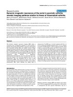
Figure 1
Dynamic gadolinium-diethylenetriamine pentaacetic acid (Gd-DTPA)-enhanced MRI of the wrist in a patient with psoriatic arthritisDynamic gadolinium-diethylenetriamine pentaacetic acid (Gd-DTPA)-
enhanced MRI of the wrist in a patient with psoriatic arthritis. Sequence
(a) shows the precontrast image; sequences (b–d) show images
acquired after 36, 90, and 180 s, respectively. The region of interest on
which the enhancement curve has been calculated is outlined. Gd-
DTPA, dynamic gadolinium-diethylenetriamine pentaacetic acid; MRI,
magnetic resonance image.
Figure 2
Individual values of REE (left) and RE (right) in patients with arthritis and controlsIndividual values of REE (left) and RE (right) in patients with arthritis and
controls. (A) Patients with psoriatic arthritis, (B) group-I rheumatoid
arthritis, or (C) groupII rheumatoid arthritis; (D) controls. Triangles indi-
cate the mean values. Vertical bars indicate standard deviations. RE,
relative enhancement; REE, rate of early enhancement.
Available online />R729
tics were only slightly higher. We had the opportunity to match
each patient with PsA with two RA patients for number of
tender and swollen joints. As a result, a new RA group (group
II) was formed that included some of the consecutive RA
patients and several new patients drawn from ongoing follow-
up studies. The two groups of patients were clinically fully
comparable. Of course, the percentage of patients with IgM
rheumatoid factor could not be easily matched in this type of
study. Another difference between groups included the more
frequent administration of prednisone and methotrexate in RA
patients. In fact, matching also for treatment was not possible
due to the relatively small number of RA patients. It could be
argued that prednisone and methotrexate could affect
dynamic MRI per se by directly acting on neovascularization.
However, these two drugs were used more frequently in both
RA groups, which behave differently from PsA as far as REE
and RE are concerned. It is therefore unlikely that their direct
effect on dynamic MRI could have influenced our results,
which are, rather, explained by a difference in inflammation.
After matching, the REE and RE were not significantly different
in the two groups of patients (Fig. 3). This finding indicates
that, at comparable levels of disease severity, synovitis
revealed by dynamic MRI presents a pattern in PsA that is sim-
ilar to that of RA. This finding contradicts the common belief
that PsA, on the whole, is a mild form of arthritis. The amount
of contrast agent transported to the inflamed synovial mem-
brane is probably a result of the number, size, and permeability
of vessels and volume of the synovial membrane itself. A
greater number of synovial vessels per squaremillimetre of tis-
sue has been demonstrated in PsA than in RA [12]. Con-
versely, significantly less lining-layer hyperplasia was
demonstrated in PsA in the same study [12]. The net effect of
these two contrasting features on Gd-DTPA diffusion is not
known and could be assessed only by comparing dynamic
MRI and synovial membrane histology in the same joints. Other
vessel characteristics of PsA synovial membrane that could
play a role in contrast agent diffusion are the marked thicken-
ing of the vessel wall [13] and the peculiar, tortuous vascular
pattern [14].
Dynamic MRI highlights the similarity of the synovial membrane
in PsA and RA and supports the view that the two conditions
may be more similar than is usually believed, at least as far as
disease activity is concerned. This observation is in keeping
with the fact that the same types of treatment, including sul-
phasalazine, methotrexate, leflunomide, and anti-tumor-necro-
sis-factor-α compounds, are effective in RA and PsA. As a
result, dynamic MRI cannot be used to differentiate the two
diseases. However, both REE and RE data were significantly
higher in PsA than in healthy controls, with only one case of
overlap between the two conditions.
A more efficient way of differentiating PsA from RA by MRI is
to study the pattern of joint involvement. Jevtic and colleagues
[15] showed that inflammation is localized within the joint
capsule in the small joints of the hand of RA patients, whereas
PsA patients also show extracapsular involvement, with thick-
ened collateral ligaments and oedema of the neighbouring soft
tissues. In another study of the knee, focal perientheseal high
signal outside the joint, and bone marrow oedema at entheseal
insertions were typical features of patients with spondyloar-
Figure 3
Slope of the mean enhancement curves in patients with arthritis and in controlsSlope of the mean enhancement curves in patients with arthritis and in controls. Patients with psoriatic arthritis (squares) or with rheumatoid arthritis
(diamonds) matched for demographic characteristics and disease severity; controls (triangles). The arrow indicates the time of gadolinium-diethylen-
etriamine pentaacetic acid infusion.
Arthritis Research & Therapy Vol 7 No 4 Cimmino et al.
R730
thropathy [16]. Our study, in which we aimed to compare the
degree of synovitis in the two forms of arthritis, did not take
into consideration damage to entheses and synovial sheaths,
areas that could be more effective in the differential diagnosis
and deserve a separate investigation.
Our results were obtained with a low-field extremity-dedicated
MRI device. The lack of discrimination between RA and PsA
with a 0.2-T MRI device could be overcome with other MRI
protocols, such as different pulse sequences, field strengths,
or magnet types. High-field MRI machines have a better signal-
to-noise ratio and could be hypothetically more sensible in the
evaluation of enhancement. Evaluation of the sensitivity of
dynamic MRI is still in its early stages. Within RA, we showed
that this technique can discriminate between different degrees
of clinical activity [7]. These considerations suggest that
differences in dynamic MRI between RA and PsA, if present,
should be relatively small. There are no papers directly com-
paring dynamic MRI obtained with low- and high-field
machines. Results of another recent study by Palosaari and
colleagues on wrist RA [17], made using a low-field MRI, are
difficult to compare with ours, because technical features such
as type of sequence, imaging parameters, acquisition plane,
number of sequences, and amount of contrast agent were dif-
ferent. In addition, the cohort of RA patients in that study,
being affected by early disease, was different. The absolute
values of signal enhancement were higher in Palosaari's study.
By contrast, a third study [18] on the rheumatoid knee per-
formed with a 1.5-T unit showed absolute enhancement values
similar to those that we obtained.
The intraobserver/interobserver agreement in evaluating
dynamic enhancement was very high. This may be surprising,
in view of the fact that the examination process included selec-
tion of slice, of maximal enhancing area, and of the size of the
ROI. We feel that high reproducibility may have been facili-
tated by a significant association between enhancement fig-
ures of elliptical areas in the three sequentially acquired wrist
slices [unpublished observations in [7]]. This makes the
choice of the slice less important. In addition, the area of max-
imum enhancement, exclusive of entheses and tendon
sheaths, is very often located on the dorsal side of the wrist
and is relatively small, another constraint of choice for the
examiner. Elliptical ROIs, although apparently less logical from
a pathophysiological point of view than ROIs outlining the
enhanced synovial membrane, were chosen to improve repro-
ducibility and standardization. A recent paper on contrast-
enhanced dynamic MRI of coronal slices of the wrist also
showed high intraobserver reliability [17].
Only one PsA patient was positive for rheumatoid factor. She
did not show a rheumatoid-like pattern of joint involvement, but
had monoarthritis of the right wrist and dactylitis, which is not
typical of RA. Of the 15 PsA patients, 8 had a rheumatoid-like
pattern of arthritis and 7 had oligomonoarthritis. Dynamic MRI
was not significantly different between these two groups, rein-
forcing the suggestion that severity of the arthritis, and not its
type or subtype, is associated with MRI findings.
The patients were selected on the basis of clinical involvement
of the wrist. This prerequisite was set because, if all consecu-
tive patients had been enrolled, many more RA patients than
PsA patients would have had wrist involvement, thus making
the two groups more difficult to compare. In a previous study
by our team [7], RA patients in remission and without wrist
arthritis had dynamic MRI values significantly lower than those
with active disease and wrist involvement. We do not know if
the results obtained in unaffected wrists of otherwise active
arthritis patients reflect local (wrist) or general disease activity.
No correlation was found between indexes of severity and
dynamic MRI in either PsA and RA. This last finding is surpris-
ing in view of our previous results on the high correlation
between clinical and laboratory indexes of RA inflammation
and dynamic MRI. However, the inclusion of severely active
arthritis only – with exclusion of patients in remission – and the
relatively low mean number of affected joints in the patients
with PsA and in the matched RA controls may account for the
difference. Nonetheless, a tendency to correlation between
number of swollen joints and REE, which in our experience is
the more sensitive measure, was observed.
Conclusion
We have shown that dynamic MRI gives similar results in PsA
and RA, suggesting that the type and degree of inflammatory
process is similar in the two diseases.
Competing interests
SI is an employee of ESAOTE, the manufacturer of the mag-
netic resonance device.
Authors' contributions
MAC and MP contributed to the conception and design of the
study, to the clinical and MRI evaluation of the patients, to the
analysis and interpretation of data, to the drafting of the article,
and to the critical revision of the article for important intellec-
tual content; SI contributed to the analysis and interpretation
of data and to the critical revision of the article for important
intellectual content; ES and GG contributed to the conception
and design of the study, to the analysis and interpretation of
data, and to the critical revision of the article for important intel-
lectual content; GS and SB contributed to the conception and
design of the study, to the clinical and MRI evaluation of the
patients, to the analysis and interpretation of data, and to the
critical revision of the article for important intellectual content.
All authors read and approved the final manuscript.
Acknowledgements
This work was supported in part by a grant from the University of Genoa,
Italy.
Available online />R731
References
1. Moll JMH, Wright V: Psoriatic arthritis. Semin Arthritis Rheum
1973, 3:55-78.
2. Helliwell P, Marchesoni A, Peters M, Barker M, Wright V: A re-
evaluation of the osteoarticular manifestations of psoriasis. Br
J Rheumatol 1991, 30:339-345.
3. Marsal S, Armadans-Gil L, Martinez M, Gallardo D, Ribera A,
Lience E: Clinical, radiographic and HLA associations as mark-
ers for different patterns of psoriatic arthritis. Rheumatology
1999, 38:332-337.
4. Helliwell PS, Hetthen J, Sokoll K, Green M, Marchesoni A, Lubrano
E, Veale D, Emery P: Joint symmetry in early and late rheuma-
toid and psoriatic arthritis. Arthritis Rheum 2000, 43:865-871.
5. Shbeeb M, Uramoto KM, Gibson LE, O'Fallon WM, Gabriel SE:
Epidemiology of psoriatic arthritis in Olmsted County, Minne-
sota, USA, 1982–1991. J Rheumatol 2000, 27:1247-1250.
6. McGonagle D, Conaghan PG, Emery P: Psoriatic arthritis: a uni-
fied concept twenty years on. Arthritis Rheum 1999,
42:1080-1086.
7. Cimmino MA, Innocenti S, Livrone F, Magnaguagno F, Silvestri E,
Garlaschi G: Dynamic gadolinium-enhanced magnetic reso-
nance imaging of the wrist in patients with rheumatoid arthritis
can discriminate active from inactive disease. Arthritis Rheum
2003, 48:1207-1213.
8. Arnett FC, Edworthy SM, Bloch DA, McShane DJ, Fries JF, Cooper
NS, Healey LA, Kaplan SR, Liang MH, Luthra HS, et al.: The Amer-
ican Rheumatism Association 1987 revised criteria for the
classification of rheumatoid arthritis. Arthritis Rheum 1988,
31:315-324.
9. Østergaard M, Lorenzen I, Henriksen O: Dynamic gadolinium-
enhanced MR imaging in active and inactive immunoinflam-
matory gonarthritis. Acta Radiol 1994, 35:275-281.
10. Cimmino MA, Bountis C, Silvestri E, Garlaschi G, Accardo S: An
appraisal of magnetic resonance imaging of the wrist in rheu-
matoid arthritis. Semin Arthritis Rheum 2000, 30:180-195.
11. Gaffney K, Cookson J, Blades S, Coumbe A, Blake D: Quantita-
tive assessment of the rheumatoid synovial microvascular
bed by gadolinium-DTPA enhanced magnetic resonance
imaging. Ann Rheum Dis 1998, 57:152-157.
12. Veale D, Yanni G, Rogers S, Barnes L, Bresnihan B, Fitzgerald O:
Reduced synovial membrane macrophage numbers, ELAM-1
expression, and lining layer hyperplasia in psoriatic arthritis as
compared with rheumatoid arthritis. Arthritis Rheum 1993,
36:893-900.
13. Espinoza LR, Vasey FB, Espinoza CG, Bocanegra TS, Germain
BF: Vascular changes in psoriatic synovium. A light and elec-
tron microscopic study. Arthritis Rheum 1982, 25:677-684.
14. Cañete JD, Rodríguez JR, Salvador G, Gómez-Centeno A, Muñoz-
Gómez J, Sanmartí R: Diagnostic usefulness of synovial vascu-
lar morphology in chronic arthritis. A systematic survey of 100
cases. Semin Arthritis Rheum 2003, 32:378-387.
15. Jevtic V, Watt J, Rozman B, Kos-Golja M, Demsar F, Jarh O: Dis-
tinctive radiological features of small hand joints and seroneg-
ative spondyloarthritis demonstrated by contrast-enhanced
(Gd-DTPA) magnetic resonance imaging. Skeletal Radiol 1995,
24:351-355.
16. McGonagle D, Gibbon W, O'Connor P, Green M, Pease C, Emery
P: Characteristic magnetic resonance imaging entheseal
changes of knee synovitis in spondyloarthropathy. Arthritis
Rheum 1998, 41:694-700.
17. Palosaari K, Vuotila J, Takalo R, Jartti A, Niemelä R, Haapea M,
Soini I, Tervonen O, Hakala M: Contrast-enhanced dynamic and
static MRI correlates with quantitative 99Tcm-labelled nano-
colloid scintigraphy. Study of early rheumatoid arthritis
patients. Rheumatology 2004, 43:1364-1373.
18. Østergaard M, Stoltenberg M, Henriksen O, Lorenzen I: Quantita-
tive assessment of synovial inflammation by dynamic gadolin-
ium-enhanced magnetic resonance imaging. A study of the
effect of intra-articular methylprednisolone on the rate of early
synovial enhancement. Br J Rheumatol 1996, 35:50-59.