báo cáo hóa học:" Dynamic magnetic resonance imaging in assessing lung function in adolescent idiopathic scoliosis: a pilot study of comparison before and after posterior spinal fusion" doc
Bạn đang xem bản rút gọn của tài liệu. Xem và tải ngay bản đầy đủ của tài liệu tại đây (492.76 KB, 7 trang )
BioMed Central
Page 1 of 7
(page number not for citation purposes)
Journal of Orthopaedic Surgery and
Research
Open Access
Research article
Dynamic magnetic resonance imaging in assessing lung function in
adolescent idiopathic scoliosis: a pilot study of comparison before
and after posterior spinal fusion
Winnie CW Chu*
1
, Bobby KW Ng
2
, Albert M Li
3
, Tsz-ping Lam
2
,
Wynnie WM Lam
1
and Jack CY Cheng
2
Address:
1
Departments of Diagnostic Radiology and Organ Imaging, The Chinese University of Hong Kong, Prince of Wales Hospital, Shatin, Hong
Kong, China,
2
Departments of Orthopaedics and Traumatology, The Chinese University of Hong Kong, Prince of Wales Hospital, Shatin, Hong
Kong, China and
3
Departments of Paediatrics, The Chinese University of Hong Kong, Prince of Wales Hospital, Shatin, Hong Kong, China
Email: Winnie CW Chu* - ; Bobby KW Ng - ; Albert M Li - ; Tsz-
ping Lam - ; Wynnie WM Lam - ; Jack CY Cheng -
* Corresponding author
Abstract
Background: Restrictive impairment is the commonest reported pulmonary deficit in AIS, which improves following surgical
operation. However, exact mechanism of how improvement is brought about is unknown. Dynamic fast breath-hold (BH)-MR
imaging is a recent advance which provides direct quantitative visual assessment of pulmonary function. By using above
technique, change in lung volume, chest wall and diaphragmatic motion in AIS patients before and six months after posterior
spinal fusion surgery were measured.
Methods: 16 patients with severe right-sided predominant thoracic scoliosis (standing Cobb's angle 50° -82°, mean 60°)
received posterior spinal fusion without thoracoplasty were recruited into this study. BH-MR sequences were used to obtain
coronal images of the whole chest during full inspiration and expiration. The following measurements were assessed: (1)
inspiratory, expiratory and change in lung volume; (2) change in anteroposterior (AP) and transverse (TS) diameter of the chest
wall at two levels: carina and apex (3) change in diaphragmatic heights. The changes in parameters before and after operation
were compared using Wilcoxon signed ranks test. Patients were also asked to score their breathing effort before and after
operation using a scale of 1–9 with ascending order of effort. The degree of spinal surgical correction at three planes was also
assessed by reformatted MR images and correction rate of Cobb's angle was calculated.
Results: The individual or total inspiratory and expiratory volume showed slight but insignificant increase after operation. There
was significantly increase in bilateral TS chest wall movement at carina level and increase in bilateral diaphragmatic movements
between inspiration and expiration. The AP chest wall movements, however, did not significantly change.
The median breathing effort after operation was lower than that before operation (p < 0.05).
There was significant reduction in coronal Cobb's angle after operation but the change in sagittal and axial angle at scoliosis apex
was not significant.
Conclusion: There is improvement of lateral chest wall and diaphragmatic motions in AIS patients six months after posterior
spinal fusion, associated with subjective symptomatic improvement. Lung volumes however, do not significantly change after
operation. BH-MR is novel non-invasive method for long term post operative assessment of pulmonary function in AIS patients.
Published: 19 November 2007
Journal of Orthopaedic Surgery and Research 2007, 2:20 doi:10.1186/1749-799X-2-20
Received: 27 January 2007
Accepted: 19 November 2007
This article is available from: />© 2007 Chu et al; licensee BioMed Central Ltd.
This is an Open Access article distributed under the terms of the Creative Commons Attribution License ( />),
which permits unrestricted use, distribution, and reproduction in any medium, provided the original work is properly cited.
Journal of Orthopaedic Surgery and Research 2007, 2:20 />Page 2 of 7
(page number not for citation purposes)
Background
Adolescent idiopathic scoliosis (AIS) is the most common
form of idiopathic scoliosis, typically affecting growing
adolescent girls, 10–16 years of age. Untreated scoliosis
has an increased risk of developing respiratory failure and
premature mortality[1]. Pulmonary function impairment
in AIS patients might be related to restricted lung volume,
poor chest wall expansibility or impaired diaphragmatic
motion. Little is known about which of the above factors
is more significantly correlated with the pulmonary deficit
in AIS. Restrictive impairment is the commonest reported
pulmonary deficit in AIS, which improves following sur-
gical operation[2,3]. However, the exact mechanism of
how the improvement is brought about is unknown.
We have previously reported a validated novel imaging
technique for assessment of pulmonary function in AIS
subjects. Kotani and colleagues have also investigated on
chest wall and diaphragmatic movement of scoliosis
patients using dynamic breathing MRI [4]. With the appli-
cation of ultrafast dynamic breath-hold (BH) MR imaging
and multiplanar reformat technique, the lung volume,
chest wall, and diaphragmatic motions between inspira-
tion and expiration can be accurately measured with high
reproducibility in both AIS subjects and normal con-
trols[5].
The aim of this pilot study was to evaluate the change in
lung volume, chest wall and diaphragmatic motion in AIS
patients before operation and six months after posterior
spinal fusion surgery.
Methods
Subjects
The study included 16 idiopathic scoliosis girls with a pre-
dominant right-sided thoracic curve (Standing Cobb's
angle ranged from 50°–82°, mean 60°). The distribution
of curves by Lenke classification is given in Table 1. Their
age ranged from 11–18 (mean 14.4). They were consecu-
tively included for scheduled posterior spinal fusion with-
out thoracoplasty from 2002 to 2004. The surgical
procedure was performed under SSEP monitoring. It con-
sisted of a standard midline posterior exposure, subperio-
steal dissection made from spinous process to the tips of
each transverse process. The facet capsules were meticu-
lously elevated till the superior rib surface was reached.
Partial excision of the ligamentum flavum was made
between spinous processes for the lordotic thoracic seg-
ments typically from T5 to T11. The exposure was made to
preserve vascularity to paraspinal muscles. Instrumenta-
tion consisted of three types. (a) 3 cases of Harrington rod
on concave side, Luque rod on convex side and Wisconsin
wire for the instrumented spinous processes. (b) 8 cases of
ISOLA instrumentation consisted of pedicle screws to
lumbar segments to build a base, proximal claw hook
construct at two segments above upper end vertebra on
both sides followed by pedicle hook at upper end vertebra
on the concave side and transverse process hook on con-
vex side. Wisconsin wires were placed for the mid-thoracic
segments. (c) 5 cases of CDM8 instrumentation were
essentially same as the ISOLA constructs except the lum-
bar pedicle screws used were top loading monoaxial
screws instead of vertebral screws connecting to rod with
slotted connectors as in the ISOLA system. After place-
ment of the fixation devices, curve reduction was made by
manual pressure at apex and counter pressures at opposite
ends under SSEP monitoring. Rod estimation and prelim-
inary contouring was then made. Decortication of facet
joints and transverse processes was made and bone grafts
were placed at each decorticated facet joint. Instrumenta-
tion was then performed and final rod contouring made
with insitu benders. Cell saver was used to retrieve blood
and transfused back to patient intra-operatively. There
were no neurological or wound complications. All AIS
subjects were neurologically normal on detail clinical
examination. Exclusion criteria included history of back
injury, weakness or numbness in one or more limbs, uri-
nary incontinence or nocturnal enuresis. None of the sub-
jects had any history of pulmonary diseases and they were
free from any respiratory symptoms or acute respiratory
infection at the time of the MR studies before and after
operation.
Ethical approval and informed consent for dynamic
breath-hold MR imaging have been obtained from all the
subjects and their parents.
MRI assessment
Pre operative MRI examination of the chest was per-
formed in all subjects within two weeks before the opera-
tion, while the post operative MRI was performed after 6
months from the date of surgery. MRI examination was
performed using a 1.5T MR scanner (Sonata, Siemens,
Erlangen, Germany). The protocol of BH-MR imaging of
the chest has been reported in previous published study
[5]. In brief, fast gradient-recalled echo pulse sequence
was used to obtain coronal images of the whole chest dur-
ing full inspiration and expiration with parameters. The
Table 1: Frequency of curve types by Lenke classification in 16
subjects included in this study
Lenke's classification Frequency
1A- 1
1AN 6
1BN 5
1C- 1
2A- 1
3AN 1
3BN 1
Journal of Orthopaedic Surgery and Research 2007, 2:20 />Page 3 of 7
(page number not for citation purposes)
images were acquired in the supine position. All subjects
were given clear instruction by experienced radiographers
and practice took place before actual imaging. The sub-
jects were instructed to fully inspire/fully expire and then
hold their breaths at either full inspiration or full expira-
tion. Scanning repeated three times for full inspiration
and another three times for full expiration. Each breath-
hold scan took about 15–20 seconds, which was well tol-
erated by all subjects. The images with the maximum
inspiratory and expiratory effort out of the three attempts
were chosen for analysis.
Post-processing of the MR images was performed using a
workstation (EasyVision, Philips Medical Systems, Best,
the Netherlands). Volumetric measurements of total
inspiratory and expiratory lung volume were determined
by a semi-automated computerized segmentation method
[6] (Fig 1). The MR images were also reformatted into
axial and coronal planes so that motions of the chest wall
and diaphragm could be assessed. The chest wall and dia-
phragmatic motions were measured in antero-posterior,
left-right and cranio-caudal directions respectively. The
chest wall diameters were measured at the level of the car-
ina (Figure 2a) and at apex of the vertebral curve (Figure
2b) respectively. The chest wall dimensions were then
measured as the largest anteroposterior (AP) and trans-
verse (TS) dimensions on either side of the scoliosis sepa-
rately. The diaphragmatic heights were taken as the
vertical distance between the line drawn tangential to the
highest point of the diaphragm and a line parallel to the
lung apex (Fig 3a and 3b). All the lung volume, chest wall
and diaphragmatic dimensions in the right and left
hemithorax were measured separately, during both inspi-
ration and expiration, and the differences were recorded.
Three measurements were made for each parameter by the
same observer and the average value was taken for the
analysis. Our previous studies showed high intraobserver
and interobserver reliability on the MR measurements [5].
Breathing Effort Assessment
Patients were also asked to score their breathing effort
before and after operation using a scale of 1–9 with
ascending order of effort. Score 0 was equivalent to non-
awareness of breathing effort at rest. Score 1–3 was equiv-
alent of awareness of gentle breathing effort at rest with
ascending order of effort. Score 4–6 was equivalent to feel-
ing of breathlessness during exercise activities in ascend-
ing order of severity. Score 7–9 was equivalent to feeling
of breathlessness at gentle exertion such as walking in
ascending order of severity.
Statistics
The parameters in each group of subjects were expressed
as median with inter-quartile range. Non-parametric Wil-
coxon signed ranks test was used to compare the MR
measurements before and after operation in all subjects.
Two-tailed probability values <0.05 were considered sig-
nificant. SPSS for Windows statistical software (Release
13, SPSS Inc., Chicago, Illinois) was used in the analysis.
Results
The spatial resolution of the reformatted axial and coronal
images of the chest was considered to be of diagnostic
quality in all subjects. The fast image acquisition time
allows good delineation of the lung volume, the chest
wall and the diaphragm.
Surgical procedures in all patients were uneventful with-
out significant complications. All patients were dis-
charged home fully mobilized on average 11 days (range
9–15) from admission. Six month after operation, the
median correction rate of Cobb's angle was 60% (range
42% to 72%). The median Cobb's angle changed from
59° to 24° while the sagittal Cobb's angle changed from
19.1° to 23.1°.
The lung volumes, chest wall and diaphragmatic parame-
ters before and after operation in all subjects are summa-
rized in Table 2.
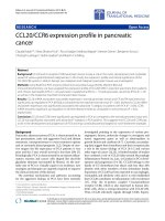
Measurement of lung volumes by a semi-automated compu-terized method of delineating the lungs and summing cross-sectional areasFigure 1
Measurement of lung volumes by a semi-automated compu-
terized method of delineating the lungs and summing cross-
sectional areas. (a) On the coronal image of the lung, thresh-
old signal intensity is selected to highlight the air in green
(lung). (b) The total lung volume is calculated by summating
the volume of all coronal sections of the lungs from the front
to the back of the body.
Journal of Orthopaedic Surgery and Research 2007, 2:20 />Page 4 of 7
(page number not for citation purposes)
For lung volumes, the right lung (on the convexity side of
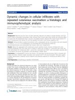
Measurement of diaphragmatic heights on the reformatted coronal imageFigure 3
Measurement of diaphragmatic heights on the reformatted coronal image. (a) At maximal inspiratory image and (b) maximal
expiratory image The diaphragmatic heights were taken as the vertical distance in between the line drawn tangent to the high-
est point of the diaphragm and a parallel line to the lung apex. The diaphragmatic motion is calculated as the difference
between inspiration and expiration.
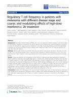
Measurement of AP and TS diameter of the chest wall on the reformatted axial imageFigure 2
Measurement of AP and TS diameter of the chest wall on the reformatted axial image. (a) Upper level at the carina (C), maxi-
mal inspiratory image. (b) Lower level at the apical vertebra (A), maximal inspiratory image. Tangential lines are drawn to the
anterior, posterior and lateral lung surfaces. The chest wall dimensions are then measured as the largest anteroposterior (AP,
thick solid lines) and transverse (TS, dotted lines) dimensions on either side of the scoliosis separately. The chest wall motion
is calculated as the difference between inspiration and expiration.
Journal of Orthopaedic Surgery and Research 2007, 2:20 />Page 5 of 7
(page number not for citation purposes)
Table 2: Lung, chest wall and diaphragmatic parameters, spinal curvatures in 16 subjects before and six months after corrective
posterior spinal fusion. Figures are expressed as median (interquartile range)
Before surgery After surgery P value (* significant at 0.05 level)
Inspiratory volume (cc)
Right lung 1719 (1232, 1858) 1764 (1362, 1938) 0.57
Left lung 1392 (1113, 1607) 1381 (1152, 1642) 0.35
Total 3127(2364, 3464) 3152 (2491, 3529) 0.50
Expiratory volume (cc)
Right lung 662 (579, 817) 716 (586, 814) 0.61
Left lung 598 (501, 700) 601 (464, 707) 0.96
Total 1269 (1083, 1498) 1361 (1038, 1477) 0.64
Change in lung volume (cc)
Right lung 996 (645, 1126) 969 (698, 1167) 0.68
Left lung 762 (535, 962) 736 (630, 1018) 0.22
Total 1770 (1206, 2089) 1730 (1362, 2203) 0.30
Right lung AP diameter at carina (mm)
Inspiratory 117 (100, 123) 118 (107, 130) 0.07
Expiratory 84 (75, 92) 88 (82, 96) 0.06
Change 29 (21, 42) 29 (21, 42) 0.98
Left lung AP diameter at carina (mm)
Inspiratory 122 (106, 133) 119 (108, 129) 0.18
Expiratory 89 (80,99) 93 (87, 101) 0.41
Change 29 (16, 46) 27 (18,33) 0.14
Right lung AP diameter at apex (mm)
Inspiratory 135 (123, 151) 137 (116, 155) 0.50
Expiratory 1114 (99, 126) 114 (100, 133) 0.88
Change 21 (15,31) 20 (13, 25) 0.20
Left lung AP diameter at apex (mm)
Inspiratory 126 (118, 145) 126 (121, 138) 0.92
Expiratory 104 (94, 114) 104 (97, 111) 0.72
Change 28 (17, 37) 23 (12, 33) 0.30
Right lung TS diameter at carina (mm)
Inspiratory 97 (80, 116) 111 (106, 116) 0.013*
Expiratory 85 (75, 93) 87 (75, 96) 0.30
Change 15 (3, 27) 28 (17, 33) 0.006*
Left lung TS diameter at carina (mm)
Inspiratory 92 (79, 103) 104 (99, 110) 0.002*
Expiratory 79 (59, 84) 86 (76, 93) 0.044*
Change 14 (11, 23) 20 (13, 28) 0.056
Right lung TS diameter at apex (mm)
Inspiratory 106 (85, 126) 114 (105, 120) 0.039*
Expiratory 87 (71, 92) 91 (84, 100) 0.034*
Change 16 (10, 31) 24 (13, 32) 0.796
Left lung TS diameter at apex (mm)
Inspiratory 112 (93, 117) 113 (104, 122) 0.088
Expiratory 98 (68, 113) 100 (83, 112) 0.163
Change 9 (6, 13) 12 (7, 17) 0.301
Right Diaphragmatic height (mm)
Inspiratory 185 (172, 202) 201 (182, 223) <0.01*
Journal of Orthopaedic Surgery and Research 2007, 2:20 />Page 6 of 7
(page number not for citation purposes)
the scoliotic curve) and total inspiratory and expiratory
lung volume showed slight but insignificant increase after
operation. Neither right/left lung nor the total vital lung
capacity (defined as the difference in lung volume
between inspiration and expiration) differ significantly
when comparing the pre-operative with the post-opera-
tive study.
For the TS diameter of bilateral chest wall, there was sig-
nificant increase in baseline value on both right and left
lung at either carina level or apical vertebral level during
both inspiration and expiration. When the difference
between inspiration and expiration was considered, the
lateral chest wall movement at carina level was improved
significantly on right/convex side (p = 0.013) while mar-
ginally significant on left/concave side (p = 0.056). but
there was no statistically significant change at the apical
vertebra level.
For the AP diameter of bilateral chest wall, there was no
significant difference on either right lung or left lung in
between operation. The AP chest wall movement also
showed no significant interval change.
For the diaphragmatic heights, they were significantly
increased on both right and left sides during inspiration
while the absolute value during expiration was unchanged
and hence the motion of the diaphragms, defined as the
difference in diaphragmatic height between inspiration
and expiration, was significantly increased after operation
when compared with the pre-operative stage.
The median breathing effort reported by the subjects
before operation was 4 (range 1–9) while the median
breathing effort after operation was 1.75 (range 1–5).
In summary, there was improvement of lateral chest wall
and diaphragmatic motions in AIS subjects six months
after posterior spinal fusion. Lung volumes however, did
not show significant increase after operation at this stage.
Discussion
Though pulmonary function impairment has long been
reported in AIS patients, it is difficult to measure and
describe respiratory motions because of the limited meth-
ods available for real time dynamic assessment. In the
present and previous published study, we have demon-
strated that by using multi-planar reformat technique,
coronal and axial sectional planes could be obtained
simultaneously with measurement of lung volumes dur-
ing a single inspiration and expiration movement. This
MR technique has also been validated which showed sig-
nificant positive correlations with plethysmography
parameters[6]. We therefore propose the use of dynamic
breath-hold MR as a novel non-invasive tool for clinical
analysis of lung volume, diaphragmatic and chest wall
motion in AIS patients.
In this study, dynamic BH-MR imaging has been used to
compare the pre and post operative lung function changes
in AIS patients after spinal fusion. We found that the
metallic implants only caused mild distortion artefacts on
the immediate adjacent structures such as the vertebral
column and the central canal; while the visualization of
the lungs, the chest walls and diaphragms were not
affected.
In the literature, there has been debate about the effect of
surgical correction on lung function. Many authors have
written that scoliosis correction definitely improves meas-
ured pulmonary functions [7], such as vital capacity. Oth-
ers disagree and declare that pulmonary functions remain
essentially unchanged [8-10] or even becoming
worsen[11].
In the previous published study, we found that the chest
wall and diaphragmatic motion in AIS patients were not
restricted, i.e. the chest wall and the diaphragmatic
motions were as mobile as those in the normal subjects
and therefore there was no suggestion of neuromuscular
dysfunction in AIS [5].
Expiratory 136 (129, 150) 146 (135, 156) 0.196
Change 49 (25, 65) 53 (44, 66) 0.039*
Left Diaphragmatic height (mm)
Inspiratory 185 (172, 206) 198 (186, 215) 0.01*
Expiratory 142 (129, 159) 148 (136, 161) 0.234
Change 44 (33, 57) 53 (44, 66) 0.039*
Breathing effort 4 (1,4) 1.75 (1, 3) 0.012*
Coronal Cobb angle (MR) 59 (53, 64) 24 (21, 28) <0.01*
Axial angle (MR) 9.9 (4.7, 13.0) 9.6 (4.0, 14.7) 0.215
Sagittal angle (MR) 19.1 (13.1, 28.8) 23.1 (17.2, 31.2) 0.056
Table 2: Lung, chest wall and diaphragmatic parameters, spinal curvatures in 16 subjects before and six months after corrective
posterior spinal fusion. Figures are expressed as median (interquartile range) (Continued)
Publish with BioMed Central and every
scientist can read your work free of charge
"BioMed Central will be the most significant development for
disseminating the results of biomedical research in our lifetime."
Sir Paul Nurse, Cancer Research UK
Your research papers will be:
available free of charge to the entire biomedical community
peer reviewed and published immediately upon acceptance
cited in PubMed and archived on PubMed Central
yours — you keep the copyright
Submit your manuscript here:
/>BioMedcentral
Journal of Orthopaedic Surgery and Research 2007, 2:20 />Page 7 of 7
(page number not for citation purposes)
In the current study, we found that the chest wall motion
and diaphragmatic motions could be further improved
after posterior spinal surgery. This was consistent with the
subjective feeling of reducing breathing effort after opera-
tion as reported in this group of patients. The reason for
increased amplitude of lateral chest wall movement could
be explained by the less distorted chest wall configuration
after surgery, while the improvement of diaphragmatic
excursion could be the combined effect of chest wall
remodeling, lessen compressive effect by both the scoli-
otic spine and the rotated mediastinum as well as
improvement in degree of hypokyphosis after surgery.
The absolute lung volumes however, did not show signif-
icant increase in this cohort. As the current study was car-
ried out shortly after the operation within six months,
longer follow up study is warranted which might show a
change in pulmonary volume.
Dynamic MR is considered as a promising investigation
tool in assessing respiratory mechanism in the AIS group.
It might be useful for both short and long term follow up
of pulmonary function of AIS patients, in particular, for
assessing post operative changes.
Conclusion
With the application of ultrafast dynamic BH-MR imaging
and multi-planar reformat technique, the lung volume,
chest wall and diaphragmatic motion between inspiration
and expiration could now be accurately measured with
high reproducibility in AIS patients. Improvement of lat-
eral chest wall and diaphragmatic motions are evident in
AIS patients six months after posterior spinal fusion. BH-
MR might be sued for long term post operative assessment
of pulmonary function in AIS patients.
Abbreviations
AIS: Adolescent Idiopathic Scoliosis
BH: breath-hold
MR: Magnetic Resonance
Competing interests
The author(s) declare that they have no competing inter-
ests.
Authors' contributions
All the authors have contributed to conception and design
of the manuscript. Particularly, WCWC and BKWN carried
out the analysis of data and wrote the manuscript; AML
and TPL contributed to the editing of the manuscript, clin-
ical recruitment and assessment; WWML participated in
the editing of the manuscript; JCYC contributed to the
editing of the manuscript and secured funding. All
authors have read and approved the final manuscript.
References
1. Leaver JM, Alvik A, Warren MD: Prescriptive screening for ado-
lescent idiopathic scoliosis: a review of the evidence. Int J Epi-
demiol 1982, 11(2):101-111.
2. Gagnon S, Jodoin A, Martin R: Pulmonary function test study and
after spinal fusion in young idiopathic scoliosis. Spine 1989,
14(5):486-490.
3. Muirhead A, Conner AN: The assessment of lung function in
children with scoliosis. J Bone Joint Surg Br 1985, 67(5):699-702.
4. Kotani T, Minami S, Takahashi K, Isobe K, Nakata Y, Takaso M, Inoue
M, Maruta T, Akazawa T, Ueda T, Moriya H: An analysis of chest
wall and diaphragm motions in patients with idiopathic scol-
iosis using dynamic breathing MRI. Spine 2004, 29(3):298-302.
5. Chu WC, Li AM, Ng BK, Chan DF, Lam TP, Lam WW, Cheng JC:
Dynamic magnetic resonance imaging in assessing lung vol-
umes, chest wall, and diaphragm motions in adolescent idio-
pathic scoliosis versus normal controls. Spine 2006,
31(19):2243-2249.
6. Gierada DS, Hakimian S, Slone RM, Yusen RD: MR analysis of lung
volume and thoracic dimensions in patients with emphy-
sema before and after lung volume reduction surgery. AJR Am
J Roentgenol 1998, 170(3):707-714.
7. Kinnear WJ, Johnston ID: Does Harrington instrumentation
improve pulmonary function in adolescents with idiopathic
scoliosis? A meta-analysis. Spine 1993, 18(11):1556-1559.
8. Shneerson JM, Edgar MA: Cardiac and respiratory function
before and after spinal fusion in adolescent idiopathic scolio-
sis. Thorax 1979, 34(5):658-661.
9. Upadhyay SS, Ho EK, Gunawardene WM, Leong JC, Hsu LC:
Changes in residual volume relative to vital capacity and
total lung capacity after arthrodesis of the spine in patients
who have adolescent idiopathic scoliosis. J Bone Joint Surg Am
1993, 75(1):46-52.
10. Zorab PA, Prime FJ, Harrison A: Lung function in young persons
after spinal fusion for scoliosis. Spine 1979,
4(1):22-28.
11. Kinnear WJ, Kinnear GC, Watson L, Webb JK, Johnston ID: Pulmo-
nary function after spinal surgery for idiopathic scoliosis.
Spine 1992, 17(6):708-713.