Báo cáo y học: "levels of soluble receptor for advanced glycation end products in patients with rheumatoid arthritis indicating deficient inflammatory control" docx
Bạn đang xem bản rút gọn của tài liệu. Xem và tải ngay bản đầy đủ của tài liệu tại đây (207.52 KB, 8 trang )
Open Access
Available online />R817
Vol 7 No 4
Research article
Decreased levels of soluble receptor for advanced glycation end
products in patients with rheumatoid arthritis indicating deficient
inflammatory control
Rille Pullerits
1
, Maria Bokarewa
1
, Leif Dahlberg
2
and Andrej Tarkowski
1
1
Department of Rheumatology and Inflammation Research, University of Göteborg, Göteborg, Sweden
2
Joint and Soft Tissue Unit, Department of Clinical Sciences, Lund University, Department of Orthopaedics, Malmö University Hospital, Malmö,
Sweden
Corresponding author: Rille Pullerits,
Received: 8 Dec 2004 Revisions requested: 6 Jan 2005 Revisions received: 4 Mar 2005 Accepted: 16 Mar 2005 Published: 25 Apr 2005
Arthritis Research & Therapy 2005, 7:R817-R824 (DOI 10.1186/ar1749)
This article is online at: />© 2005 Pullerits et al, licensee BioMed Central Ltd.
This is an Open Access article distributed under the terms of the Creative Commons Attribution License ( />2.0), which permits unrestricted use, distribution, and reproduction in any medium, provided the original work is cited.
Abstract
The receptor for advanced glycation end products (RAGE) is a
member of the immunoglobulin superfamily being expressed as
a cell surface molecule and binding a variety of ligands. One of
these ligands is high-mobility group box chromosomal protein 1,
a potent proinflammatory cytokine, expression of which is
increased in synovial tissue and in synovial fluid of rheumatoid
arthritis (RA) patients. The interaction of high-mobility group box
chromosomal protein 1 with cell-surface RAGE leads to an
inflammatory response. In contrast, the presence of soluble
RAGE (sRAGE) may abrogate cellular activation since the
ligand is bound prior to interaction with the surface receptor.
Our aim was to analyse to what extent sRAGE is present in
patients with chronic joint inflammation (RA) as compared with
patients with non-inflammatory joint disease and with healthy
subjects, and to assess whether there is an association between
sRAGE levels and disease characteristics.
Matching samples of blood and synovial fluid were collected
from 62 patients with RA with acute joint effusion. Blood from
45 healthy individuals, synovial fluid samples from 33 patients
with non-inflammatory joint diseases and blood from six patients
with non-inflammatory joint diseases were used for comparison.
sRAGE levels were analysed using an ELISA.
RA patients displayed significantly decreased blood levels of
sRAGE (871 ± 66 pg/ml, P < 0.0001) as compared with
healthy controls (1290 ± 78 pg/ml) and with patients with non-
inflammatory joint disease (1569 ± 168 pg/ml). Importantly,
sRAGE levels in the synovial fluid of RA patients (379 ± 36 pg/
ml) were lower than in corresponding blood samples and
correlated significantly with blood sRAGE. Interestingly, a
significantly higher sRAGE level was found in synovial fluid of
RA patients treated with methotrexate as compared with
patients without disease-modifying anti-rheumatic treatment.
We conclude that a decreased level of sRAGE in patients with
RA might increase the propensity towards inflammation,
whereas treatment with methotrexate counteracts this feature.
Introduction
Rheumatoid arthritis (RA) is a chronic inflammatory synovitis
that is dominated by the presence of macrophages, lym-
phocytes and synovial fibroblasts, which leads to the destruc-
tion of bone and cartilage. The pathogenesis of the disease is
complex, involving a wide range of molecules.
The receptor for advanced glycation end products (RAGE) is
a multi-ligand member of the immunoglobulin superfamily
being expressed as a cell surface molecule and interacting
with a diverse class of ligands [1,2]. RAGE is expressed by
many of the cells that participate in the development of RA,
including macrophages, neutrophils and T cells. RAGE is
expressed on macrophages and T cells within synovial tissues
of RA patients as well as on synovial fluid macrophages [3].
DMARD = disease-modifying anti-rheumatic drug; ELISA = enzyme-linked immunosorbent assay; EN-RAGE = extracellular newly identified RAGE-
binding protein; HMGB1 = high-mobility group box chromosomal protein 1; IL = interleukin; NID = non-inflammatory joint disease; RA = rheumatoid
arthritis; RAGE = receptor for advanced glycation end products; sRAGE = soluble receptor for advanced glycation end products.
Arthritis Research & Therapy Vol 7 No 4 Pullerits et al.
R818
Moreover, synovial fibroblasts that account for about 50% of
the cellular constituents of the synovial lining layer constitu-
tively express RAGE [4].
The RAGE protein is composed of three immunoglobulin-like
regions, a transmembrane domain and a highly charged short
cytosolic tail that is essential for post-RAGE signalling. One of
the features of the receptor is its recognition of families of lig-
ands, rather than a single protein. The RAGE repertoire of lig-
ands includes products of non-enzymatic glycoxidation
(advanced glycation end products), the amyloid-β protein, the
S100/calgranulin family of proinflammatory cytokine-like medi-
ators, β2-integrin Mac-1 on leukocytes and the high mobility
group box chromosomal protein 1 (HMGB1), all of which are
associated with inflammation [2]. Studies have shown that
engagement of RAGE by a ligand results in a rapid and sus-
tained cellular activation and gene transcription [1]. Sustained
receptor expression leads to a positive feedback loop in which
the ligand–receptor interaction increases expression of the
receptor itself on the cell surface, leading to further amplifica-
tion of inflammatory response.
Soluble RAGE (sRAGE), a truncated form of the receptor, is
composed of only the extracellular ligand-binding domain lack-
ing the cytosolic and transmembrane domains (i.e. the part
that transfers a signal into the cell). This soluble form of the
receptor has the same ligand binding specificity and therefore
competes with cell-bound RAGE for ligand binding, therefore
functioning as a 'decoy' abrogating cellular activation, since
the cell surface receptor remains unoccupied. Indeed, it has
been demonstrated in a number of experimental animal models
that treatment of animals with sRAGE prevents cell-bound
RAGE signalling. For example, in a mouse model of collagen-
induced arthritis, treatment of mice with sRAGE significantly
reduced synovial inflammation, as well as cartilage and bone
destruction [5].
In humans, sRAGE is produced by alternative splicing of
RAGE mRNA [6-8]. In addition, it has also been shown that
pericytes and endothelial cells produce and release sRAGE
extracellularly, suggesting the presence of a negative feed-
back mechanism in RAGE signalling [7]. The proportion and
production of the soluble form of the endogenous receptor
may therefore influence the regulation of RAGE-mediated
functions in various tissues and inflammatory conditions,
including RA.
Since sRAGE acts as a competitive receptor for cellular
RAGE, the balance between these two types of receptors
might be of importance in the pathogenesis of RA. Our aim
was to evaluate the levels of sRAGE in patients with RA and
to assess whether there is an association between sRAGE
levels and disease characteristics. As a comparison, we ana-
lysed sRAGE levels in patients with non-inflammatory joint dis-
eases (NIDs) and in healthy subjects.
Materials and methods
Patients and controls
Blood and synovial fluid samples were collected from 62 RA
patients (mean age 62 ± 13 years, mean disease duration 10
± 8 years) who met the American College of Rheumatology
criteria for RA [9]. Synovial fluids from 33 patients (mean age
43 ± 18 years) with NID were used as controls. In addition,
paired blood samples from six NID patients (mean age 58 ±
12 years) were available for analysis. Patients in the NID group
were diagnosed to have the following diseases: osteoarthritis,
six patients (two blood samples); anterior cruciate ligament
rupture, 21 patients; rupture of meniscus, four patients (three
blood samples); and knee joint contusion, two patients (one
blood sample). All NID patients were examined by an ortho-
paedic surgeon and a rheumatologist, and chronic inflamma-
tory joint diseases were excluded.
Blood samples from 45 healthy adults with no history of diabe-
tes mellitus or renal disease (mean age 54 ± 9 years) who
underwent routine blood testing at the Sahlgrenska University
Hospital as blood donors or volunteered in our laboratories
were collected to determine serum sRAGE levels in a healthy
population. Thirty-six out of 62 RA patients received disease-
modifying anti-rheumatic drugs (DMARDs). Methotrexate pre-
dominated and was used by 27 patients, either as a mono-
therapy (19 patients) or in combination with biological agents
(six patients [anti-tumour necrosis factor alpha targeted ther-
apy, five patients; anti-IL-1 therapy, one patient]) or sul-
phasalazin (two patients). One patient was receiving anti-
tumour necrosis factor alpha targeted agent in combination
with azathioprin and cyclosporin A, while eight patients
received monotherapy with other DMARDs (parenteral or oral
gold salt compounds, three patients; cyclosporin A, one
patient; sulphasalazin, three patients; leflunomide, one
patient). The remaining 26 patients, receiving non-steroidal
anti-inflammatory drugs or on monotherapy with corticoster-
oids, were considered as having no DMARD treatment.
The clinical investigation was approved by the Ethical Commit-
tee of Göteborg University, and informed consent was
obtained from all patients.
Clinical and laboratory assessment
Clinical examinations were performed by the rheumatologist in
all RA patients, and disease activity variables were recorded.
The serum concentration of C-reactive protein was measured
with a standard nephelometric assay, with normal range 0–5
mg/l. White blood cell counts in the blood were assessed
using a microcell counter (F300; Sysmex, Norderstedt, Ger-
many). The white blood cell count in the synovial fluid was also
assessed in 24 RA patients.
A murine hybridoma cell line (B13.29, subclone B9), which is
dependent on IL-6 for its growth, was employed for the
Available online />R819
measurement of IL-6 in synovial fluid as previously described
in detail [10].
Radiographs of the hands and feet were obtained from all RA
patients. Criteria for the erosive disease were the presence of
one or more bone erosions, defined as loss of cortical defini-
tion of the joint and recorded in proximal interphalangeal joints,
metacarpophalangeal joints, carpal joints, wrist joints and met-
atarsophalangeal joints. Thirty-nine patients out of 62 had ero-
sive disease. The presence of rheumatoid factor of any of the
immunoglobulin isotypes was considered positive. Thirty-eight
patients had seropositive RA.
Data of patients and healthy controls are summarized in Table
1.
Collections and preparation of patient samples
Synovial fluid was collected from RA patients who attended
the Department of Rheumatology at Sahlgrenska University
Hospital in Göteborg with acute knee joint effusion. Synovial
fluids were aseptically aspirated and immediately transferred
into sodium citrate solution (0.129 mol/l, pH 7.4). Blood sam-
ples from the same patients were simultaneously obtained
from the cubital vein into the sodium citrate containing tubes.
Synovial fluid from NID patients who attended the Department
of Orthopaedics at Malmö University Hospital in Malmö was
obtained by arthrocentesis.
The collected blood and synovial fluid samples were centri-
fuged at 2000 × g for 10 min, aliquoted, and stored at -70°C
until use.
Reagents
The levels of sRAGE in sera and synovial fluid were deter-
mined using a specific sandwich ELISA kit (R&D Systems,
Minneapolis, MN, USA) according to the manufacturer's pro-
tocol. ELISA plates coated with mouse monoclonal antibody
against RAGE were used for quantitative detection of sRAGE.
After incubation with blood or synovial fluids, polyclonal cap-
ture antibody against the extracellular portion of RAGE was
used. The minimum detectable dose of sRAGE was 4 pg/ml.
According to the manufacturer, no significant cross-reactivi-
ties to EN-RAGE, HMGB1, S100A10 or S100Baa were
observed.
Recombinant human HMGB1 was purchased from Sigma (St
Louis, MO, USA).
Statistical analysis
Non-parametric methods were used for statistical compari-
sons since data showed a non-normal distribution. Statistical
differences with respect to sRAGE levels between independ-
ent groups were calculated using the Kruskall–Wallis test fol-
lowed by the Mann–Whitney U test. The Wilcoxon signed rank
test for paired samples was used to compare differences
between variables in matched samples. Correlations between
different variables in patients were assessed with the Spear-
man rank correlation test. Fisher's exact probability test was
used to assess differences between groups with regard to dis-
ease characteristics. All sRAGE values are expressed show-
ing the median and the mean ± standard error of the mean.
Patients' age and disease duration are reported as the mean
± standard deviation. P < 0.05 is considered significant.
Results
The levels of sRAGE in blood and synovial fluid
We investigated sRAGE levels in the synovial fluid and in the
bloodstream of 62 patients who had RA. Blood samples from
45 healthy controls, synovial fluid from 33 patients with NID
and paired blood samples from six patients with NID were
assessed as controls.
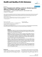
RA patients displayed significantly decreased (P < 0.0001)
blood levels of sRAGE (872 ± 65 pg/ml) as compared with
healthy controls (1290 ± 78 pg/ml) and with NID patients
(1569 ± 168 pg/ml). The sRAGE levels in synovial fluid of RA
patients (379 ± 36 pg/ml) were two times lower than in corre-
sponding blood samples (P < 0.0001), and were in the same
Table 1
Clinical and demographic characteristics of patients and healthy controls
Rheumatoid arthritis patients Non-inflammatory joint disease patients Healthy controls
Patients 62 33 45
Age (years ± standard deviation) 61.8 ± 13.9 43.0 ± 18.0 54.4 ± 9.0
Sex (male/female) 18/44 20/13 2/43
Disease duration (years ± standard deviation) 10.1 ± 8.5
Rheumatoid factor (+/-) 38/24
Radiographic changes (erosive/non-erosive) 39/23
Treatment (DMARD/no DMARD) 36/26
DMARD, disease-modifying anti-rheumatic drug.
Arthritis Research & Therapy Vol 7 No 4 Pullerits et al.
R820
range as in the synovial fluid of patients with NID (364 ± 30
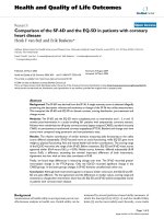
pg/ml) (Fig. 1). There was a significant positive correlation
between sRAGE levels in the matching samples of blood and
synovial fluid (r
s
= 0.48, P = 0.0002) (Fig. 2).
Patients who had RA were significantly older than healthy con-
trols and patients with NID (mean age 61.8 ± 13.9 years ver-
sus 54.4 ± 9.0 years and 43.0 ± 18.0 years, respectively).
However, no correlation with age was found in any of the
groups with respect to synovial fluid and blood sRAGE levels.
Indeed, when RA patients were stratified into younger (≤ 65
years) and older (>65 years) subgroups, no statistically signif-
icant difference was found between these groups with respect
to sRAGE levels. Our results indicate, however, that within the
age-matched groups (mean age 52.7 ± 10.2 years for RA ver-
sus 54.4 ± 9.1 years for controls) up to 65 years of age there
was still a major statistical significance regarding circulating
sRAGE levels (873 ± 72 pg/ml versus 1290 ± 78 pg/ml, P =
0.0001) (Fig. 3). Synovial sRAGE level was in the same range
in both younger RA patients (≤ 65 years, 345 ± 36 pg/ml) and
in older RA patients (>65 years old, 430 ± 73 pg/ml), and in
patients with NID (364 ± 30 pg/ml).
Correlation between sRAGE levels and clinical features
of RA
We investigated further the association between sRAGE lev-
els with main characteristics of the disease. Stratification of
patient data by radiological imaging showed that 39 patients
fulfilled the criteria for erosive disease, and 23 patients had no
erosions on recent radiographs. There was no difference in
patients' age between these two radiographic groups (61.3 ±
12.6 years versus 62.6 ± 16.1 years, respectively). No statis-
tically significant differences in synovial fluid and blood
sRAGE levels were found between these two groups (Table
2). However, patients with seropositive RA had a tendency
towards lower serum sRAGE levels than patients with seron-
egative disease (Fig. 4). Blood and synovial levels of sRAGE
were not associated with disease duration or acute-phase
reactant C-reactive protein. In contrast, the synovial sRAGE
levels in RA patients with erosive disease correlated signifi-
cantly with synovial white blood cell counts (r
s
= 0.53, P <
0.04), whereas no association was found between synovial
fluid sRAGE and synovial IL-6 levels in RA patients.
The effect of the treatment on sRAGE levels in RA
patients
At the time of sampling all patients were receiving anti-inflam-
matory treatment. Since methotrexate is the most used
DMARD in RA treatment and was predominant in our patient
population, we decided to investigate whether this treatment
had an effect of sRAGE levels in RA patients. A subgroup of
patients (n = 19) receiving monotherapy with methotrexate
was analysed and compared with patients without DMARD
treatment (n = 26). The patients' data are presented in Table
3.
The baseline characteristics of patients in both groups were
similar with respect to age and sex of patients and the pres-
ence of rheumatoid factor. However, as expected, patients
receiving DMARD treatment had significantly longer disease
duration than patients who did not take disease-modifying
drugs (13.1 ± 9.6 years versus 8.2 ± 8.3 years, P < 0.04).
Figure 1
Levels of soluble receptor for advanced glycation end products (soluble RAGE) in blood and synovial fluid (SF) of rheumatoid arthritis (RA) patients and in patients with degenerative/traumatic joint diseases (non-inflammatory joint disease [NID])Levels of soluble receptor for advanced glycation end products (soluble
RAGE) in blood and synovial fluid (SF) of rheumatoid arthritis (RA)
patients and in patients with degenerative/traumatic joint diseases
(non-inflammatory joint disease [NID]). In addition, blood levels of solu-
ble RAGE were assessed in healthy controls. Box plots show the 25th
and 75th percentiles. Horizontal lines in bold within boxes indicate
medians, and dashed lines indicate means. Vertical bars indicate the
5th and 95th percentiles. Statistical differences with respect to soluble
RAGE levels between groups were calculated using the Mann–Whit-
ney U test, and differences between paired samples were calculated by
the Wilcoxon signed rank test. Mean ± standard error of the mean
(median) values are shown. NS, not significant.
0
200
400
600
800
1000
1200
1400
1600
1800
2000
2200
Healthy
sera
NID
SF
RA
SF
RA
sera
P < 0.0001
NS
Soluble RAGE (pg/ml)
P < 0.0001
1569
±
168
(1437)
364
±
30
(375)
379
±
36
(328)
871
±
66
(770)
NID blood
NID SF
RA SF
RA blood
1290
±
78
(1227)
Healthy
blood
NS
P = 0.0025
Figure 2
Scattergram showing an association between blood and synovial solu-ble receptor for advanced glycation end products (sRAGE) levels in rheumatoid arthritis patientsScattergram showing an association between blood and synovial solu-
ble receptor for advanced glycation end products (sRAGE) levels in
rheumatoid arthritis patients. The Spearman rank correlation coefficient
(r
s
) and P value are given.
0
200
400
600
800
1000
1200
1400
1600
Synovial sRAGE
(pg/ml)
0
500
1000 1500 2000 2500
Blood
sRAGE
(pg/ml)
r
s
=0.48
P = 0.0002
Available online />R821
Also, erosive disease was more common in this group (15/19
[79%] versus 10/26 [39%], P < 0.02).
Importantly, significantly higher sRAGE levels were found in
the synovial fluid of RA patients treated with methotrexate (Fig.
5) as compared with non-treated patients. Even in this case,
the synovial fluid sRAGE displayed significant correlation (r
s
=
0.47, P < 0.05) with blood levels.
HMGB1 expression does not influence sRAGE detection
by ELISA
One of the high-affinity binding ligands for RAGE is HMGB1.
Previous studies have shown that high (microgram) levels of
HMGB1 are found in the synovial fluid and sera of RA patients
[11,12]. In addition, we demonstrated (results not shown) that
blood sRAGE in RA patients may be found on Western blot
examination at 60–80 kDa, indicating in vivo or in vitro com-
plex formation or dimerization. The complex formation between
these two proteins could possibly affect the measurement of
sRAGE by ELISA.
This prompted us to test whether HMGB1 binding to sRAGE
influenced the detection of the latter in our experimental set-
tings. If it were the case, the decreased sRAGE levels found in
our RA patient population would be explained by in vivo or ex
vivo HMGB1 interaction. Recombinant human RAGE in
concentrations of 500 pg/ml and 2000 pg/ml was incubated
with different concentrations (0, 0.1, 1 and 10 µg/ml) of
recombinant human HMGB1, and a standard ELISA analysis
was performed. Our results showed that HMGB1 did not
affect the sRAGE detection by ELISA (data not shown),
indicating that lower sRAGE levels measured in RA patients
are not due to soluble receptor engagement with HMGB1.
Discussion
This is the first study examining sRAGE levels in patients with
RA. Cell surface RAGE expression is largely dictated by the
interaction with its ligands. The expression of cellular RAGE is
rather low in mature animals and in human adults. Accumula-
tion of RAGE ligands results in increased expression of the
cell surface receptor itself [13]. Furthermore, the receptor–lig-
and interaction leads to increased RAGE-mediated signalling,
Table 2
Levels of soluble receptor for advanced glycation end products (sRAGE) in sera and in synovial fluid of rheumatoid arthritis patients
according to different disease characteristics
Disease characteristic n Blood sRAGE Synovial sRAGE
Erosive rheumatoid arthritis 39
Rheumatoid factor-positive 33 832 ± 87 (771) 345 ± 39 (323)
Rheumatoid factor-negative 6 1105 ± 209 (935) 447 ± 171 (273)
Non-erosive rheumatoid arthritis 23
Rheumatoid factor-positive 5 582 ± 141 (602) 463 ± 153 (498)
Rheumatoid factor-negative 18 945 ± 128 (772) 397 ± 79 (281)
Data presented as the mean ± standard error of the mean (median).
Figure 3
Blood soluble receptor for advanced glycation end products (sRAGE) levels in age-matched groups of rheumatoid arthritis (RA) patients and healthy controlsBlood soluble receptor for advanced glycation end products (sRAGE)
levels in age-matched groups of rheumatoid arthritis (RA) patients and
healthy controls. Box plots show the 25th and 75th percentiles. Hori-
zontal lines within boxes in bold indicate medians, and dashed lines
indicate means. Vertical bars indicate the 5th and 95th percentiles. Sta-
tistical differences with respect to sRAGE levels between groups were
calculated using the Mann–Whitney U test.
0
250
500
750
1000
1250
1500
1750
2000
2250
Blood
sRAGE
levels (pg/ml)
Healthy controls
RA patients
>65years
≤ 65 years
n =37 n =45 n =25
P = 0.0001
Arthritis Research & Therapy Vol 7 No 4 Pullerits et al.
R822
resulting in an activation of several intracellular pathways
including NF-κB [14].
sRAGE, a truncated form of the receptor, binds ligands with
affinity equal to that of cellular RAGE. It therefore has the abil-
ity to prevent RAGE signalling acting as a decoy by binding lig-
ands and preventing them from reaching cell surface RAGE.
sRAGE has successfully been used in variety of animal dis-
ease models to antagonize RAGE-mediated pathologic proc-
esses [5,14-16]. Experiments to date have shown that
pericytes and endothelial cells produce and release RAGE
extracellularly, suggesting the presence of a negative
feedback mechanism and immune surveillance mechanisms in
RAGE signalling [7].
In our study, we found that RA patients have significantly
decreased blood levels of sRAGE as compared with the
healthy population and patients with NID. Why do RA patients
display low levels of sRAGE? In the case of RA, there is a wide
diversity of RAGE ligands present in the inflamed joints, as
well as in the circulation, that could lead to the binding and
consumption of sRAGE during the inflammatory process. One
of the high-affinity ligands for RAGE/sRAGE is HMGB1, a
Table 3
Clinical and demographic characteristics of patients receiving disease-modifying anti-rheumatic treatment with methotrexate or
having no disease-modifying anti-rheumatic drug (DMARD) treatment
Characteristic Methotrexate treated No DMARD
Patients (n)19 26
Age (years ± standard deviation) 63.8 ± 14.3 60.8 ± 14.0
Sex (male/female) 5/14 8/18
Disease duration (years ± standard deviation) 13.1 ± 9.6* 8.2 ± 8.3
Rheumatoid factor (+/-) 12/7 14/12
Radiographic data (erosive/non-erosive) 15/4* 10/16
* P < 0.05 as compared with patients without DMARD treatment.
Figure 4
Blood soluble receptor for advanced glycation end products (sRAGE) levels of rheumatoid arthritis patients stratified with respect to seropos-itivity and erosivity in comparison with healthy controlsBlood soluble receptor for advanced glycation end products (sRAGE)
levels of rheumatoid arthritis patients stratified with respect to seropos-
itivity and erosivity in comparison with healthy controls. Box plots show
the 25th and 75th percentiles. Horizontal lines in bold within boxes indi-
cate medians, and dashed lines indicate means. Vertical bars indicate
the 5th and 95th percentiles. Statistical differences with respect to
sRAGE levels between groups were calculated using the Mann–Whit-
ney U test. The mean ± standard deviation (median) values are shown. *
P < 0.01 as compared with healthy controls. RF, rheumatoid factor; no
eros, no erosion.
0
582
±
141
(602)
RF +
No
eros
1290
±
78
(1227)
Healthy
945
±
128
(772)
1105
±
209
(935)
832
±
87
(771)
RF –
No
eros
RF –
Erosive
RF +
Erosive
200
400
600
800
1000
1200
1400
1600
1800
2000
Blood
sRAGE
(pg/ml)
*
*
*
Figure 5
Levels of soluble receptor for advanced glycation end products (soluble RAGE) in blood and synovial fluids of rheumatoid arthritis (RA) patients who received methotrexate treatment or were not treated with disease-modifying antirheumatic drugs (DMARDs) at allLevels of soluble receptor for advanced glycation end products (soluble
RAGE) in blood and synovial fluids of rheumatoid arthritis (RA) patients
who received methotrexate treatment or were not treated with disease-
modifying antirheumatic drugs (DMARDs) at all. Box plots show the
25th and 75th percentiles. Horizontal lines in bold within boxes indicate
medians, and dashed lines indicate means. Vertical bars indicate the
5th and 95th percentiles. Statistical differences with respect to soluble
RAGE levels between groups were calculated using the Mann–Whit-
ney U test. Mean ± standard error of the mean (median) values are
shown. NS, not significant.
No
DMARDs
Methotrexate
treatment
0
200
400
600
800
1000
1200
1400
1600
1800
2000
2200
Soluble RAGE (pg/ml)
NS
P <0.05
RA
synovial
fluid
RA blood
976
±
142
(787)
777
±
81
(699)
501
±
81
(414)
306
±
39
(229)
Available online />R823
potent cytokine playing an important role in the pathogenesis
of chronic inflammation. HMGB1 is a potent trigger of arthritis
and its expression is increased in synovial tissue of RA
patients as well as in experimental arthritis [12,17]. HMGB1
levels in the synovial fluid and sera of RA patients are signifi-
cantly elevated as compared with levels in osteoarthritis
patients [11,18]. It is thus probable that sRAGE may form in
vivo complexes with HMGB1 in the sera/synovial fluid of RA
patients, leading to inaccurately low levels of sRAGE. Upon
co-incubation of these two proteins, however, HMGB1 bind-
ing to sRAGE did not affect the detection of the latter, indicat-
ing that lower sRAGE levels measured in RA patients are not
due to neutralization by HMGB1.
An alternative explanation for the decreased sRAGE levels in
RA might be a true consumption of this molecule. In the inflam-
matory milieu, such as in the rheumatoid joint, other sRAGE
ligands also exist. Foell and colleagues have recently reported
that extracellular newly identified RAGE-binding protein (EN-
RAGE), a member of the S100/calgranulin family, was
strongly expressed in inflamed synovial tissue. Furthermore,
highly increased serum and synovial fluid levels of EN-RAGE
were found in arthritic patients in comparison with control sub-
jects [19]. Finally, raised advanced glycation end product lev-
els have been found in serum and synovial fluid of patients with
RA [20]. The presence of high levels of these soluble ligands
in RA patients provides a basis for increased consumption of
the sRAGE by interaction, followed by elimination of such
sRAGE–ligand complexes via the reticuloendothelial system
[21].
In addition, cell-bound RAGE functions as a counter-receptor
for leukocyte integrins, thereby being directly involved in
leukocyte recruitment, especially in inflammatory conditions
when the receptor expression increases [22]. Also, in this con-
text, sRAGE has been suggested to function as a potential
inhibitor of leukocyte recruitment [22]. In RA patients with ero-
sive disease, we observed a positive correlation between the
white blood cell count and synovial sRAGE levels, indicating
that endothelial cells in the synovial blood secrete sRAGE
extracellularly as a negative feedback mechanism to limit the
inflammation. Alternatively, MMP-9 has been found to shed
cell-bound RAGE into the culture medium in mice [23]. It is
possible that in the rheumatoid joint, where expression of
MMP-8 and MMP-9 is increased [24], sRAGE levels are regu-
lated by matrix metalloproteinases in a similar manner.
Taken together, we suggest that soluble RAGE may block the
ligand–RAGE interaction on the cell surface by directly bind-
ing leukocyte β2-integrin Mac-1 and thereby decreasing influx
of inflammatory cells into the joint cavity, functioning as an
immune surveillance mechanism. Lower levels of sRAGE
detected in RA patients might thus increase the propensity
towards inflammation since RAGE ligands have better access
to cell membrane-bound receptor, the binding of which leads
to the activation of inflammatory pathways.
Consistent with this concept, RA patients treated with meth-
otrexate, one of the most efficient DMARDs, displayed
increased sRAGE as compared with RA patients with no
immunosuppressive treatment. It is known that methotrexate
induces an increase of extracellular adenosine, which further
downregulates the expression of adhesion molecules includ-
ing β2-integrin Mac-1, a ligand for RAGE/sRAGE [25,26].
Methotrexate is also known to downregulate EN-RAGE
expression in the synovium of arthritis patients [19] and to sup-
press activity of tumour necrosis factor alpha [25,27], the
cytokine that has been shown to upregulate cellular RAGE
[28]. Hypothetically, as the level of membrane-bound receptor
and its ligands declines with treatment, less sRAGE is con-
sumed and the balance is restored.
We found that sRAGE levels in RA patients' synovial fluid and
sera displayed strong correlation on an individual level. Diverse
splicing variants of RAGE have been found in many tissues
and the proportion seems to differ between individuals [6-8].
The proportion and production of the soluble form of the
endogenous receptor may therefore influence the regulation of
RAGE-mediated functions in various tissues and inflammatory
conditions, including RA. Whether low sRAGE levels in RA
patients are the consequence of the disease or a potential
contributing factor to the disease needs to be elucidated.
Conclusion
We conclude that a decreased level of sRAGE in patients with
RA might increase the propensity towards inflammation,
whereas treatment with methotrexate counteracts this feature.
Competing interests
The author(s) declare that they have no competing interests.
Authors' contributions
RP carried out all the experiments, performed the statistical
analyses and wrote the manuscript. MB and LD participated in
patients' examinations, provided samples from synovial fluid/
blood as well as collected clinical data about patient groups.
AT conceived of the study, participated in its design and
helped in the writing of the manuscript.
Acknowledgements
This work was supported by grants from the Göteborg Medical Society,
the Swedish Association against Rheumatism, the Göteborg Associa-
tion against Rheumatism, the King Gustaf V foundation, the Swedish
Medical Research Council, the Nanna Svartz Foundation, Stiftelsen
Goljes Minne, the Lundberg Foundation, the Swedish Center for
Research in Sports, Medical Faculty of Lund University and the Univer-
sity of Göteborg.
Arthritis Research & Therapy Vol 7 No 4 Pullerits et al.
R824
References
1. Schmidt AM, Yan SD, Yan SF, Stern DM: The multiligand recep-
tor RAGE as a progression factor amplifying immune and
inflammatory responses. J Clin Invest 2001, 108:949-955.
2. Schmidt AM, Yan SD, Yan SF, Stern DM: The biology of the
receptor for advanced glycation end products and its ligands.
Biochim Biophys Acta 2000, 1498:99-111.
3. Drinda S, Franke S, Ruster M, Petrow P, Pullig O, Stein G, Hein G:
Identification of the receptor for advanced glycation end prod-
ucts in synovial tissue of patients with rheumatoid arthritis.
Rheumatol Int in press. 2004. Mar 26
4. Hou FF, Jiang JP, Guo JQ, Wang GB, Zhang X, Stern DM, Schmidt
AM, Owen WF Jr: Receptor for advanced glycation end prod-
ucts on human synovial fibroblasts: role in the pathogenesis
of dialysis-related amyloidosis. J Am Soc Nephrol 2002,
13:1296-1306.
5. Hofmann MA, Drury S, Hudson BI, Gleason MR, Qu W, Lu Y, Lalla
E, Chitnis S, Monteiro J, Stickland MH, et al.: RAGE and arthritis:
the G82S polymorphism amplifies the inflammatory response.
Genes Immun 2002, 3:123-135.
6. Malherbe P, Richards JG, Gaillard H, Thompson A, Diener C,
Schuler A, Huber G: cDNA cloning of a novel secreted isoform
of the human receptor for advanced glycation end products
and characterization of cells co-expressing cell-surface scav-
enger receptors and Swedish mutant amyloid precursor
protein. Brain Res Mol Brain Res 1999, 71:159-170.
7. Yonekura H, Yamamoto Y, Sakurai S, Petrova RG, Abedin MJ, Li
H, Yasui K, Takeuchi M, Makita Z, Takasawa S, et al.: Novel splice
variants of the receptor for advanced glycation end-products
expressed in human vascular endothelial cells and pericytes,
and their putative roles in diabetes-induced vascular injury.
Biochem J 2003, 370:1097-1109.
8. Park IH, Yeon SI, Youn JH, Choi JE, Sasaki N, Choi IH, Shin JS:
Expression of a novel secreted splice variant of the receptor
for advanced glycation end products (RAGE) in human brain
astrocytes and peripheral blood mononuclear cells. Mol
Immunol 2004, 40:1203-1211.
9. Arnett FC, Edworthy SM, Bloch DA, McShane DJ, Fries JF, Cooper
NS, Healey LA, Kaplan SR, Liang MH, Luthra HS, et al.: The Amer-
ican Rheumatism Association 1987 revised criteria for the
classification of rheumatoid arthritis. Arthritis Rheum 1988,
31:315-324.
10. Pullerits R, Bokarewa M, Jonsson IM, Verdrengh M, Tarkowski A:
Extracellular cytochrome c, a mitochondrial apoptosis-related
protein, induces arthritis. Rheumatology (Oxford) 2005,
44:32-9.
11. Taniguchi N, Kawahara K, Yone K, Hashiguchi T, Yamakuchi M,
Goto M, Inoue K, Yamada S, Ijiri K, Matsunaga S, et al.: High
mobility group box chromosomal protein 1 plays a role in the
pathogenesis of rheumatoid arthritis as a novel cytokine.
Arthritis Rheum 2003, 48:971-981.
12. Andersson U, Erlandsson-Harris H: HMGB1 is a potent trigger of
arthritis. J Intern Med 2004, 255:344-350.
13. Stern D, Du Yan S, Fang Yan S, Marie Schmidt A: Receptor for
advanced glycation endproducts: a multiligand receptor mag-
nifying cell stress in diverse pathologic settings. Adv Drug
Deliv Rev 2002, 54:1615-1625.
14. Schmidt AM, Hofmann M, Taguchi A, Yan SD, Stern DM: RAGE: a
multiligand receptor contributing to the cellular response in
diabetic vasculopathy and inflammation. Semin Thromb
Hemost 2000, 26:485-493.
15. Hofmann MA, Drury S, Fu C, Qu W, Taguchi A, Lu Y, Avila C, Kam-
bham N, Bierhaus A, Nawroth P, et al.: RAGE mediates a novel
proinflammatory axis: a central cell surface receptor for S100/
calgranulin polypeptides. Cell 1999, 97:889-901.
16. Taguchi A, Blood DC, del Toro G, Canet A, Lee DC, Qu W, Tanji
N, Lu Y, Lalla E, Fu C, et al.: Blockade of RAGE-amphoterin sig-
nalling suppresses tumour growth and metastases. Nature
2000, 405:354-360.
17. Pullerits R, Jonsson IM, Verdrengh M, Bokarewa M, Andersson U,
Erlandsson-Harris H, Tarkowski A: High mobility group box chro-
mosomal protein 1, a DNA binding cytokine, induces arthritis.
Arthritis Rheum 2003, 48:1693-1700.
18. Ulloa L, Batliwalla FM, Andersson U, Gregersen PK, Tracey KJ:
High mobility group box chromosomal protein 1 as a nuclear
protein, cytokine, and potential therapeutic target in arthritis.
Arthritis Rheum 2003, 48:876-881.
19. Foell D, Kane D, Bresnihan B, Vogl T, Nacken W, Sorg C, Fitzger-
ald O, Roth J: Expression of the pro-inflammatory protein
S100A12 (EN-RAGE) in rheumatoid and psoriatic arthritis.
Rheumatology (Oxford) 2003, 42:1383-1389.
20. Drinda S, Franke S, Canet CC, Petrow P, Brauer R, Huttich C,
Stein G, Hein G: Identification of the advanced glycation end
products N(epsilon)-carboxymethyllysine in the synovial tis-
sue of patients with rheumatoid arthritis. Ann Rheum Dis 2002,
61:488-492.
21. Renard C, Chappey O, Wautier MP, Nagashima M, Lundh E,
Morser J, Zhao L, Schmidt AM, Scherrmann JM, Wautier JL:
Recombinant advanced glycation end product receptor phar-
macokinetics in normal and diabetic rats. Mol Pharmacol 1997,
52:54-62.
22. Chavakis T, Bierhaus A, Al-Fakhri N, Schneider D, Witte S, Linn T,
Nagashima M, Morser J, Arnold B, Preissner KT, Nawroth PP: The
pattern recognition receptor (RAGE) is a counterreceptor for
leukocyte integrins: a novel pathway for inflammatory cell
recruitment. J Exp Med 2003, 198:1507-1515.
23. Devaux Y, Senior RM, Ray P: RAGE: a new target for MMP-9 in
the regulation of inflammatory response in the lung during oxi-
dative stress. Am J Respir Crit Care Med 2004, 169:A456.
[Abstract]
24. Tchetverikov I, Ronday HK, Van El B, Kiers GH, Verzijl N, TeKop-
pele JM, Huizinga TW, DeGroot J, Hanemaaijer R: MMP profile in
paired serum and synovial fluid samples of patients with rheu-
matoid arthritis. Ann Rheum Dis 2004, 63:881-883.
25. Chan ES, Cronstein BN: Molecular action of methotrexate in
inflammatory diseases. Arthritis Res 2002, 4:266-273.
26. Wollner A, Wollner S, Smith JB: Acting via A2 receptors, adeno-
sine inhibits the upregulation of Mac-1 (Cd11b/CD18) expres-
sion on FMLP-stimulated neutrophils. Am J Respir Cell Mol Biol
1993, 9:179-185.
27. Sajjadi FG, Takabayashi K, Foster AC, Domingo RC, Firestein GS:
Inhibition of TNF-alpha expression by adenosine: role of A3
adenosine receptors. J Immunol 1996, 156:3435-3442.
28. Tanaka N, Yonekura H, Yamagishi S, Fujimori H, Yamamoto Y,
Yamamoto H: The receptor for advanced glycation end prod-
ucts is induced by the glycation products themselves and
tumor necrosis factor-alpha through nuclear factor-kappa B,
and by 17beta-estradiol through Sp-1 in human vascular
endothelial cells. J Biol Chem 2000, 275:25781-25790.