Báo cáo y học: "Catabolic stress induces expression of hypoxia-inducible factor (HIF)-1α in articular chondrocytes: involvement of HIF-1α in the pathogenesis of osteoarthritis" ppt
Bạn đang xem bản rút gọn của tài liệu. Xem và tải ngay bản đầy đủ của tài liệu tại đây (783.9 KB, 11 trang )
Open Access
Available online />R904
Vol 7 No 4
Research article
Catabolic stress induces expression of hypoxia-inducible factor
(HIF)-1α in articular chondrocytes: involvement of HIF-1α in the
pathogenesis of osteoarthritis
Kazuo Yudoh, Hiroshi Nakamura, Kayo Masuko-Hongo, Tomohiro Kato and Kusuki Nishioka
Department of Bioregulation, Institute of Medical Science, St. Marianna University School of Medicine, Kawasaki, Japan
Corresponding author: Kazuo Yudoh,
Received: 20 Nov 2004 Revisions requested: 16 Dec 2004 Revisions received: 23 Apr 2005 Accepted: 5 May 2005 Published: 27 May 2005
Arthritis Research & Therapy 2005, 7:R904-R914 (DOI 10.1186/ar1765)
This article is online at: />© 2005 Yudoh et al.; licensee BioMed Central Ltd.
This is an Open Access article distributed under the terms of the Creative Commons Attribution License ( />2.0), which permits unrestricted use, distribution, and reproduction in any medium, provided the original work is properly cited.
Abstract
Transcription factor hypoxia-inducible factor (HIF)-1 protein
accumulates and activates the transcription of genes that are of
fundamental importance for oxygen homeostasis – including
genes involved in energy metabolism, angiogenesis, vasomotor
control, apoptosis, proliferation, and matrix production – under
hypoxic conditions. We speculated that HIF-1α may have an
important role in chondrocyte viability as a cell survival factor
during the progression of osteoarthritis (OA). The expression of
HIF-1α mRNA in human OA cartilage samples was analyzed by
real-time PCR. We analyzed whether or not the catabolic factors
IL-1β and H
2
O
2
induce the expression of HIF-1α in OA
chondrocytes under normoxic and hypoxic conditions (O
2
<6%). We investigated the levels of energy generation, cartilage
matrix production, and apoptosis induction in HIF-1α-deficient
chondrocytes under normoxic and hypoxic conditions. In
articular cartilages from human OA patients, the expression of
HIF-1α mRNA was higher in the degenerated regions than in the
intact regions. Both IL-1β and H
2
O
2
accelerated mRNA and
protein levels of HIF-1α in cultured chondrocytes. Inhibitors for
phosphatidylinositol 3-kinase and p38 kinase caused a
significant decrease in catabolic-factor-induced HIF-1α
expression. HIF-1α-deficient chondrocytes did not maintain
energy generation and cartilage matrix production under both
normoxic and hypoxic conditions. Also, HIF-1α-deficient
chondrocytes showed an acceleration of catabolic stress-
induced apoptosis in vitro. Our findings in human OA cartilage
show that HIF-1α expression in OA cartilage is associated with
the progression of articular cartilage degeneration. Catabolic-
stresses, IL-1β, and oxidative stress induce the expression of
HIF-1α in chondrocytes. Our results suggest an important role
of stress-induced HIF-1α in the maintenance of chondrocyte
viability in OA articular cartilage.
Introduction
The breakdown or absence of oxygen homeostasis is a hall-
mark of many common diseases, such as cancer, myocardial
infarction, and arthritis. In most normal and tumor tissues,
adaptation to hypoxic conditions is critical for successful tis-
sue expansion [1,2]. In response to down-regulation of oxygen
homeostasis, cells during hypoxic challenge transiently or
chronically tolerate lowered oxygen levels by means of adap-
tive mechanisms [1]. In mitochondrial oxidative phosphoryla-
tion, oxygen is the terminal electron acceptor during ATP
production. Several enzymatic reactions require oxygen as a
substrate [3,4]. Responses to hypoxia include a metabolic
shift to anaerobic glycolysis as well as the initiation of neoan-
giogenesis via the expression of angiogenic factors to
increase the opportunity for oxygen to reach the tissue [1-5].
Oxygen homeostasis and its down-regulation are involved in
the pathogenesis of common diseases [3].
It is well known that the transcription factor hypoxia-inducible
factor 1 (HIF-1) appears to be one of the major regulators of
the hypoxic response [3,6]. HIF-1 controls hypoxic expression
of erythropoietin, as well as the expression of genes with met-
abolic functions such as glucose transport and metabolism,
and angiogenic factors such as vascular endothelial cell
DMEM = Dulbecco's modified Eagle's medium; ERK = extracellular signal-regulated kinase; GAG = glycosaminoglycan; HIF-1α = hypoxia-inducible
factor 1α; IL-1 = interleukin-1; MAPK = mitogen-activated protein kinase; MCL = medial collateral ligament; OA = osteoarthritis; ODN = oligonucle-
otide; PBS = phosphate-buffered saline; PI3K = phosphatidylinositol 3-kinase; TBST = Tris-buffered saline/Tween 20; TdT = terminal deoxynucleoti-
dyl transferase; Tris = tris(hydroxymethyl)aminomethane.
Arthritis Research & Therapy Vol 7 No 4 Yudoh et al.
R905
growth factor (VEGF) [6-8]. HIF-1 is a heterodimer of the PAS
subfamily of basic-helix-loop-helix transcription factors, and it
consists of the subunit HIF-1α (120 kDa), produced in
response to hypoxia, and the constitutively expressed HIF-1α
(91 to 94 kDa) subunit [9]. HIF-1 protein accumulates and
activates the transcription of genes that are of fundamental
importance for oxygen homeostasis, including genes involved
in energy metabolism, angiogenesis, vasomotor control, apop-
tosis, proliferation and matrix production, under hypoxic condi-
tions [6,8,9].
Articular cartilage is an avascular tissue lacking a capillary net-
work, in which oxygen is limited due to its delivery via diffusion
through the synovial fluid. It is well known that there is a phys-
iological gradient of oxygenation within articular cartilage [10-
12]. It has been reported that the partial pressure of O
2
in syn-
ovial fluid in joints affected by osteoarthritis (OA) is between
40 and 85 mmHg, corresponding to an oxygen concentration
of approximately 6 to 11% [13]. Since O
2
must enter from the
cartilage surface, the concentration of oxygen is approximately
6% at the surface zone of the articular tissue and less than 1%
in the deep zone. We histologically examined the oxygen gra-
dation in articular cartilage tissue by immunofluorescence
staining with a specific probe. We performed the analysis in
human articular cartilage tissue in patients undergoing arthro-
plastic knee surgery. The levels of immunostaining revealed an
O
2
tension (approximately 3 to 8%) at the surface of the carti-
lage similar to that in positive control tumor tissues with
already known O
2
tension. There is a general consensus that
articular chondrocytes are adapted to hypoxic conditions.
Since HIF-1α expression is associated with low O
2
, this factor
may play a role in chondrocyte survival and the maintenance of
fundamental homeostasis in the normally hypoxic articular car-
tilage. In addition, degeneration of articular cartilage may
directly influence the chondrocyte microenvironment, espe-
cially cellular adaptation to hypoxic conditions, in articular car-
tilage. Even a slight change may affect the adaptative hypoxic
conditions of chondrocytes, resulting in alteration of the cellu-
lar microenvironment that is involved in the maintenance of
articular cartilage. Indeed, more recently it has been demon-
strated that HIF-1α is expressed in OA articular cartilage [14].
However, the exact role of this factor in the pathogenesis of
OA remains unclear.
We postulated that HIF-1α could play an important role as a
survival factor protecting tissue against catabolic changes
during the progression of OA. Our data show here for the first
time a correlation between the levels of expression of HIF-1α
and degeneration of articular cartilage in patients with OA. To
clarify the role of HIF-1α in the pathogenesis of OA, we inves-
tigated whether or not hypoxia and catabolic factors (IL-1β and
H
2
O
2
) affected the expression of HIF-1α, energy generation,
cartilage matrix production, and apoptosis in OA
chondrocytes.
We also report evidence for the action of HIF-1α as a
chondrocyte survival factor in OA.
Materials and methods
Preparation of human articular cartilage samples
Donor OA cartilage samples were obtained from knee joints of
OA patients undergoing arthroplastic knee surgery (seven OA
patients) after obtaining the patients' informed consent. The
characteristics of patients are summarized in Table 1. Each
sample was cut and divided into two pieces: one was used for
histological evaluation and the other was stored at -30°C for
later analysis by real-time PCR analysis.
Each cartilage sample was evaluated histologically and mac-
roscopically for the degree of degeneration according to the
scales of Mankin and colleagues and of Collins [15,16]. Artic-
ular cartilage samples with subchondral bones were fixed for
2 days in 4% paraformaldehyde solution and then decalcified
in 4% paraformaldehyde containing 0.85% sodium chloride
and 10% acetic acid. Tissues were dehydrated in a series of
ethanol solutions and infiltrated with xylene and before being
embedded in paraffin and cut into 6-µm sections. Sections
were deparaffinized through sequential immersion in xylene
and a graded series of ethanol solutions in accordance with
conventional procedures. Sections were also stained with
safranin O-fast green to determine the loss of proteoglycans
[17].
Chondrocyte isolation and culture
Human articular cartilage samples were obtained from knee
joints during arthroplastic surgery for OA (n = 7, one male, six
females, 61, 62, 64, 66, 67, 68, 72 years old) after obtaining
the patients' informed consent. Cartilage tissues were cut into
small pieces, washed in PBS, and digested in Dulbecco's
modified Eagle's medium (DMEM; Sigma, St. Louis, MO) con-
taining 1.5 mg/ml collagenase B (Sigma). Digestion was car-
ried out at 37°C overnight on a shaking platform. Cells were
centrifuged, washed with PBS, and plated with fresh DMEM.
Chondrocytes were cultured in DMEM supplemented with
10% heat-inactivated fetal calf serum, 2 mM L-glutamine, 25
mM HEPES (2-[4-(2-hydroxyethyl)-1-piperazinyl] ethanesul-
fonic acid), and 100 units/ml penicillin and streptomycin at
37°C in a humidified 5% CO
2
atmosphere [18].
Chondrocyte culture under hypoxic conditions
Human chondrocytes were dispensed into a 10-cm culture
plate. The plates were placed in a sealed hypoxia chamber
(Billups-Rothenberg, Del Mar, CA, USA) equilibrated with a
humidified 5% CO
2
atmosphere or with certified gas contain-
ing 1% O
2
, 5% CO
2
, and 94% N
2
[19,20]. In this hypoxia
chamber system, approximately 5 to 6% O
2
tension was
observed after 15 min of gas flow (20 l/min). The O
2
tension in
the culture medium was monitored with an oxygen meter (Fuso
Rekaseihin Ltd, Tokyo, Japan) as described by the manufac-
turer. We monitored the O
2
tension with an oxygen meter to
Available online />R906
maintain the concentration of approximately 6%. When so
indicated, recombinant human IL-1β (10 ng/ml; Sigma) or
H
2
O
2
(10.0 µM; Wako Pure Industries, Tokyo, Japan) was
added, and the cells were incubated under normoxic or
hypoxic culture conditions at 37°C. As a positive control,
COCl
2
(150 µM; Sigma), a chemical inducer of HIF-1, was
added to the cells during the incubation time in normoxia or
hypoxia [20].
In other experiments, human chondrocytes were cultured in
the presence or absence of a phosphatidylinositol 3-kinase
(PI3K) inhibitor LY294002 (Sigma), a p38 mitogen-activated
protein kinase (MAPK) inhibitor SB203580 (Sigma), and
extracellular signal-regulated kinase (ERK 1/2) inhibitor
PD98059 (Wako).
Immunoblotting
Cells were lysed in boiling sample buffer as suggested by the
manufacturer (Sigma). Samples were then homogenized by
repeated aspiration through a 26-gauge needle. Cellular pro-
teins were resolved by SDS-PAGE (12.5% acrylamide) and
were transferred to nitrocellulose membranes. Blots were
incubated for 2 hours in Tris-buffered saline/Tween 20 (TBST;
10 mM Tris/HCL, pH 8.0, 150 mM NaCl, and 0.2% Tween 20)
containing 2% powdered skimmed milk and 1% bovine serum
albumin. After three washes with TBST, membranes were
incubated for 2 hours with the primary antibody to HIF-1α
(diluted 1000-fold in TBST) (Santa Cruz Biotechnology Inc,
Santa Cruz, CA) and for 1 hour with horseradish-peroxidase-
conjugated goat antimouse IgG (diluted 1000-fold) (DAKO,
Glostrup, Denmark). Bound antibodies were detected using
an ECL detection kit (Amersham Bioscience KK, Tokyo,
Japan). Densitometry of the signal bands was analyzed with
Image Gauge version 4.0 (FUJI Photo Film, Tokyo, Japan).
Proteoglycan production in chondrocytes
Chondrocyte activity was measured by the production of gly-
cosaminoglycan (GAG) from cultured chondrocytes.
Chondrocytes were cultured under either normoxic or hypoxic
conditions using the sealed hypoxia chamber. After 24 hours
of incubation, we collected the cells and supernatant. The
amount of GAG in the supernatant was measured by using a
spectrophotometric assay with dimethylmethylene blue
(Aldrich Chemical, Milwaukee, WI, USA) measured at 540 nm
using shark chondroitin sulfate (Sigma) as a standard [19].
Measurement of lactic acid in cultured chondrocytes
Supernatants from chondrocyte cultures were collected after
24 hours under normoxic or hypoxic conditions. Lactic acid
was determined by a colorimetric assay (Sigma) at 540 nm in
accordance with the manufacturer's instructions. Lactic acid
levels were normalized to total protein content as measured by
the Bradford assay (Bio-Rad, Hercules, CA, USA) [21].
ATP levels in cultured chondrocytes
Chondrocytes were collected after a 24-hour incubation under
normoxic or hypoxic conditions. The ATP Bioluminescence
assay kit CLS II (Roche, Heidelberg, Germany) was used. The
assay is based on the light-emitting oxidation of luciferin by
luciferase in the presence of extremely low levels of ATP. After
collecting the chondrocytes by scraping, cells were centri-
fuged for 10 min at 500 × g in the cold. Chondrocytes pellets
were extracted by boiling 100 mM Tris (tris(hydroxyme-
thyl)aminomethane) buffer containing 4 mM EDTA (ethylenedi-
aminetetraacetic acid) for 2 min in order to inactivate
NTPases. Cell remnants were removed by centrifugation at
1000 × g. Supernatants were removed and placed on ice.
Determination of free ATP was as outlined in the manufac-
turer's protocol. Light emission was measured at 562 nm
using a luminometer. ATP levels were normalized to protein
content as measured by the Bradford assay (Bio-Rad) [19].
RT-PCR
Total RNA was extracted from articular cartilage by acid gua-
nidine–phenol–chloroform extraction using ISOGEN
®
(Nip-
pon Gene Inc, Tokyo, Japan). First-strand complementary
Table 1
Characteristics of patients with osteoarthritis
Mankin grade
Donor Age (y) Sex Disease duration (y) Intact region Degenerated region
161Female5.537
262Female6.049
364Male8.438
466Female11.529
567Female9.629
668Female8.428
772Female9.039
Arthritis Research & Therapy Vol 7 No 4 Yudoh et al.
R907
DNA (cDNA) was synthesized with Superscript II reverse tran-
scriptase. PCR amplification was performed using specific
primers (Table 2). The PCR products were analysed by elec-
trophoresis in 2% agarose gels stained with ethidium bromide,
and bands were visualized and photographed under ultraviolet
excitation.
Real-time PCR
For PCR analyses, cDNA from triplicate dishes from four inde-
pendent experiments (24 hours of hypoxia or normoxia) were
diluted to a final concentration of 10 ng/ µl. Quantitative real-
time RT-PCR was performed with a TaqMan Universal Master-
mix (Biosystems Inc, Foster City, CA). cDNA (50 ng) was used
as template to determine the relative amounts of mRNA by
real-time PCR (ABI 7700 sequence detection system) using
specific primers and probes (Table 2). The reaction was con-
ducted as follows: 95°C for 4 min, and 40 cycles of 15s at
95°C and 1 min at 60°C (21). To standardize mRNA levels, we
amplified 18S rRNA as an internal control and calculated
using Microsoft Excel.
Antisense oligonucleotide treatment of chodrocytes
HIF-1α depletion in chondrocytes was accomplished by using
antisense oligonucleotide (ODN) loading using phospho-
rothioate derivatives of antisense (5'-GCCGGCGCCCTC-
CAT-3') or control sense (5'-ATGGAGGGCGCCGGC-3')
oligonucleotides. Antisense HIF-1α ODN and control ODN
were designed and synthesized by BIOGNOSTIK (Göttingen,
Germany). Scrambled oligonucleotide was used as control.
Chondrocytes were washed in serum-free medium and then in
medium containing 20 mg/ml transfection reagent (Qiagen
Inc, Valencia, CA, USA) with 2 µM HIF-1α antisense or control
ODN. Cells were incubated for 4 hours at 37°C and then
replaced with medium containing growth factors. The cellular
uptake efficiency was monitored by fluorescein-isothiocy-
anate-labeled ODN (transfection efficiency approximately 60
to 70% after 4 hours of treatment). The transfection efficiency
detected by fluorescein-isothiocyanate-labeled ODN was
maintained after a further 24 hours of incubation. Treated cells
were cultured in hypoxic or normoxic conditions for the indi-
cated periods of time (24 hours) in each experiment. HIF-1α
mRNA was quantified by RT-PCR and western blotting analy-
sis as described above. Data were analyzed for four independ-
ent experiments.
Apoptosis
Human subconfluent chondrocytes were cultured in the pres-
ence of 10 ng/ml IL-1β for 24 hours under the normoxic or
hypoxic conditions described above. Cellular apoptosis was
detected using the Apoptosis detection kit (TdT in situ apop-
tosis detection kit: R&D systems Inc., MN, USA) in chondro-
cyte cell cultures in accordance with the manufacturer's
protocol. The kit was used to identify apoptotic cells by detect-
ing DNA fragmentation through a combination of enzymology
and immunohistochemistry techniques. Biotinylated nucle-
otides are incorporated into the 3'-OH ends of the DNA frag-
ments by terminal deoxynucleotidyl transferase (TdT). Cells
containing fragmented nuclear chromatin characteristic of
apoptosis exhibit a brown nuclear staining. Apoptosis was
assessed by measuring the percentage of apoptotic nuclei in
each sample [22,23].
Statistical analysis
Results were expressed as means ± standard deviations. Data
were analyzed by a nonparametric statistical analysis. An anal-
ysis resulting in value of P < 0.05 was considered statistically
significant.
Results
HIF-1α mRNA expression in articular cartilage from
patients with OA
To clarify the expression of HIF-1α mRNA in human OA carti-
lage, the real-time PCR analysis for HIF-1α was performed
with donor-matched pairs of intact and degenerated articular
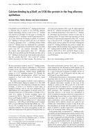
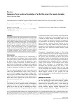
cartilage isolated from the same OA sample. Fig. 1a shows a
representative safranin-O staining in the degenerated region
and intact region of articular cartilage from OA patients. The
levels of HIF-1α mRNA in all seven donor articular cartilage
samples were higher in the degenerated regions than in the
intact regions (Fig. 1b).
Table 2
Sequences of PCR primers and probes
Primer (5'-3') Probe
c-Jun fw: TGCATGCTATCATTGGCTCATAC CCCGGCAACACACA-MGB
rv: CACACCATCTTCTGGTGTACAGTCT
HIF-1α fw:CTATGGAGGCCAGAAGAGGGTAT AGATCCCTTGAAGCTAG-MGB
rv:CCCACATCAGGTGGCTCATAA
Glucose transporter-1 fw:GGGCATGTGCTTCCAGTATGT CAACTGTGCGGCCCCTACGTCTTC
rv:ACGAGGAGCACCGTGAAGAT
fw, forward; rv, reverse
Available online />R908
Catabolic factors induce the expression of HIF-1α in
human articular cartilage
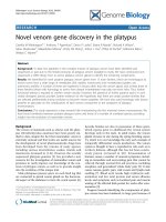
To clarify the effect of catabolic factors on HIF-1α expression
in human articular cartilage, the quantitative real-time PCR and
western blotting analysis were performed under normoxic and
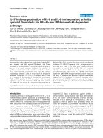
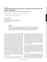
hypoxic culture conditions. In normoxic culture conditions,
mRNA levels of HIF-1α were observed in cultured chondro-
cytes, whereas HIF-1α protein was undetected regardless of
stimulation of IL-1β and H
2
O
2
(Fig. 2). Under hypoxic culture
conditions, both HIF-1α mRNA and protein were detected in
cultured chondrocytes (Figs 2, 3). The expression of HIF-1α
was significantly accelerated by the chondrocyte catabolic
factors IL-1β and H
2
O
2
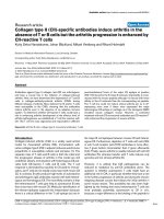
(Figs 2, 3). Under hypoxic conditions,
the inhibitors of PI3K and p38 kinase caused a significant
decrease in the catabolic-factor-induced HIF-1α expression
(Fig. 3a, b). Data from four independent experiments were
analyzed.
Role of HIF-1α in free ATP production in human articular
chondrocytes
To study the role of HIF-1α in chondrocyte energy production,
we measured the free ATP levels of cultured chondrocytes
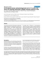
under normoxic and hypoxic culture conditions. In control
chondrocytes, the levels of free ATP in hypoxia were signifi-
cantly higher than in normoxia. Under hypoxic conditions, HIF-
1α-deficient chondrocytes showed a significant decrease of
free ATP in comparison with control ODN-treated chondro-
cytes (Fig. 4b). In HIF-1α-deficient chondrocytes, free ATP
production was approximately 20% of control cells under
hypoxic conditions. In contrast, although HIF-1α-deficient
chondrocytes showed a slight decrease of energy generation
under normoxic conditions, there was no statistically signifi-
cant difference in energy generation between the three groups
(normal chondrocytes, antisense ODN-treated chondrocytes,
and chondrocytes treated with the scrambled ODN). Data for
four independent experiments were analyzed.
Influence of HIF-1α deficiency on glycolysis in human
chondrocytes
As shown in Fig. 4c, d, significant increases of lactic acid (c)
and glucose transporter-1 (d) were observed in control ODN-
treated chondrocytes under hypoxic culture conditions
compared with normoxic culture conditions. In contrast, HIF-
1α-deficient chondrocytes showed a complete loss of the
induced increases in glycolytic activities even under hypoxic
culture conditions.
Figure 1
Levels of HIF-1α mRNA in the articular cartilage from patients with osteoarthritis (OA)Levels of HIF-1α mRNA in the articular cartilage from patients with osteoarthritis (OA). (a)Representative x-ray film of knee joint and safranin-O stain-
ing for hypoxia-inducible factor 1α(HIF-1α) in the degenerated region and intact region of articular cartilage from a 66-year-old woman with OA.
Original magnification of histological sections × 200. (b) The mRNA levels of HIF-1α were higher in the degenerated regions than in the intact
regions from the same OA sample.
Arthritis Research & Therapy Vol 7 No 4 Yudoh et al.
R909
Under normoxic conditions, HIF-1α-deficient chondrocytes
showed a slight decrease of energy generation; however,
there was no statistically significant difference in energy gen-
eration between control chondrocytes and antisense HIF-1α-
treated chondrocytes. Data for four independent experiments
were analyzed.
Proteoglycan production from chondrocytes in different
oxygen tension
To test whether HIF-1α-mediated alteration affects the poten-
tial to produce matrix proteins in chondrocytes, we determined
the amount of GAG produced by cultured chondrocytes. We
observed large increases in the concentration of GAG in con-
trol ODN-treated cultures under hypoxia compared with nor-
moxia. GAG levels were decreased in HIF-1α-deficient
chondrocytes under hypoxia to approximately 35% of control
levels (Fig. 5). Data for four independent experiments were
analyzed.
Apoptosis induction by catabolic factors in HIF-1α-
deficient chondrocytes
Under hypoxic conditions, IL-1β-induced apoptosis was
increased in hypoxic chondrocytes lacking HIF-1α, to twice
that of control ODN-treated chondrocytes (Fig. 6). Even under
normoxic conditions, HIF-1α-deficient chondrocytes showed
significantly increased levels of apoptosis compared with their
control counterparts. Data for four independent experiments
were analyzed.
Discussion
Our findings show the potential involvement of HIF-1α expres-
sion in the progression of articular cartilage degeneration. In
patients with OA, stronger expression of HIF-1α mRNA in
chondrocytes was observed in degenerating regions than in
intact regions from the same articular cartilage samples. Our
findings in human articular cartilage tissues indicate for the
first time that expression of HIF-1α mRNA is closely involved
in the progression of articular cartilage degeneration.
The HIF-1 complex is ubiquitous, and the presence of this
complex in growth-plate chondrocytes has been documented
recently [24-26]. Schipani and colleagues reported that in
HIF-1α-null mice, hypoxic chondrocytes showed massive cell
death in cartilaginous elements such as the chondrosternal
junction of the ribs and growth plate, suggesting that HIF-1α
is not only crucial for survival of hypoxic chondrocytes, but also
Figure 2
IL-1β and H
2
O
2
induce the expression of HIF-1α mRNA in human articular chondrocytesIL-1β and H
2
O
2
induce the expression of HIF-1α mRNA in human articular chondrocytes. Under normoxic culture conditions, mRNA levels of
hypoxia-inducible factor 1α (HIF-1α) were observed in cultured chondrocytes, whereas HIF-1α protein was undetected in the cells. HIF-1α mRNA
was accelerated by IL-1β or H
2
O
2
in cultured chondrocytes under hypoxic conditions. Cobalt chloride (CoCl
2
), chemical inducer of HIF-1, was used
for the positive control. *P < 0.05, **P < 0.01 compared with the control. Cont., control.
Available online />R910
modulates the process of chondrocyte proliferation, differenti-
ation, and growth arrest in growth-plate chondrocytes [26].
More recently, Coimbra and colleagues also showed that HIF-
1α is expressed in cultured cartilage and chondrocytes under
both normoxic and hypoxic conditions [14]. Their findings of
HIF-1α expression in chondrocytes are basically consistent
with our results from both human and rat OA cartilages. How-
ever, from their data, it remained unclear whether HIF-1α
expression in chondrocytes is related to the degeneration of
articular cartilage in vivo. Indeed, Coimbra and colleagues
showed that HIF-1α was expressed not only in normal
chondrocytes and cartilage but also in OA chondrocytes,
under both hypoxic and normoxic conditions in vitro. In our
present study, HIF-1α protein was undetected in chondro-
cytes under normoxic conditions. It is well known that cellular
HIF-1α is not detected in normoxia [27-29]. Under normoxic
conditions, the HIF-1α protein undergoes ubiquitination and
rapid degeneration in proteasomes [30].
Our data suggest that chondrocyte catabolic factors IL-1β and
oxidative stress (oxidative free radicals) may induce the
expression of HIF-1α in articular chondrocytes. IL-1 has been
shown both to inhibit chondrocyte anabolic activity, including
the down-regulation of proteoglycan synthesis, and to stimu-
late catabolic activity, including production of metalloprotein-
ases [31,32]. IL-1 also stimulates chondrocyte expression of
inducible nitric oxide synthesis, iNOS, which results in an
increase in NO production [33]. Numerous reports have
already demonstrated that oxidative stress acts as a catabolic
factor in articular cartilage [34-38]. Articular chondrocytes
actively produce endogenous reactive oxygen species, O
2
-
[35], NO [36],
-
HO [37], and H
2
O
2
[3]). Oxidative damage in
cartilage may affect chondrocyte function, resulting in
changes in cartilage homeostasis that are relevant to cartilage
aging and the development of OA. Our data indicated that in
cultured chondrocytes, both mRNA and protein levels of HIF-
1α were up-regulated by both IL-1β and H
2
O
2
under hypoxic
but not normoxic conditions. These findings suggest that OA-
related catabolic stresses (IL-1β, H
2
O
2
) induce the expression
of HIF-1α in the degenerated articular cartilages as degenera-
tion progresses.
Interestingly, besides hypoxia, many cytokines and growth fac-
tors have been shown to be capable of stabilizing and activat-
ing HIF-1α under normoxic conditions. Stimulation of cultured
synovial fibroblasts with IL-1β and TNFα increases levels of
HIF-1α mRNA. Moreover, incubation with IL-1β leads to stabi-
lization of HIF [39]. Our results of catabolic stress-induced
expression of HIF-1α in chondrocytes are consistent with
these findings. These findings suggest that HIF-1α may, at
least in part, have some role in the pathogenesis of inflamma-
tory arthritis even under normoxic conditions, although further
studies are needed to clarify this issue. Also, these findings,
including our results, provide evidence to support the idea that
Figure 3
Catabolic factors induce the expression of HIF-1α protein in human articular cartilageCatabolic factors induce the expression of HIF-1α protein in human articular cartilage. (a)Hypoxia-inducible factor 1α (HIF-1α) protein was acceler-
ated by IL-1β or H
2
O
2
in cultured chondrocytes under hypoxic conditions. (b)Under hypoxic conditions, the inhibitors of PI3K and p38 mitogen-acti-
vated protein kinase (MAPK) reduced protein levels of IL-1β-induced HIF-1α expression. Cobalt chloride (CoCl
2
), chemical inducer of HIF-1, was
used for the positive control. *P < 0.05, **P < 0.01 compared with the control. LY294002: phosphatidylinositol 3-kinase inhibitor; SB203580: p38
mitogen-activated protein kinase inhibitor; PD98059: extracellular signal-regulated kinase inhibitor.
Arthritis Research & Therapy Vol 7 No 4 Yudoh et al.
R911
OA-related catabolic factors (IL-1β etc.) induce HIF-1α during
the progression of cartilage degeneration.
In this context, we also studied the signal transduction path-
ways involved in stress-induced HIF-1α expression in
chondrocytes. It has been reported that p38 MAPK, PI3K, and
ERK MAPK pathways are responsible for the stress-induced
responses in a variety of cells [40-42]. We found that both IL-
1β and H
2
O
2
induced a prolonged activation of p38 in
chondrocytes (data not shown). Under hypoxic conditions, the
inhibitors of PI3K and p38 kinase caused a significant
decrease in catabolic-factor-induced HIF-1α expression; this
finding supports the idea that PI3K and p38 kinase, but not
ERK, activation are required for catabolic stress-induced HIF-
1α expression in chondrocytes in hypoxia. Local accumulation
of a regulating protein to adapt to hypoxia may be mediated, at
least in part, by p38 and PI3K in articular chondrocytes. In
addition, we have focused on the redox factor 1 (Ref-1, also
known as APE, HAP1, and APEX), a ubiquitous multifactorial
protein that is a redox-sensitive regulator of mutifactorial tran-
scription factors, including nuclear factor κB, c-myc gene, acti-
vating protein-1, and HIF-1α. Ref-1 may play a critical role in
the regulation of endothelial cell fate in response to patho-
physiological stimuli such as hypoxia [43]. We have studied
the interactions between Ref-1 and HIF-1 activity in OA
chondrocytes (data not shown).
Figure 4
Effect of HIF-1α on ATP production and glycolysis in human articular cartilageEffect of HIF-1α on ATP production and glycolysis in human articular cartilage. (a)Hypoxia-inducible factor 1α (HIF-1α) depletion by antisense oligo-
nucleotide was assessed by RT-PCR and western blotting analyses. HIF-1α mRNA and protein expressions were reduced in antisense HIF-1α-
treated chondrocyte populations. Scrambled oligonucleotide was used as control oligonucleotide. Representative data from four independent exper-
iments are shown. (b)In hypoxia, HIF-1α-deficient chondrocytes showed a significant decrease of free ATP in comparison with control oligonucle-
otide-treated chondrocytes. Statistical differences were calculated using data from four independent experiments. (c, d) The levels of lactate (c) and
glucose transporter-1 (Glu-1) (d) were increased in the scrambled ODN-treated chondrocytes under hypoxic culture conditions compared with nor-
moxic culture condition. In HIF-1α-deficient chondrocytes, both glycolytic activities were reduced under hypoxic conditions. Statistical differences
were calculated using data from four independent experiments.
a
P < 0.01, control oligonucleotide hypoxia vs HIF-1α-deficient hypoxia; *P < 0.05,
**P < 0.01.
Available online />R912
Our in vitro data clearly indicate that expression of HIF-1α is
responsible for the energy generation and cellular survival of
hypoxic chondrocytes. We have shown that HIF-1α activity is
essential for regulation of glycolysis, energy generation,
synthesis of cartilage matrix proteins, and cell survival in OA
chondrocytes under hypoxic conditions. HIF-1α-null chondro-
cytes did not maintain their viability; energy generation, and
matrix production under normoxic and hypoxic conditions. In
addition, HIF-1α-null chondrocytes showed accelerated apop-
tosis induction by IL-1β, suggesting that HIF-1α has an impor-
tant role in the survival of tissues that lack a functional
vasculature, such as articular cartilage.
Articular cartilage adapts to hypoxic conditions, since the car-
tilage is an avascular tissue. Nutrition and oxygen for articular
cartilage are supplied from the synovial fluid. Even a surface
zone of articular cartilage has lower oxygen tension
(approximately 6%) than synovial fluid (approximately 6 to
15%) [10-13]. There is an oxidative gradient in articular carti-
lage. Oxygen homeostasis in normal articular cartilage is main-
tained under hypoxic conditions. During the progression of
cartilage degeneration, OA-related catabolic stresses,
mechanical and chemical, including IL-1β and oxidative stress,
could induce the degradation of the extracellular matrix and
decrease chondrocyte viability, resulting in the down-regula-
tion of chondrocyte environment and the further degeneration
of articular cartilage. The OA-related changes may also affect
oxygen tension and the hypoxic conditions in articular carti-
lage. Breakdown of oxygen homeostasis in articular cartilage
may influence the chondrocytes adapted to hypoxic conditions
within the articular cartilage. Although further studies are
Figure 5
Glycosaminoglycan production and apoptosis induction in HIF-1α-defi-cient chondrocytesGlycosaminoglycan production and apoptosis induction in HIF-1α-defi-
cient chondrocytes. In the scrambled oligonucleotide-treated groups,
the amount of glycosaminoglycan (GAG) produced by cultured
chondrocytes was higher under hypoxic conditions than under nor-
moxic conditions. Under hypoxic conditions, GAG levels decreased in
chondrocytes deficient in hypoxia-inducible factor 1α (HIF-1α). Statisti-
cal differences were calculated using data from four independent
experiments.
a
P < 0.01, control oligonucleotide hypoxia vs HIF-1α-anti-
sense hypoxia; *P < 0.05; **P < 0.01.
Figure 6
Apoptosis induction in HIF-1α-deficient chondrocytesApoptosis induction in HIF-1α-deficient chondrocytes. IL-1β was used for apoptosis induction under hypoxic or normoxic conditions in chondrocytes
lacking hypoxia-inducible factor 1α (HIF-1α), treated with oligonucleotide, and cultured without oligonucleotide, and also not exposed to the oligonu-
cleotide or HIF-1α antisense nucleotides. IL-1β-induced apoptosis was significantly increased in HIF-1α-deficient chondrocytes compared with
chondrocytes treated with scrambled oligonucleotide under both normoxic and hypoxic culture conditions. Statistical differences were calculated
using data from four independent experiments. **P < 0.01, control oligonucleotide group vs HIF-1α-antisense group.
Arthritis Research & Therapy Vol 7 No 4 Yudoh et al.
R913
needed to clarify the exact mechanism of HIF-1α expression,
HIF-1α may be expressed in response to change of cellular
microenvironment, especially O
2
tension, in the tissue. Jewell
and colleagues reported that HIF-1α was up-regulated by
reoxygenation [44]. Their findings suggest that expression of
HIF-1α may be influenced by the cellular microenvironment.
When there is deviation from the stable condition in terms of
O
2
tension, HIF-1α expression may be influenced to maintain
the cellular homeostasis. Articular chondrocytes that are well
adapted to hypoxia into the tissue may express HIF-1α with the
deviating from adaptation to hypoxia. We postulated that HIF-
1α is expressed in response to catabolic change in articular
cartilage to maintain the cell viability and readaptation to
hypoxia. Catabolic stress during the development of OA could
influence the chondrocyte adaptation to hypoxia in the tissue.
Our findings of catabolic-factor-induced HIF-1α expression in
chondrocytes provide evidence to support the idea that HIF-
1α expression is up-regulated in response to catabolic degen-
eration of articular cartilage. Although genes under the control
of HIF-1α have not yet been analyzed in OA, stress-induced
HIF-1α may lead to the expression of anti-apoptotic factors or
act as a chondroprotective factor to maintain chondrocyte via-
bility in the face of catabolic changes in articular cartilage.
Many molecular aspects of HIF-1 signaling should be also
studied to further clarify the role of HIF-1 in the pathogenesis
of OA.
Conclusion
Taken together, our findings show for the first time that expo-
sure to IL-1β and oxidative stress induce HIF-1α expression in
degenerated articular cartilage, which is mediated, at least in
part, by activation of PI3K and p38 kinase. Our data suggest
a potential for HIF-1α in the maintenance of chondrocyte via-
bility in articular cartilage challenged by progressive articular
cartilage degeneration, and they provide new insights into
pathogenesis of OA.
Competing interests
The authors declare that they have no competing interests.
Authors' contributions
KY carried out in vitro studies (cell culture), participated in the
design of the study, conducted sequence alignment, and
drafted the manuscript. HN, K H-M, TK, and KN conceived the
study, participated in its design and coordination, and helped
to draft the manuscript. All authors read and approved the final
manuscript.
Acknowledgements
This study was supported by grants from the Ministry of Education, Cul-
ture, Sports, Science and Technology of Japan, the Ministry of Health,
Labour and Welfare of Japan, and the Japan Rheumatism Foundation.
References
1. Maxwell PH, Ratcliffe PJ: Oxygen sensors and angiogenesis.
Semin Cell Dev Biol 2002, 13:29-37.
2. Maxwell PH: HIF-1's relationship to oxygen: simple yet
sophisticated. Cell Cycle 2004, 3:156-159.
3. Distler JHW, Wenger RH, Gassmann M, Kurowska M, Hirth A, Gay
S, Distler O: Physiologic responses to hypoxia and implica-
tions for hypoxia-inducible factors in the pathogenesis of
rheumatoid arthritis. Arthritis Rheum 2004, 50:10-23.
4. Semenza GL, Roth PH, Fang HM, Wang GL: Transcriptional reg-
ulation of genes encoding glycolytic enzymes by hypoxia-
inducible factor 1. J Biol Chem 1994, 269:23757-23763.
5. Kung AL, Wang S, Klco JM, Kaelin WG, Livingston DM: Suppres-
sion of tumor growth through disruption of hypoxia-inducible
transcription. Nat Med 2000, 6:1335-1340.
6. Semenza GL: Regulation of mammalian O
2
homeostasis by
hypoxia-inducible factor 1. Annu Rev Cell Dev Biol 1999,
15:551-578.
7. Richard DE, Berra E, Pouyssegur J: Angiogenesis: how a tumor
adapts to hypoxia. Biochem Biophys Res Commun 1999,
266:718-722.
8. Wenger RH: Mammalian oxygen sensing, signaling and gene
regulation. J Exp Biol 2000, 203:1253-1263.
9. Wang GL, Jiang BH, Rue EA, Semenza GL: Hypoxia-inducible
factor 1 is a basic-helix-loop-helix-PAS heterodimer regulated
by cellular O2 tension. Proc Natl Acad Sci USA 1995,
92:5510-5514.
10. Silver IA: Measurement of pH and ionic composition of pericel-
lular sites. Philos Trans R Soc Lond B Biol Sci 1975,
271:261-272.
11. Cernanec J, Guilak F, Weinberg JB, Pisetsky DS, Fermor B: Influ-
ence of hypoxia and reoxygenation on cytokine-induced pro-
duction of proinflammatory mediators in articular cartilage.
Arthritis Rheum 2002, 46:968-975.
12. Treuhaft PS, McCarty DJ: Synovial fluid pH, lactate, oxygen and
carbon dioxide partial pressure in various joint diseases.
Arthritis Rheum 1971, 14:475-484.
13. Grimshaw MJ, Mason RM: Bovine articular chondrocyte func-
tion in vitro depends upon oxygen tension. Osteoarthritis
Cartilage 2000, 8:386-392.
14. Coimbra IB, Jimenez SA, Hawkins DF, Piera-Velazquez S, Stokes
DG: Hypoxia inducible factor-1 alpha expression in human
normal and osteoarthritic chondrocytes. Osteoarthritis
Cartilage 2004, 12:336-345.
15. Collins DH: The Pathology of Articular and Spinal Diseases Lon-
don: Edward Arnold and Co; 1949:76-79.
16. Mankin HJ, Dorfman H, Lippiello L, Zarins A: Biochemical and
metabolic abnormalities in articular cartilage from osteo-
arthritic human hips. II. Correlation of morphology with bio-
chemical and metabolic data. J Bone Joint Surg Am 1971,
53:523-537.
17. Dudler J, Renggli-Zulliger N, Busso N, Lotz M, So A: Effect of
interleukin 17 on proteoglycan degradation in murine knee
joints. Ann Rheum Dis 2000, 59:529-532.
18. Piret JP, Lecocq C, Toffoli S, Ninane N, Raes M, Michiels C:
Hypoxia and CoCl
2
protect HepG2 cells against serum depri-
vation- and t-BHP-induced apoptosis: a possible anti-apop-
totic role for HIF-1. Exp Cell Res 2004, 295:340-349.
19. Farndale RW, Buttle DJ, Barrett AJ: Improved quantitation and
discrimination of sulphated glycosaminoglycans by use of
dimethylmethylene blue. Biochim Biophys Acta 1986,
883:173-177.
20. Parsch D, Brummendorf TH, Richter W, Fellenberg J: Replicative
aging of human articular chondrocytes during ex vivo
expansion. Arthritis Rheum 2002, 46:2911-2916.
21. Pfander D, Cramer T, Schipani E, Johnson RS: HIF-1alpha con-
trols extracellular matrix synthesis by epiphyseal
chondrocytes. J Cell Sci 2003, 116:1819-1826.
22. Hall JL, Matter CM, Wang X, Gibbons GH: Hyperglycemia inhib-
its vascular smooth muscle cell apoptosis through a protein
kinase C-dependent pathway. Circ Res 2000, 87:574-580.
23. Hall JL, Wang X, Van Adamson, Zhao Y, Gibbons GH: Overex-
pression of Ref-1 inhibits hypoxia and tumor necrosis factor-
induced endothelial cell apoptosis through nuclear factor-
kappab-independent and -dependent pathways. Circ Res
2001, 88:1247-1253.
Available online />R914
24. Wiener CM, Booth G, Semenza GL: In vivo expression of
mRNAs encoding hypoxia-inducible factor 1. Biochem Biophys
Res Commun 1996, 225:485-488.
25. Jain S, Maltepe E, Lu MM, Simon C, Bradfield CA: Expression of
ARNT, ARNT2, HIF1 alpha, HIF2 alpha and Ah receptor mRNAs
in the developing mouse. Mech Dev 1998, 73:117-123.
26. Schipani E, Ryan HE, Didrickson S, Kobayashi T, Knight M, John-
son RS: Hypoxia in cartilage: HIF-1alpha is essential for
chondrocyte growth arrest and survival. Genes Dev 2001,
15:2865-2876.
27. Jeong JW, Bae MK, Ahn MY, Kim SH, Sohn TK, Bae MH, Yoo MA,
Song EJ, Lee KJ, Kim KW: Regulation and destabilization of
HIF-1alpha by ARD1-mediated acetylation. Cell 2002,
111:709-720.
28. Ivan M, Kondo K, Yang H, Kim W, Valiando J, Ohh M, Salic A, Asara
JM, Lane WS, Kaelin WG Jr: HIFalpha targeted for VHL-medi-
ated destruction by proline hydroxylation: implications for O
2
sensing. Science 2001, 292:464-468.
29. Huang J, Zhao Q, Mooney SM, Lee FS: Sequence determinants
in hypoxia-inducible factor-1alpha for hydroxylation by the
prolyl hydroxylases PHD1, PHD2, and PHD3. J Biol Chem
2002, 277:39792-39800.
30. Maxwell PH, Wiesener MS, Chang GW, Clifford SC, Vaux EC,
Cockman ME, Wykoff CC, Pugh CW, Maher ER, Ratcliffe PJ: The
tumour suppressor protein VHL targets hypoxia-inducible fac-
tors for oxygen-dependent proteolysis. Nature 1999,
399:271-275.
31. Benton HP, Tyler JA: Inhibition of cartilage proteoglycan syn-
thesis by interleukin I. Biochem Biophys Res Commun 1988,
154:421-428.
32. Gowen M, Wood DD, Ihrie EJ, Meats JE, Russell RG: Stimulation
by human interleukin 1 of cartilage breakdown and production
of collagenase and proteoglycanase by human chondrocytes
but not by human osteoblasts in vitro. Biochim Biophys Acta
1984, 797:186-193.
33. Stadler J, Stefanovic-Racic M, Billiar TR, Curran RD, McIntyre LA,
Georgescu HI, Simmons RL, Evans CH: Articular chondrocytes
synthesize nitric oxide in response to cytokines and
lipopolysaccharide. J Immunol 1991, 147:3915-3920.
34. Loeser RF, Carlson CS, Del Carlo M, Cole A: Detection of nitro-
tyrosine in aging and osteoarthritic cartilage: Correlation of
oxidative damage with the presence of interleukin-1beta and
with chondrocyte resistance to insulin-like growth factor 1.
Arthritis Rheum 2002, 46:2349-2357.
35. Hiran TS, Moulton PJ, Hancock JT: Detection of superoxide and
NADPH oxidase in porcine articular chondrocytes. Free Radic
Biol Med 1997, 23:736-743.
36. Stefanovic-Racic M, Morales TI, Taskiran D, McIntyre LA, Evans
CH: The role of nitric oxide in proteoglycan turnover by bovine
articular cartilage organ cultures. J Immunol 1996,
156:1213-1220.
37. Tiku ML, Liesch JB, Robertson FM: Production of hydrogen per-
oxide by rabbit articular chondrocytes: enhancement by
cytokines. J Immunol 1990, 145:690-696.
38. Tiku ML, Yan YP, Chen KY: Hydroxyl radical formation in
chondrocytes and cartilage as detected by electron paramag-
netic resonance spectroscopy using spin trapping reagents.
Free Radic Res 1998, 29:177-187.
39. Thornton RD, Lane P, Borghaei RC, Pease EA, Caro J, Mochan E:
Interleukin 1 induces hypoxia-inducible factor 1 in human gin-
gival and synovial fibroblasts. Biochem J 2000, 350:307-312.
40. Iwasa H, Han J, Ishikawa F: Mitogen-activated protein kinase
p38 defines the common senescence-signalling pathway.
Genes Cells 2003, 8:131-144.
41. Lin AW, Barradas M, Stone JC, van Aelst L, Serrano M, Lowe SW:
Premature senescence involving p53 and p16 is activated in
response to constitutive MEK/MAPK mitogenic signaling.
Genes Dev 1998, 12:3008-3019.
42. Geng Y, Valbracht J, Lotz M: Selective activation of the mitogen-
activated protein kinase subgroups c-Jun NH2 terminal kinase
and p38 by IL-1 and TNF in human articular chondrocytes. J
Clin Invest 1996, 98:2425-2430.
43. Hall JL, Wang X, Adamson V, Zhao Y, Gibbons GH: Overexpres-
sion of Ref-1 inhibits hypoxia and tumor necrosis factor-
induced endothelial cell apoptosis through nuclear factor-kB-
independent and -dependent pathways. Circ Res 2001,
88:1247-1253.
44. Jewell UR, Kvietikova I, Scheid A, Bauer C, Wenger RH, Gas-
smann M: Induction of HIF-1a in response to hypoxia is
instantaneous. FASEB J 2001, 15:1312-1314.