Báo cáo y học: "Relationship between radiographic grading of osteoarthritis and the biochemical markers for arthritis in knee osteoarthritis" doc
Bạn đang xem bản rút gọn của tài liệu. Xem và tải ngay bản đầy đủ của tài liệu tại đây (150.68 KB, 5 trang )
R208
Introduction
New approaches in the treatment of osteoarthritis (OA),
including new drug development, are hindered by the lack
of objective and measurable standards for disease progres-
sion by which such treatments can be evaluated. Current
methods of evaluating disease progression, including
radiographs and biochemical markers, are not accurate
enough to be used in clinical trials of potential treatments.
Generally, an interval of a year or two is needed to
observe any significant change in radiographic grading,
whereas only a few months may be sufficient with
biochemical markers to observe changes in the joint,
which is most advantageous for monitoring treatment
efficacy in arthritis [1]. There is a great potential in the use
of biochemical markers of arthritis to diagnose the disease
at an earlier stage, assess the severity of the disease and
monitor the effect of any treatment. However, few
sufficiently sophisticated biochemical markers are currently
used in clinical applications.
If such markers were to become available, effective drug
treatment would be possible or the timing and choice of
surgery could be improved. It is therefore very important to
make progress in the study of imaging and biochemical
markers currently available. The aim of this study is to
investigate the relationship between radiographic grading
and biochemical markers for arthritis.
Materials and methods
Subjects
The present study is based on 71 postmenopausal women
aged 49–85 years (mean of 68.5) with OA of the knee,
CRP = C-reactive protein; GOA = generalized osteoarthritis; MMP = matrix metalloproteinase; OA = osteoarthritis; TIMP = tissue inhibitor of
metalloproteinases.
Arthritis Research & Therapy Vol 6 No 3 Takahashi et al.
Research article
Relationship between radiographic grading of osteoarthritis and
the biochemical markers for arthritis in knee osteoarthritis
Masaaki Takahashi, Kenichi Naito, Masashi Abe, Tomokazu Sawada and Akira Nagano
Department of Orthopaedic Surgery, Hamamatsu University School of Medicine, Hamamatsu, Japan
Corresponding author: Masaaki Takahashi (e-mail: )
Received: 10 Dec 2003 Revisions requested: 14 Jan 2004 Revisions received: 12 Feb 2004 Accepted: 24 Feb 2004 Published: 12 Mar 2004
Arthritis Res Ther 2004, 6:R208-R212 (DOI 10.1186/ar1166)
© 2004 Takahashi et al., licensee BioMed Central Ltd. This is an Open Access article: verbatim copying and redistribution of this article are
permitted in all media for any purpose, provided this notice is preserved along with the article's original URL.
Abstract
The aim of this study was to investigate the relationship
between the biochemical markers of arthritis and the radio-
graphic grading of osteoarthritis (OA) in knees. Seventy-one
women aged 49–85 years with knee OA were studied.
Anterior–posterior knee radiographs and hand radiographs
were taken in all patients. The radiographic grading of OA in
the knee was performed by using the Kellgren–Lawrence
criteria and the joint space width. The 71 patients with knee
OA were divided into two groups: 37 patients exhibiting
generalized osteoarthritis (GOA) and 34 non-GOA patients,
according to the grading of their hand radiograph. C-reactive
protein (CRP), urinary pyridinoline, YKL-40, plasma matrix
metalloproteinase (MMP)-3, MMP-9 and tissue inhibitor of
metalloproteinases (TIMP)-1 were measured as the biochemical
markers of arthritis. The radiographic grading with the
Kellgren–Lawrence scale revealed a significant relationship to
the joint space width (P = 0.003): the joint space width
decreased with increasing Kellgren–Lawrence grade. All
biochemical markers had negative correlations with the joint
space width, but only urinary pyridinoline had a significant
correlation (P = 0.039). Pyridinoline (P = 0.034) and TIMP-1
(P = 0.017) also exhibited a significant relationship to the
Kellgren–Lawrence grade. In GOA evaluations, the joint space
width did not differ between GOA and non-GOA patients.
CRP, pyridinoline, YKL-40 and MMP-3 levels were significantly
greater in GOA patients than in non-GOA patients. CRP,
pyridinoline, YKL-40, MMP-3 and TIMP-1 levels each related to
at least one of the radiographic gradings. Furthermore,
pyridinoline related to every type of radiographic grading
examined in the present study.
Keywords: generalized osteoarthritis, markers, osteoarthritis, radiographic grading
Open Access
Available online />R209
which was diagnosed from clinical symptoms, examina-
tions and radiographic findings. Secondary OA patients,
such as post-traumatic OA cases, were excluded from the
study. All patients fulfilled the ACR criteria for knee OA
[2]. The procedures followed were in accordance with the
principles of the Declaration of Helsinki in 1975, as
revised in 1983.
Grading of OA
Antero-posterior weight-bearing radiographs of both
knees and postero-anterior hand radiographs were taken
[3]. The bilateral weight-bearing antero-posterior knee radio-
graph was taken with the patient standing with toes
pointed straight ahead, knees fully extended, and weight
equally distributed on both feet. The X-ray beam was
aimed at the lower pole of the patella and kept parallel to
the joint surface. The target–film distance was 36 inches
[3]. The grading of radiographs was scored by an
experienced observer (KN) who was blinded to the source
of subjects. The joint space width of the medial and lateral
compartments of antero-posterior films of the knee was
measured in millimetres. A vertical line was drawn from the
midfemoral medial and lateral condyles to the tibial
plateau, and the lesser of the two measurements was
taken as the joint space width [4].
Knee radiographs were evaluated with the Kellgren–
Lawrence grading scale: grade 1, doubtful narrowing of
joint space and possible osteophytic lipping; grade 2,
definite osteophytes and possible narrowing of joint
space; grade 3, moderate multiple osteophytes, definite
narrowing of joints space, some sclerosis and possible
deformity of bone contour; grade 4, large osteophytes,
marked narrowing of joint space, severe sclerosis and
definite deformity of bone contour [5]. The grade used for
analysis was the higher of the two knees.
We define generalized OA (GOA) by the hypothesis that OA
found in the hand is an indicator of disease in other large
joints, including the spine. An individual was considered to
have GOA if more than three interphalangeal joints scored at
grades 2–4 on the Kellgren–Lawrence grading scale [5,6].
With these criteria, 71 patients were divided into two
groups: 37 GOA and 34 non-GOA patients.
Measurements of biochemical markers
Blood and urine samples were collected from all
participants on the same day. Informed consent was
obtained from all participants.
C-reactive protein (CRP)
CRP was assayed by latex photometric immunoassay as
an in-hospital routine laboratory procedure. The assay
detects CRP concentrations in the range 1–400 mg/l. The
intra-assay and interassay coefficients of variance were
below 10%.
Urinary pyridinoline
Aliquots of urine sample were hydrolysed with an equal
volume of 12 M HCl for 20 hours at 110°C. Pyridinoline
was measured with high-performance liquid chromato-
graphy (HPLC) directly linked to an ASPEC (Automated
Sample Preparation with Extraction Columns) system [7].
The values of pyridinoline were corrected by urinary
creatinine. The intra-assay and interassay coefficients of
variance were 6.4% and 5.9%, respectively.
Serum YKL-40
Serum YKL-40 was measured with an enzyme-linked
immunosorbent assay (ELISA) kit, a YKL-40™ (Metra
Biosystems Inc, Mountain View, CA, USA), in accordance
with the manufacturer’s instructions [8]. The intra-assay
variation of the method was 6.5% and the interassay
variation was 12%.
Matrix metalloproteinases (MMPs) and tissue inhibitor
of metalloproteinases (TIMP)
The plasma levels of MMP-3, MMP-9 and TIMP-1 were
measured with enzyme immunoassay kits (Fuji Chemical
Industries, Toyama, Japan) [9]. The intra-assay and
interassay variations in MMP-3, MMP-9 and TIMP-1 were
less than 8.9%.
Statistical analysis
The statistical significance between the two groups was
determined by the Mann–Whitney U-test, and the
statistical significance between three or more groups was
determined with the Kruskal–Wallis test. Significant
correlation was determined by the Spearman rank
correlation test. P < 0.05 was considered significant.
Results
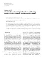
The relationship between the Kellgren–Lawrence grading
and the joint space width is shown in Fig. 1. They were
significantly related (P = 0.003): the joint space width
decreased with increasing Kellgren–Lawrence grade. The
relationship between the Kellgren–Lawrence grading and
the biochemical markers is also shown in Fig. 1. A signifi-
cant relationship was noted for pyridinoline (P = 0.034)
and TIMP-1 (P = 0.017).
Table 1 shows the correlations between the
biochemical markers in all of the subjects as determined
by the Spearman rank test. There were significant
correlations between CRP and YKL-40, between
pyridinoline and MMP-3, and between YKL-40 and
MMP-3.
Table 2 shows the correlations between the joint space
width and the biochemical markers. All markers had
negative correlations with the joint space width, but only
urinary pyridinoline had a significant correlation
(P = 0.039).
Arthritis Research & Therapy Vol 6 No 3 Takahashi et al.
R210
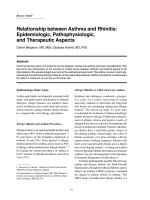
Figure 2 shows the comparison of the joint space width
and biochemical markers between patients with and
without GOA. The joint space width did not differ
between GOA and non-GOA. CRP (P = 0.043),
pyridinoline (P = 0.046), YKL-40 (P < 0.0001) and MMP-3
(P = 0.008) were significantly greater in GOA than in
non-GOA.
Discussion
We studied two radiographic grading criteria: Kellgren–
Lawrence grading and joint space width. These two
methods for evaluating the degree of OA are widely used.
In the present study they were significantly related to each
other: the Kellgren–Lawrence grade varied inversely with
joint space width. We also used the concept of GOA as a
means of radiographic grading of OA [10]. Several
disease subsets in OA are recognized clinically. These
subsets of OA fall into two broad groups: those causing
biomechanical loading or instability at a specific joint, and
those influencing generalized and systemic susceptibility
to the process at multiple joints. The concept of
generalized and systemic susceptibility is supported by
studies indicating a subset of patients with polyarticular
disease known as GOA. Earlier GOA is characterized by
hand OA as a generalized nodal OA, which is a familial
disease affecting mostly women and characterized by the
development of Heberden nodes and a specific pattern of
OA showing multiple joint involvement. Because levels of
serum biochemical markers for arthritis depend on the
circulating concentrations of molecules derived from the
affected joints, the marker level is expected to be higher in
patients with multiple joint involvement than in patients
with a specific joint site. Therefore, in this study, to
ascertain the ability of joint markers we used two OA
populations: a knee OA group and a GOA group. The
results obtained showed that CRP, pyridinoline, YKL-40
and MMP-3 were significantly greater in the GOA group
than in the non-GOA group. In contrast, there was no
significant difference in joint space width between the
GOA and non-GOA groups.
Figure 1
Relationship between Kellgren–Lawrence grading (KL), joint space
width (JSW) and biochemical markers. Pyridinoline (Pyr) level is
expressed as nmol/mmol creatinine. There were significant
relationships between joint space width (P = 0.003), pyridinoline (P =
0.034) and TIMP-1 (P = 0.017) determined with the Kruskal–Wallis
test. Bars indicate maximum and minimum values within the
observation range. P values are shown in each panel; N.S. indicates
not significant. CRP, C-reactive protein; MMP, matrix
metalloproteinase; TIMP, tissue inhibitor of metalloproteinases.
Table 1
Correlations between biochemical markers
Marker Pyridinoline YKL-40 MMP-3 MMP-9 TIMP-1
CRP 0.027 (0.2184) 0.103 (0.0009) 0.050 (0.4047) –0.007 (0.8462) 0.300 (0.0528)
Pyridinoline 0.090 (0.0198) 0.490 (0.0272) 0.186 (0.0554) 0.206 (0.0973)
YKL-40 0.382 (0.0046) 0.154 (0.1027) 0.083 (0.0682)
MMP-3 0.081 (0.0938) –0.001 (0.6267)
MMP-9 –0.282 (0.1033)
Correlation coefficients and P values (in parenthesis) were determined with the Spearman rank test. CRP, C-reactive protein; MMP, matrix
metalloproteinase; TIMP, tissue inhibitor of metalloproteinases.
There are two major categories of substances that are
currently being investigated as potential biochemical
markers for arthritis. One includes constituents of the
extracellular matrix of the joint tissues; the other includes
enzymes or cytokines that metabolize the molecules of the
joint tissues. Among the biochemical markers investigated
in the present study, pyridinoline is a major crosslink of
collagen in the joint tissues, which is abundant both in
cartilage and bone, and belongs to the former group.
MMPs and TIMP are proteolytic enzymes that belong to
the latter group [11]. We do not know which group
YKL-40 belongs to, because the function and origin of this
substance is still not clear [12]. Although the function of
YKL-40 is not yet known, several studies have suggested
that YKL-40 might be a useful new marker for patients
with OA and rheumatoid arthritis [13]. However, the
biochemical markers of bone metabolism are also
proposed as indicators of disease progress of OA
[14,15]. Spector and colleagues demonstrated that bone
resorption is increased in patients with progressive knee
OA and is not increased in those with nonprogressive
knee OA. Altered bone turnover might be a diagnostic or
therapeutic target in patients with progressive OA [16].
Urinary pyridinoline has the most consistent relationship
with radiographic grades of OA among the biochemical
markers studied here. Pyridinium crosslinks consist of two
major molecules, namely pyridinoline and its analogue
deoxypyridinoline. Although both crosslinks are located in
several tissues, deoxypyridinoline is more specifically
located in bone, whereas pyridinoline is most abundant in
cartilage and bone. Urinary excretion of deoxypyridinoline
is therefore used clinically as a marker of bone metabo-
lism, whereas urinary excretion of pyridinoline is considered
to be a biochemical marker for cartilage destruction and
metabolism as well as bone metabolism [17]. However,
because pyridinoline locates in several tissues of the joint
in a significant amount [18], urinary pyridinoline might be
affected by the synthesis of osteophytes, sclerosis of
subchondral bone and synovial degeneration as well as
cartilage degeneration in the joints of OA.
Conclusion
In conclusion, CRP, pyridinoline, YKL-40, MMP-3 and
TIMP-1 levels were each related to at least one of the
radiographic gradings. Furthermore, pyridinoline was related
to every type of radiographic grading examined in the
present study.
Competing interests
None declared.
References
1. Garnero P, Landewe R, Boers M, Verhoeven A, Van Der Linden S,
Christgau S, Van Der Heijde D, Boonen A, Geusens P: Associa-
Available online />R211
Table 2
Correlations between joint space width and biochemical
markers
Biochemical marker rP
CRP –0.212 0.751
Pyridinoline –0.282 0.039
YKL-40 –0.146 0.725
MMP-3 –0.241 0.466
MMP-9 –0.088 0.971
TIMP-1 –0.189 0.368
CRP, C-reactive protein; MMP, matrix metalloproteinase; TIMP, tissue
inhibitor of metalloproteinases.
Figure 2
Comparison of joint space width (JSW) and the biochemical markers
between generalized osteoarthritis (GOA) and non-GOA. Joint space
width did not differ between GOA and non-GOA. C-reactive protein
(CRP; P = 0.043), pyridinoline (Pyr; P = 0.046), YKL-40 (P < 0.0001)
and matrix metalloproteinase-3 (MMP-3; P = 0.008) were significantly
greater in GOA than in non-GOA determined with the Mann–Whitney
U-test. Bars indicate maximum and minimum values within the
observation range. P values are shown in each panel; N.S. indicates
not significant. TIMP, tissue inhibitor of metalloproteinases.
Arthritis Research & Therapy Vol 6 No 3 Takahashi et al.
R212
tion of baseline levels of markers of bone and cartilage
degradation with long-term progression of joint damage in
patients with early rheumatoid arthritis: the COBRA study.
Arthritis Rheum 2002, 46:2847-2856.
2. Altman RD: Criteria for classification of clinical osteoarthritis. J
Rheumatol 1991, 18 (suppl 27):10-12.
3. Leach RE, Gregg T, Siber FJ: Weight-bearing radiography in
osteoarthritis of the knee. Radiology 1970, 97:265-268.
4. Altman RD, Fries JF, Bloch DA, Carstens J, Cooke TD, Genant H,
Gofton P, Groth H, McShane DJ, Murphy WA, et al.: Radi-
ographic assessment of progression in osteoarthritis. Arthritis
Rheum 1987, 30:1214-1225.
5. Kellgren JH, Lawrence JS: Radiological assessment of osteo-
arthrosis. Ann Rheum Dis 1957, 16:494-502.
6. Doherty M, Watt I, Diepple P: Influence of primary generalized
osteoarthritis on development of secondary osteoarthritis.
Lancet 1983, ii:8-11.
7. Pratt DA, Daniloff Y, Duncan A, Robins SP: Automated analysis
of the pyridinium crosslinks of collagen in tissue and urine
using solid-phase extraction and reversed-phase high-perfor-
mance liquid chromatography. Anal Biochem 1992, 207:168-
175.
8. Harvey S, Weisman M, O’Dell J, Scott T, Krusemeier M, Visor J,
Swindlehurst C: YKL-40: new marker of joint disease. Clin
Chem 1998, 44:509-516.
9. Obata K, Iwata K, Okada Y, Kohrin Y, Ohuchi E, Yoshida S,
Shinmei M, Hayakawa T: A one-step sandwich enzyme immuno-
assay for human matrix metalloproteinase 3 (stromelysin-1)
using monoclonal antibodies. Clin Chim Acta 1992, 211:59-72.
10. Cooper C, Egger P, Coggon D, Hart DJ, Masud T, Cicuttini F,
Doyle DV, Spector TD: Generalized osteoarthritis in women:
pattern of joint involvement and approaches to definition for
epidemiological studies. J Rheumatol 1996, 23:1938-1942.
11. Hasty KA, Reife RA, Kang AH, Stuart JM: The role of stromelysin
in the cartilage destruction that accompanies inflammatory
arthritis. Arthritis Rheum 1990, 33:388-397.
12. Steck E, Breit S, Breusch SJ, Axt M, Richter W: Enhanced
expression of the human chitinase 3-like 2 gene (YKL-39) but
not chitinase 3-like 1 gene (YKL-40) in osteoarthritic cartilage.
Biochem Biophys Res Commun 2002, 299:109-115.
13. Johansen JS, Kirwan JR, Price PA, Sharif M: Serum YKL-40 con-
centrations in patients with early rheumatoid arthritis: relation
to joint destruction. Scand J Rheumatol 2001, 30:297-304.
14. Naitou K, Kushida K, Takahashi M, Ohishi T, Inoue T: Bone
mineral density and bone turnover in patients with knee
osteoarthritis compared with generalized osteoarthritis. Calcif
Tissue Int 2000, 66:325-329.
15. Hunter DJ, Spector TD: The role of bone metabolism in
osteoarthritis. Curr Rheumatol Rep 2003, 5:15-19.
16. Bettica P, Cline G, Hart DJ, Meyer J, Spector TD: Evidence for
increased bone resorption in patients with progressive knee
osteoarthritis: longitudinal results from the Chingford study.
Arthritis Rheum 2002, 46:3178-3184.
17. Robins SP, Stewart P, Astbury C, Bird HA: Measurement of the
cross linking compound, pyridinoline, in urine as an index of
collagen degeneration in joint disease. Ann Rheum Dis 1986,
45:969-973.
18. Takahashi M, Kushida K, Hoshino H, Suzuki M, Sano M, Miyamoto
S, Inoue T: Concentrations of pyridinoline and deoxypyridino-
line in joint tissue from patients with osteoarthritis or rheuma-
toid arthritis. Ann Rheum Dis 1996, 55:324-327.
Correspondence
Masaaki Takahashi MD PhD, Department of Orthopedic Surgery,
Hamamatsu University School of Medicine, 1-20-1 Handayama,
Hamamatsu, 431-3129, Japan. Tel: +81 53 435 2299; fax: +81 53
435 2296; e-mail: