Báo cáo y học: "Absence of autoantibodies against correctly folded recombinant fibrillin-1 protein in systemic sclerosis patients" pps
Bạn đang xem bản rút gọn của tài liệu. Xem và tải ngay bản đầy đủ của tài liệu tại đây (346.24 KB, 6 trang )
Open Access
Available online />R1221
Vol 7 No 6
Research article
Absence of autoantibodies against correctly folded recombinant
fibrillin-1 protein in systemic sclerosis patients
Jürgen Brinckmann
1
, Nico Hunzelmann
2
, Ehab El-Hallous
3
, Thomas Krieg
2
, Lynn Y Sakai
4
,
Sven Krengel
1
and Dieter P Reinhardt
5
1
Department of Dermatology, University of Lübeck, Lübeck, Germany
2
Department of Dermatology, University of Cologne, Cologne, Germany
3
Department of Medical Molecular Biology, University of Lübeck, Lübeck, Germany
4
Department of Biochemistry and Molecular Biology and Shriners Hospital for Children, Oregon Health and Science University, Portland, OR, USA
5
Department of Anatomy and Cell Biology and Faculty of Dentistry, McGill University, Montreal, Canada
Corresponding author: Jürgen Brinckmann,
Received: 1 Jun 2005 Revisions requested: 23 Jun 2005 Revisions received: 11 Jul 2005 Accepted: 8 Aug 2005 Published: 6 Sep 2005
Arthritis Research & Therapy 2005, 7:R1221-R1226 (DOI 10.1186/ar1813)
This article is online at: />© 2005 Brinckmann et al.; licensee BioMed Central Ltd.
This is an Open Access article distributed under the terms of the Creative Commons Attribution License ( />2.0), which permits unrestricted use, distribution, and reproduction in any medium, provided the original work is properly cited.
Abstract
Autoantibodies against short recombinant fragments of fibrillin-
1 produced in bacterial expression systems have been found in
tight-skin mouse, systemic sclerosis, mixed connective tissue
disease, and primary pulmonary hypertension syndrome. In
patients with scleroderma, the frequency of anti-fibrillin-1
antibodies was 42% in Caucasians. Until now it has been
unclear whether this immune response has a primary function in
disease pathogenesis or is a secondary phenomenon. In the
present study we analyzed the frequency of autoantibodies
against two overlapping recombinant polypeptides spanning the
N-terminal and C-terminal halves of human fibrillin-1, which were
produced in human embryonic kidney (HEK-293) cells. Correct
three-dimensional structures of the recombinant fibrillin-1
polypeptides were shown by electron microscopy and
immunoreactivity with antibodies. Screening of fibrillin-1
antibodies was performed in 41 sera from systemic sclerosis
patients and in 44 healthy controls with a Caucasian
background. Microtiter plates were coated with the recombinant
polypeptides of fibrillin-1 and incubated with 1:100 diluted sera.
Positive binding was defined as being more than 2 SD above the
mean of the control group. ELISAs showed that none of the sera
of patients with systemic sclerosis contained autoantibodies
against the N-terminal or C-terminal recombinant fibrillin-1
polypeptide. The data show the absence of autoantibodies
against recombinant fibrillin-1 protein in Caucasian systemic
sclerosis patients. Because the correct three-dimensional
folding of the recombinant proteins has been substantiated by
several independent methods, we conclude that autoantibodies
against correctly folded fibrillin are not a primary phenomenon in
the pathogenesis of systemic sclerosis.
Introduction
Systemic sclerosis (SSc) is a connective tissue disease char-
acterized by an excess deposition of collagen in skin and/or
internal organs leading to malfunction and organ failure. The
extent and progression of the fibrotic process presumably
caused by the imbalance between extracellular matrix synthe-
sis and degradation largely determines the prognosis of the
disease. One hallmark of the disease is the presence of circu-
lating autoantibodies against non-organ-specific nuclear and
nucleolar antigens, which can be detected in at least 95% of
patients. They include anti-centromere, anti-topoisomerase I
and anti-RNA polymerase antibodies and are associated with
distinct disease subtypes [1].
Heterozygous tight-skin mice (Tsk/+) are characterized by a
phenotype of skin thickening and visceral fibrosis due to an
increased deposition of extracellular matrix proteins in skin and
organs. Furthermore, Tsk/+ mice develop lung emphysema
and cardiac hypertrophy and have therefore been adopted as
a potential genetic model of human SSc, cardiac hypertrophy
and hereditary emphysema [2]. In a similar manner to human
SSc, Tsk/+ mice produce autoantibodies against SSc-spe-
BSA = bovine serum albumin; DMEM = Dulbecco's modified Eagle's medium; ELISA = enzyme-linked immunosorbent assay; kDa = kilodaltons; mAb
= monoclonal antibody; SSc = systemic sclerosis; TBS = Tris-buffered saline; Tsk = tight-skin mouse.
Arthritis Research & Therapy Vol 7 No 6 Brinckmann et al.
R1222
cific antigens such as topoisomerase I and RNA polymerase
[3].
A duplication in the mouse fibrillin-1 gene was described for
the Tsk/+ mouse, which is associated with premature death in
utero for homozygous Tsk/Tsk animals [4]. Fibrillin-1 is one of
the major structural components of microfibrils, which are
extracellular supramolecular aggregates found in many elastic
and non-elastic tissues (reviewed in [5]). Microfibrils are
thought to be important in the assembly and organization of
the elastic fibers by mediating tropoelastin deposition [6].
Fibrillin-1 and other members of the fibrillin family are repeti-
tively aligned within microfibrils and constitute their structural
backbone [7,8]. Murai and colleagues found that Tsk/+ mice
spontaneously produce autoantibodies against a small recom-
binant protein spanning the proline-rich region of human fibril-
lin-1 [9]. This recombinant fragment comprises about 2% of
the total fibrillin-1 molecule. Recently, the presence of autoan-
tibodies against the same recombinant fibrillin-1 fragment has
also been shown for sera from patients with SSc, localized
scleroderma, mixed connective tissue disease and primary
pulmonary hypertension syndrome [10-12]. Frequencies of
autoantibodies showed remarkable differences between the
ethnic groups studied. Choctaw American Indians and Japa-
nese patients with SSc exhibited the highest frequency, with
81% and 78% respectively, whereas Caucasians with SSc
were positive to a smaller extent with 34% [10].
In the present study we analyzed the autoantibody titer in Cau-
casian SSc patients against two overlapping recombinant
fragments spanning the entire human fibrillin-1. One fragment
constitutes the amino-terminal half of fibrillin-1 (amino acid res-
idues 19 to 1,527) and the other fragment its carboxy-terminal
half (residues 1,487 to 2,725). Before the analysis of antibody
titers by ELISA, the proper folding of both recombinant pro-
teins was shown by electron microscopy after rotary shadow-
ing and binding of monoclonal and polyclonal antibodies by
dot-blotting with or without previous reduction of the recom-
binant proteins.
Materials and methods
Patients and tissue specimens
Sera from Caucasian patients with SSc (n = 41; 29 female, 12
male; mean age 58.2 ± 14.3 years) and from healthy Cauca-
sian controls (n = 44; 31 female, 13 male; mean age 46.9 ±
19.8 years) were studied. Patients with SSc were diagnosed
in accordance with the American College of Rheumatology
preliminary criteria for the classification of SSc [13]. Limited
systemic sclerosis was present in 25 patients, and diffuse sys-
temic sclerosis in 16. The range of disease duration was
between 6 months and 27 years. The antibody profile showed
positive titers of anti-nuclear antibodies for all patients. Of
these, 16 had SCL-70, 13 anti-centromere, 1 RNA polymer-
ase and 11 undifferentiated antibodies. Antibody testing con-
sisted of the determination of the fluorescence pattern and
titer on HEP2 cells (Viramed, Germany) as well as subsequent
testing by a commercial ELISA for U1-RNP, Sm, Ro-SSA, La-
SSB, Scl-70 and centromere reactivity (Orgentec, Germany).
All samples were obtained after obtaining written consent from
the donors under protocols approved by the local ethical
committee.
Expression and production of recombinant fibrillin-1
polypeptides
The expression plasmids to express the N-terminal half
(pDNSP-rF16) and the C-terminal half (pcDNA-rF6H) of
human fibrillin-1 have previously been described in detail [14].
On the basis of SDS-PAGE and electron microscopy after
rotary shadowing (see below), the purity of the recombinant
fragments was more than 90%. Stable clones with these
expression plasmids were obtained with human embryonic
kidney (HEK-293) cells as described in detail [15]. The
expression of pDNSP-rF16 in eukaryotic cells produces a
secreted polypeptide (rF16) with the sequence Ala-Pro-Leu-
Ala-Ser
19
-Val
1,527
-(His)
6
. The expression of pcDNA-rF6H in
eukaryotic cells produces a secreted polypeptide (rF6H) with
the sequence Ala-Pro-Leu-Ala-Asp
1,487
-Lys
2,725
-(His)
6
. Pro-
duction and purification of rF16 and rF6H were performed as
in the procedures described elsewhere [16].
Electron microscopy after rotary shadowing
The purified proteins were adjusted to a concentration of 0.25
mg/ml and dialyzed against 100 mM NH
4
HCO
3
. The samples
were diluted with 0.05% (v/v) acetic acid to a final protein con-
centration of 60 µg/ml and mixed with glycerol to a final con-
centration of 50% (v/v) glycerol. Then 80 µl of the samples
was sprayed onto freshly cleaved mica from a distance of 25
cm and dried under high vacuum (about 9 nbar) for about 2 to
3 hours in an Edwards Auto 306 vacuum coater. Rotary shad-
owing was performed by platinum evaporation for 15 s at 50
mA and 2.5 kV at an angle of 5° and a distance of 12 cm. The
samples were rotated at 120 r.p.m., followed by coating with
coal for stabilization for 2 s at 100 mA and 2.5 kV at an angle
of 90°. The replicas were floated onto a very clean surface of
distilled water and then supported with 400-mesh copper
grids. Replicas were examined at 100 kV in a transmission
electron microscope (Zeiss TEM 109).
Cell culture
Human dermal fibroblasts were derived from explant cultures
of dissected tissues obtained from surgical samples after
informed consent had been obtained. The cells were cultured
in DMEM supplemented with 10% fetal bovine serum and
penicillin/streptomycin (Invitrogen). Cells (10
6
) were plated in
a 60 mm dish and grown for 72 hours. The cell layers were
washed with phosphate-buffered saline and then incubated
for 24 hours in 3 ml of DMEM without serum. The conditioned
medium was harvested and treated with 1 mM phenylmethyl-
sulfonyl fluoride.
Available online />R1223
Dot-blot assay
Either 2 µg of purified recombinant proteins rF16 and rF6H or
1 ml of conditioned medium were transferred to nitrocellulose
membranes using a dot-blot apparatus (Bio-Rad) with or with-
out previous reduction of the proteins with 0.05 M dithiothrei-
tol. After staining with Ponceau S, non-specific binding sites
on the nitrocellulose membrane were blocked for 1 hour with
Tris-buffered saline (TBS) containing 5% (w/v) non-fat milk.
Nitrocellulose membranes were probed with a polyclonal
antiserum against rF6H (diluted 1:500 [17]) and with mono-
clonal antibodies directed against rF6H (mAb 69, about 4 µg/
ml) and rF16 (mAb 201 and mAb 26, both about 4 µg/ml [18])
followed by peroxidase-conjugated anti-rabbit or anti-mouse
secondary antibody (diluted 1:800; Bio-Rad). Bound antibod-
ies were revealed in accordance with the manufacturer's
instructions by using the horseradish peroxidase developer 4-
chloronaphthol (Bio-Rad).
ELISA assay
Microtiter plates were coated with 100 µl of 10 µg/ml purified
recombinant human fibrillin-1 fragments rF16 and rF6H or
BSA overnight at 4°C. After being washed three times with
TBS containing 0.05% Tween 20 (TBS/Tween), the plates
were blocked for 1 hour with 200 µl of 5% non-fat milk powder
in TBS at room temperature (20°C). After being washed with
TBS, the plates were incubated for 2 hours with 100 µl of test
sera diluted 1:100 with TBS containing 5% non-fat milk pow-
der at room temperature. After being washed three times with
TBS/Tween, the plates were incubated for 1.5 hours with 100
µl of the horseradish peroxidase-conjugated secondary anti-
body (diluted 1:800) at room temperature (goat anti-rabbit for
positive control sera, and goat anti-human for human sera;
Sigma, Germany). After three washings with TBS/Tween,
color development was achieved with 100 µl of 1 mg/ml 5-
aminosalicylic acid in 0.02 M phosphate buffer (pH 6.8) and
1.5 µl/ml H
2
O
2
. Color development was stopped after 1 hour
by the addition of 100 µl of 2 M NaOH. Absorbance was
measured at 492 nm with an ELISA reader (Anthos, Austria).
All experiments were run in parallel triplicates; the ELISA test
was performed twice. The background binding of serum anti-
bodies to BSA-coated wells was subtracted from the binding
of serum to the respective rF16-coated and rF6H-coated wells
after subtraction of the respective background of rF16-coated,
rF6H-coated and BSA-coated blanks. Positive binding was
defined as more than 2 SD above the mean of the control sera.
The coefficient of variation was 7.4% (n = 10).
Results
Ultrastructural analysis of recombinant fibrillin-1
polypeptides
To analyze the molecular shape of the recombinant polypep-
tides, they were revealed by electron microscopy after rotary
shadowing (Fig. 1). These results showed thread-like
extended molecules for the recombinant polypeptides rF16
and rF6H representing the N-terminal and C-terminal halves of
human fibrillin-1. At the termini of rF16 and rF6H the molecules
occasionally adopted a curved shape.
The analysis of molecular dimensions revealed that the length
of rF16 (73.1 ± 5.7 nm, n = 75) and rF6H (64.2 ± 5.9 nm, n
= 56) corresponded well to the lengths for very similar con-
structs described previously [16] as well to the respective
parts in full-length fibrillin-1 [19]. The extended shape of the
recombinant proteins is a very good indicator of correct fold-
ing, because the molecular shape is determined by numerous
intramolecular disulfide bridges stabilizing this extended struc-
ture [20,21].
Immunoreactive analysis of recombinant fibrillin-1
polypeptides
To analyze the immunoreactive properties of native fibrillin-1
synthesized by human dermal fibroblasts and the recombinant
polypeptides rF16 and rF6H, dot-blotting under reducing and
non-reducing conditions was performed (Fig. 2) Native fibrillin-
1 reacts with monoclonal antibodies mAb 26 or mAb 201 or
with polyclonal antibody anti-rF6H only under non-reducing
conditions (not under reducing conditions). These data show
that the antibodies primarily recognize epitopes in the cor-
rectly folded fibrillin-1 molecule but not in the denatured fibril-
lin-1 molecule. When the recombinant fibrillin-1 polypeptides
rF16 and rF6H were tested in this assay, they showed much
more reactivity in the non-reduced conformation than in the
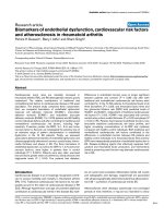
Figure 1
Recombinant amino-terminal (rF16) and carboxy-terminal (rF6H) halves of human fibrillin-1 were analyzed by electron microscopy after rotary shadowingRecombinant amino-terminal (rF16)(a) and carboxy-terminal (rF6H)(b)
halves of human fibrillin-1 were analyzed by electron microscopy after
rotary shadowing. Representative images and histrograms of the meas-
ured lenghs of the recombinant fragments are shown. Note that both
fragments showed thread-like extended molecules. The measurements
are plotted as number of measurements, in 5 nm windows. The average
length of rF16 was 73.1 ± 5.7 nm (mean ± SD; n = 75) and the aver-
age length of rF6H was 64.2 ± 5.9 nm (mean ± SD; n = 56).
Arthritis Research & Therapy Vol 7 No 6 Brinckmann et al.
R1224
reduced conformation, showing that the corresponding
epitopes are present in the same correct conformation as in
native fibrillin-1. These data substantiate that the recombinant
polypeptides are correctly folded.
ELISA analysis of sera from patients and controls by
using rF16 and rF6H
A cutoff value was established for each ELISA as a value of 2
SD above the mean of 44 control sera. For rF16 the cutoff
ELISA score was 0.072 and for rF6H it was 0.1. The analysis
of 41 sera from Caucasian patients with systemic sclerosis
showed that none of the sera exceeded the cutoff value for the
N-terminal half of fibrillin-1. Furthermore, the ELISA score of all
sera tested for the presence of antibodies against the C-termi-
nal half of fibrillin-1 was in the normal range of the controls
(Fig. 3).
Discussion
Mutations in the gene encoding fibrillin-1 have been docu-
mented for Marfan syndrome and some related disorders in
humans, and for Tsk in animals [22,4]. The Tsk mutation in the
fibrillin-1 gene, a 30-kilobase gene duplication of exons 17 to
40 containing a long centrally located stretch of calcium-bind-
ing epidermal growth factor-like domains, is accompanied by
premature death in utero in homozygous mice, whereas mice
heterozygous for the duplication are viable and show the tight-
Figure 2
Immunoreactive analysis of fibrillin-1 antibodies against recombinant fibrillin-1 polypeptides and against native fibrillin-1Immunoreactive analysis of fibrillin-1 antibodies against recombinant fibrillin-1 polypeptides and against native fibrillin-1. Purified recombinant amino-
terminal (rF16) and carboxy-terminal (rF6H) halves of human fibrillin-1 (2 µg of each) or 1 ml of conditioned medium (containing less than about 0.2
µg of fibrillin-1) produced by human dermal fibroblasts were transferred to nitrocellulose membranes with (upper panel) or without (lower panel) pre-
vious reduction by dithiothreitol. Nitrocellulose membranes were probed with a polyclonal antibody against rF6H (anti-rF6H) or with monoclonal anti-
bodies (mAbs) 26, 201, and 69 directed against rF16 and rF6H. The dot-blots show that the binding of all antibodies depends markedly on the
presence of disulfide bonds, which are crucial for the proper folding of epitopes in both native fibrillin-1 and the recombinant fragments.
Figure 3
Analysis of immunoreactivity of sera from systemic sclerosis patients and healthy controls of Caucasian originAnalysis of immunoreactivity of sera from systemic sclerosis patients and healthy controls of Caucasian origin. ELISA assays with the recombinant
amino-terminal (rF16 (a)) and carboxy-terminal (rF6H (b)) halves of human fibrillin-1 are shown. Microtiter plates were coated with purified rF16 and
rF6H or bovine serum albumin. The plates were incubated with test sera diluted 1:100 for 2 hours at room temperature. After incubation with horse-
radish peroxidase-conjugated secondary antibody and color development, the absorbance was determined by an ELISA reader. Positive binding
was defined as more than 2 SD above the mean (dashed line) of the control sera. If the blank value exceeded the sample value the absorbance was
set to zero in the figure. None of the sera showed a positive reactivity to rF16 or rF6H.
Available online />R1225
skin phenotype. The mutation results in a larger protein (418
kDa, as compared with 350 kDa in normal animals) which after
incorporation along with wild-type fibrillin-1 seems to render all
microfibrils more susceptible to proteolysis [23]. In a similar
manner to SSc in humans, Tsk mice develop autoimmunity
with antibodies against topoisomerase I and RNA polymerase.
Recently, autoantibodies against a small 30 kDa human
recombinant fibrillin-1 polypeptide covering the proline-rich
region (residues 395 to 446) have been detected in 41% of
Tsk mice [9].
Autoantibodies against the same recombinant fibrillin-1
polypeptide were also found in humans affected by SSc or pri-
mary pulmonary hypertension syndrome [10,12]. Especially in
SSc, the frequency of anti-fibrillin-1 antibodies and the recog-
nized epitopes differ according to the ethnic background of
patients, as shown in a subsequent study [24]. In that study,
reactivity against recombinant polypeptides covering the N-
terminal end (residues 15 to 193), the proline-rich region (res-
idues 367 to 425), and a stretch of calcium-binding epidermal
growth factor-like domains (residues 1,326 to 1,549) was
tested. Taking the different epitopes tested in that study
together, Choctaw Native Americans, Japanese patients and
African Americans revealed the highest levels with 100% and
80%, respectively. In the same study, sera from Caucasian
SSc patients showed the presence of anti-fibrillin-1 antibodies
in 42% of patients. Whether the occurrence of these autoan-
tibodies has a primary role in the pathogenesis of SSc or is a
secondary phenomenon is open to discussion.
In our study of 41 Caucasian patients with SSc, none of the
sera showed positive reactivity against the recombinant
polypeptide spanning either the N-terminal half or the C-termi-
nal half of fibrillin-1. Structural studies by rotary shadowing and
evaluation of molecular lengths showed that the recombinant
fibrillin-1 polypeptides used resemble native molecules. They
adopt the correct dimensions and extended conformations
similar to regions observed in whole molecules of native fibril-
lin-1 purified from cell culture medium [19]. Various mono-
clonal and polyclonal antibodies recognize native fibrillin-1
only in a non-reduced (correctly folded) conformation but not
in the reduced (misfolded) conformation because numerous
intramolecular disulfide bonds stabilize the native conforma-
tion of fibrillins [20,21]. Similar binding properties of these
monoclonal and polyclonal antibodies to the recombinant
polypeptides rF16 and rF6H strongly support the notion that
these polypeptides are folded correctly. Our data clearly show
that SSc in Caucasians is not characterized by the presence
of autoantibodies against properly folded fibrillin-1. This obser-
vation indicates that the presence of autoantibodies against
fibrillin-1 does not have a primary role in the pathogenesis of
the disease.
The recombinant fibrillin-1 antigens used in other studies
showing a positive binding of antibodies obtained from SSc
patients were relatively small (59, 179 and 224 residues) and
were produced in bacterial expression systems [10,24]. No
structural or functional characterization for these recombinant
polypeptides is available to determine whether they adopt
native or misfolded conformations. It is possible that the anti-
fibrillin-1 autoantibodies detected with such recombinant
polypeptides recognize cryptic or misfolded antigenic
epitopes for example, which may become available after prote-
olytic fragmentation of fibrillin-1 in SSc or may be antibodies
against cross-reacting antigens. In this light, one can specu-
late that these autoantibodies are a secondary phenomenon in
SSc. This interpretation is further substantiated by a metabolic
analysis of fibrillin-1 synthesized by SSc fibroblasts in cell cul-
ture, which revealed decreased amounts of abnormal microfi-
brils [25]. Furthermore, in the same study in vitro, data
indicated that the amount of fibrillin-1 in the extracellular matrix
produced by SSc cells diminished faster than in the matrix of
control cells, arguing for a higher susceptibility to proteolytic
degradation.
Conclusion
Our data clearly show that sera from 41 Caucasian SSc
patients contained no autoantibodies against properly folded
recombinant human fibrillin-1. These data therefore provide
evidence that autoimmunity against fibrillin-1 is a secondary
phenomenon in the pathogenesis of SSc in Caucasians.
Competing interests
The author(s) declare that they have no competing interests.
Authors' contributions
Expression and production of recombinant polypeptides were
performed by EE and DPR. Electron microscopy was per-
formed by EE and DPR. ELISA analysis was performed JB,
DPR, SK and NH. Immunoreactive analysis was performed by
JB, LYS and DPR. Study design and coordination were per-
formed by JB, NH and DPR. Editing of the manuscript was per-
formed by JB, NH, DPR, TK and LYS. All authors read and
approved the final manuscript.
Acknowledgements
We are grateful to Martina Alexander for excellent technical assistance.
The work was supported by grants by the Deutsche Forschungsgemein-
schaft (SFB367-A1, Br 1146/3-3), the Bundesminsterium für Bildung
und Forschung (BMBF, German Network for Systemic Scleroderma),
the Köln Fortune Program and the Canadian Institutes of Health
Research (MOP-68836).
References
1. Harris ML, Rosen A: Autoimmunity in scleroderma: the origin,
pathogenetic role, and clinical significance of autoantibodies.
Curr Opin Rheumatol 2003, 15:778-784.
2. Jimenez SA, Christner PJ: Murine animal models of systemic
sclerosis. Curr Opin Rheumatol 2002, 14:671-680.
3. Bona C, Rothfield N: Autoantibodies in scleroderma and tight-
skin mice. Curr Opin Immunol 1994, 6:931-937.
4. Siracusa LD, McGrath R, Ma Q, Moskow JJ, Manne J, Christner PJ,
Buchberg AM, Jimenez SA: A tandem duplication within the
Arthritis Research & Therapy Vol 7 No 6 Brinckmann et al.
R1226
fibrillin 1 gene is associated with the mouse tight skin
mutation. Genome Res 1996, 6:300-313.
5. Ramirez F, Pereira L: The fibrillins. Int J Biochem Cell Biol 1999,
31:255-259.
6. Mecham RP, Broekelmann T, Davis EC, Gibson MA, Brown-Augs-
burger P: Elastic fibre assembly: macromolecular interactions.
Ciba Found Symp 1995, 192:172-181.
7. Lin G, Tiedemann K, Vollbrandt T, Peters H, Batge B, Brinckmann
J, Reinhardt DP: Homo- and heterotypic fibrillin-1 and -2 inter-
actions constitute the basis for the assembly of microfibrils. J
Biol Chem 2002, 277:50795-50804.
8. Charbonneau NL, Dzamba BJ, Ono RN, Keene DR, Corson GM,
Reinhardt DP, Sakai LY: Fibrillins can co-assemble in fibrils, but
fibrillin fibril composition displays cell-specific differences. J
Biol Chem 2003, 278:2740-2749.
9. Murai C, Saito S, Kasturi KN, Bona CA: Spontaneous occurrence
of anti-fibrillin-1 autoantibodies in tight-skin mice. Autoimmu-
nity 1998, 28:151-155.
10. Tan FK, Arnett FC, Antohi S, Saito S, Mirarchi A, Spiera H, Sasaki
T, Shoichi O, Takeuchi K, Pandey JP, et al.: Autoantibodies to the
extracellular matrix microfibrillar protein, fibrillin-1, in patients
with scleroderma and other connective tissue diseases. J
Immunol 1999, 163:1066-1072.
11. Arnett FC, Tan FK, Uziel Y, Laxer RM, Krafchik BR, Antohi S, Bona
C: Autoantibodies to the extracellular matrix microfibrillar pro-
tein, fibrillin 1, in patients with localized scleroderma. Arthritis
Rheum 1999, 42:2656-2659.
12. Morse JH, Antohi S, Kasturi K, Saito S, Fotino M, Humbert M,
Simonneau G, Basst RJ, Bona CA: Fine specificity of anti-fibril-
lin-1 autoantibodies in primary pulmonary hypertension
syndrome. Scand J Immunol 2000, 51:607-611.
13. Subcommittee for scleroderma criteria of the American Rheuma-
tism Association Diagnostic and Therapeutic Criteria Committee:
Preliminary criteria for the classification of systemic sclerosis
(scleroderma). Arthritis Rheum 1980, 23:581-590.
14. Jensen SA, Reinhardt DP, Gibson MA, Weiss AS: Protein inter-
action studies of MAGP-1 with tropoelastin and fibrillin-1. J
Biol Chem 2001, 276:39661-39666.
15. Fox JW, Mayer U, Nischt R, Aumailley M, Reinhardt D, Wiedemann
H, Mann K, Timpl R, Krieg T, Engel J, et al.: Recombinant nidogen
consists of three globular domains and mediates binding of
laminin to collagen type IV. EMBO J 1991, 10:3137-3146.
16. Reinhardt DP, Keene DR, Corson GM, Poschl E, Bachinger HP,
Gambee JE, Sakai LY: Fibrillin-1: organization in microfibrils
and structural properties. J Mol Biol 1996, 258:104-116.
17. Tiedemann K, Batge B, Muller PK, Reinhardt DP: Interactions of
fibrillin-1 with heparin/heparan sulfate, implications for micro-
fibrillar assembly. J Biol Chem 2001, 276:36035-36042.
18. Sakai LY, Keene DR, Engvall E: Fibrillin, a new 350-kD glycopro-
tein, is a component of extracellular microfibrils. J Cell Biol
1986, 103:2499-2509.
19. Sakai LY, Keene DR, Glanville RW, Bachinger HP: Purification
and partial characterization of fibrillin, a cysteine-rich struc-
tural component of connective tissue microfibrils. J Biol Chem
1991, 266:14763-14770.
20. Downing AK, Knott V, Werner JM, Cardy CM, Campbell ID, Hand-
ford PA: Solution structure of a pair of calcium-binding epider-
mal growth factor-like domains: implications for the Marfan
syndrome and other genetic disorders. Cell 1996, 85:597-605.
21. Yuan X, Downing AK, Knott V, Handford PA: Solution structure
of the transforming growth factor beta-binding protein-like
module, a domain associated with matrix fibrils. EMBO J
1997, 16:6659-6666.
22. Collod-Beroud G, Le Bourdelles S, Ades L, Ala-Kokko L, Booms P,
Boxer M, Child A, Comeglio P, De Paepe A, Hyland JC, et al.:
Update of the UMD-FBN1 mutation database and creation of
an FBN1 polymorphism database. Hum Mutat 2003,
22:199-208.
23. Gayraud B, Keene DR, Sakai LY, Ramirez F: New insights into the
assembly of extracellular microfibrils from the analysis of the
fibrillin 1 mutation in the tight skin mouse. J Cell Biol 2000,
150:667-680.
24. Tan FK, Arnett FC, Reveille JD, Ahn C, Antohi S, Sasaki T, Nishioka
K, Bona CA: Autoantibodies to fibrillin 1 in systemic sclerosis:
ethnic differences in antigen recognition and lack of correla-
tion with specific clinical features or HLA alleles. Arthritis
Rheum 2000, 43:2464-2471.
25. Wallis DD, Tan FK, Kielty CM, Kimball MD, Arnett FC, Milewicz
DM: Abnormalities in fibrillin 1-containing microfibrils in der-
mal fibroblast cultures from patients with systemic sclerosis
(scleroderma). Arthritis Rheum 2001, 44:1855-1864.