Báo cáo y học: "Plasmatic B-Type Natriuretic Peptide and C-Reactive Protein in Hyperacute Stroke as Markers of Ct-Evidence of Brain Edema." pptx
Bạn đang xem bản rút gọn của tài liệu. Xem và tải ngay bản đầy đủ của tài liệu tại đây (593.01 KB, 6 trang )
Int. J. Med. Sci. 2008, 5
18
International Journal of Medical Sciences
ISSN 1449-1907 www.medsci.org 2008 5(1):18-23
© Ivyspring International Publisher. All rights reserved
Research Paper
Plasmatic B-Type Natriuretic Peptide and C-Reactive Protein in Hyperacute
Stroke as Markers of Ct-Evidence of Brain Edema
Pedro J Modrego
1
, Beatriz Boned
2
, Juan J Berlanga
3
, Mercedes Serrano
3
1. Dept of Neurology. Miguel Servet University Hospital. Zaragoza. Spain
2. Biochemistry Unit. Hospital de Alcañiz. Spain
3. Neurology Unit. Hospital de Alcañiz. Spain
Correspondence to: Dr PJ Modrego, Dept of Neurology, Miguel Servet University Hospital, 50009 Zaragoza, Spain. E-mail:
Received: 2007.11.05; Accepted: 2008.01.08; Published: 2008.01.13
OBJECTIVE. Plasmatic B-type-natriuretic peptide (NT-PBNP) and C-reactive protein (CRP) have been reportedly
elevated in stroke patients; however their clinical significance remains uncertain. The purpose of this work is to
investigate whether elevation of these proteins at baseline predicts CT-evidence of brain edema.
METHODS. We recruited 41 consecutive patients with stroke and determined NT-PBNP and CRP at baseline
(within 5 hours after onset), after 48-72 hours, and at discharge. Stroke severity was measured by means of the
NIHS scale at baseline and at discharge. We also carried out brain CT at admittance and after 48 hours.
RESULTS. There were 29 ischemic strokes and 12 hemorrhagic strokes. Evidence of brain edema on delayed scan
was seen in 14 patients. Baseline levels of NT-PBNP did not predict CT-evidence of edema but CRP levels did so
significantly (0.7 mg/dl in patients without edema versus 4.7 mg in patients with edema; p=0.001). Both
NT-PBNP and PC levels correlated poorly to NIHSS score and increased markedly from baseline to the second
determination in patients with edema. For these patients the NT-PBNP increase was 133.6 pmol/l in comparison
to 1.58 pmol/l in patients without edema (p=0.002). Neither CRP nor NT-PBNP baseline levels were predictive of
dependency or death.
CONCLUSIONS. We conclude that CRP at baseline but not NT-PBNP predicts CT evidence of brain edema in
stroke patients. We hypothesize that NT-PBNP levels elevated in response to edema after 48 hours of admission.
Key words: Stroke; Brain edema; C-reactive protein; B-type natriuretic peptide.
INTRODUCTION
Atrial natriuretic peptide (ANP) is released into
circulation in response to atrial distention or sodium
load. This counteracts the Renin-Angiotensin
Aldosterone system and causes an increase in the
glomerular filtration rate with excretion of sodium and
water. Brain natriuretic peptide (BNP) is released from
the ventricular myocardium in response to elevation of
ventricular diastolic pressure [1]. BNP is known to
correlate with the presence and severity of congestive
heart failure [2]. Usually it is determined the
N-terminal fragment (NT-pro-BNP) of the BNP. In the
last years, the NT-pro-BNP levels in the serum have
emerged as important predictor of mortality in stable
coronary heart disease
[3]. It has also bee regarded as
predictor of mortality and cardiovascular events in
older adults [4], as predictor of sudden death in
patients with chronic heart failure [5], and as predictor
of the risk of cardiovascular events and death in
persons without heart failure [6]. BNP levels are also
predictive of heart failure
[7] and myocardial infarction
[8] in patients with stroke or TIA. Furthermore, BNP
levels are also predictive of ischemic stroke in patients
with cerebrovascular disease beyond traditional risk
factors
[9].
C-reactive protein is a well-known marker of
inflammation but the role as marker of
atherothrombosis is currently being investigated. CRP
was also related to increased mortality and
cardiovascular events in older adults
[4], to increased
risk of heart failure in subjects with cerebrovascular
disease
[7], and to increased risk of stroke and TIA in
the elderly
[10].
ANP
[11-13], BNP
[13-15], and CRP
[16-22] have
been reported to be elevated in the acute phase of
stroke but their clinical significance is mostly
unknown. Brain edema is common in acute stroke and,
many times, life-threatening. The reason for which
malignant edema develops in some patients but does
not in others is so far elusive. It becomes especially
manifest after 48 hours from stroke onset. However on
many occasions the edema is not seen until 48 hours or
later from onset.
The main goal of this work is to find accurate
Int. J. Med. Sci. 2008, 5
19
baseline markers of delayed brain edema that might be
helpful to anticipate the appropriate and early
anti-edema measures. We hypothesize that plasmatic
BNP and CRP values in hyperacute stroke might
predict the development of CT-based evidence of
delayed brain edema, and that these proteins increase
in response to edema. The second objective of the
study is to know whether the values of these proteins
change according to clinical outcome.
PATIENTS AND METHODS
In this work we enrolled 48 patients with stroke
in a consecutive manner. Stroke was defined as rapidly
developing symptoms and signs of focal cerebral
dysfunction lasting more than 24 hours with no
apparent origin other than vascular. On admission we
carried out general medical and neurological
examination, blood pressure measurement,
electrocardiogram, X-ray film of the chest, and brain
Computed Tomography. Immediately a blood sample
was drawn to determine ultrasensitive C-reactive
protein and the N-terminal fraction (NT-PBNP) of the
B-type natriuretic peptide within 5 hours after stroke
onset. Serum NT-PBNP levels were measured by
electrochemiluminiscence immunoassay on a Modular
Analytics E170 analyzer (Roche Diagnostics).
Ultrasensitive C-reactive protein (CRP-U) was
measured by quantitative immunoturbidimetric assay
on an Architect c-8000 analyzer (Abbot Diagnostics).
The normal NT-PBNP values in the serum for
healthy people are below 30 pmol/l and those of CRP
below 0.05 mg/dL.
The stroke severity was measured by means of
the National Institute of Health Stroke Scale (NIHSS).
At 48 hours after admission we also carried out a new
brain CT and determined again the CRP and NT-PBNP
in the serum because the edema tends to be more
evident after this elapse of time. By edema in ischemic
stroke we mean mass-effect and sulcal effacement. For
hemorrhagic stroke we mean the surrounding
hypodensity of the hematoma with increased
mass-effect in comparison to baseline scan. CT images
were evaluated for the presence/absence of edema
before knowing the levels of CRP and NT-PBNP.
At discharge we performed another
determination of CRP and NT-PBNP in the serum, as
well as new NIHS scale and the modified Rankin scale
for assessing independence.
For statistical analysis we divided the patients
into two groups: those who developed edema at 48
hours and those who did not. Comparisons were made
with either t-Test or Mann-Whitney U-test in
accordance with the presence/absence of normality in
the distribution of the variables. We also analyzed
correlation between stroke severity and the values of
CRP and NT-PBNP. The predictive values were
analyzed by means of the ROC curves. A model of
logistic regression was constructed to predict edema so
as to control for potential confounders such as age, sex,
atrial fibrillation, hypertension, diabetes, and
hyperlipidemia. Operations were made with the SPSS
software, version 10 (Chicago, IL).
This study was made with the informed consent
of patients and/or relatives, and the authorization of
the local review board as well.
RESULTS
Among the 48 patients recruited 4 were excluded
because of congestive heart failure (signs of
pulmonary edema on Rx film of the chest and/or
reduced ventricular function on echocardiography)
and 3 because of early death from ischemic stroke. The
cause of early death was brain herniation in two
patients, and heart failure in the other. The mean age
of the patients excluded was 84.5 years; two were
women and two were man. The four excluded patients
showed very high levels of NT-PBNP (> 400 pmol/L)
as it is expected in patients with heart failure. The
remaining 41patients underwent the complete protocol
of the study. The mean age of the 41 patients was 78.3
years (range: 54-91). There were 26 men and 15
women. None of the patients included had history of
congestive heart failure nor underwent thrombolysis.
We neither saw patients with renal failure on the basis
of plasmatic creatinine levels. In table 1 are reported
the main baseline characteristics and risk factors.
Table 1. Main baseline characteristics and risk factors. N=41
Mean age: 78.32 (SD: 6.77); range: 54-91
Sex: 26 male (634%) and 15 female.
Mean temperature on admission : 36.6 0C (SD:0.4)
Mean systolic blood pressure on admission: 164.49 (25.36); range:
107-220 mm Hg
History of hypertension: 21 (51.2%).
Atrial fibrillation: 4 (9.75%).
Diabetes mellitus: 9 (22%)
Hyperlipidemia: 7 (17%)
Trasient ischemic attack : 9 (21.9%)
Coronary Heart Disease : 4 (9.75%)
Tobacco habit: 3 (7.3%)
Obesity: 4 (9.75%)
The type of stroke was ischemic in 29 patients and
hemorrhagic of hypertensive cause in 12. Among
ischemic strokes 6 were cardioembolic, 7 were lacunar,
and the remainder (16) were ischemic
non-cardioembolic (11 atherosclerotic and 5 of
undetermined cause). The location of stroke was the
anterior circulation in 30 patients and the posterior
circulation in 11. The hemorrhagic stroke location was
as follows: basal ganglia in 8, lobar in 2, thalamic in
one patient, and cerebellar in another one.
Int. J. Med. Sci. 2008, 5
20
On delayed CT 14 patients showed evidence of
brain edema and 27 did not. The mean age for patients
with edema was 80.29 years (SD: 4.16), and 77.3 (SD:
7.67) for those without edema (difference not
significant). Among patients with edema 3 were
female and 11 male. History of hypertension was
positive in 5 patients (35.7%) who showed edema and
in 16 (59.2%) of those without edema. The mean
systolic/diastolic blood pressure at baseline for
patients with edema was 166/80 mm Hg in
comparison to 163/77 in those without edema. After 48
hours of stroke onset the blood pressure only elevated
in 3 patients with edema (from 130/75 to 160/92; from
155/85 to 170/95; and from 165/90 to 188/100); in the
remainder it was similar as at baseline. The mean
NIHSS score on admission was 7.5 (SD: 6.18) and at
discharge it was 5.94 (SD: 6.54). For patients with
ischemic stroke the baseline NIHSS was 7.6 (SD: 5.4)
and 7.3 (SD: 5.1) for those with hemorrhage. The mean
NIHSS score for patients with edema was 11.77 (SD:
6.5) and for those without edema it was 5.5 (SD: 4.2),
which was significant on t-test (p=0.007).
The mean length of stay in hospital was 8.2 days
(range: 3-30 days), and 5 patients died from stroke
after having obtained the second blood sample for
determinations of the proteins. The mean Rankin score
at discharge was 2.14 (SD: 1.83) with 22 patients being
independent. None of the patients included in the
study had overt evidence of infection on admission on
the basis of standard blood and urine tests, X-film of
the chest and temperature controls.
In table 2 are presented the values of CRP and
NT-PBNP of three determinations for all of the patients
of the study. These values were increased in the three
determinations in comparison to those of healthy
people with the highest peak in the second
determination. In table 3 are reported separately the
same values for patients with edema and for those
without edema. The 4 patients excluded because of
congestive heart failure had very high levels of
NT-PBNP (>400 pmol/l) and normal CRP values on
admission.
Table 2. Mean values of CRP and NT-PBNP of the 41 patients
VALUES MEAN SD RANGE
CRP-Baseline 2.08 4.36 0.03-20.7 mg/dl
CRP-48 hours 3.94 6.4 0.09-26.13
CRP-Discharge 3.44 5 0.04-24.8
NT-PBNP-baseline 103.1 169.7 2.8-672 pmol/l
NT-PBNP-48 hours 144.22 187.3 1.6-798.3
NT-PBNP-Discharge 111.8 171.55 1.58-736
Table 3. Values of CRP and NT-PBNP for patients with and
without brain edema. IQ range means 25-75% interquartile
range.
VALUES EDEMA- YES
(14)
EDEMA- NO
(27)
P
values
CRP-baseline Mean: 4.7 (6.76)
mg/dL
Median: 1.61
mg/dL
IQ range :
0.61-6.4
Mean: 0.78 (0.94)
mg/dL
Median: 0.37
mg/dL
IQ range :
0.19-1.16
0.001
CRP-48 hours Mean: 8.01 (8.4)
mg/dL
Median: 5.39
IQ range:
1.73-12.47
Mean: 1.76 (3.5)
mg/dL
Median: 0.53
IQ range:
0.28-1.82
0.0001
CRP-Discharge Mean: 7.25 (7.3)
mg/dL
Median: 5.94
IQ range:
1.64-10.1
Mean: 1.92 (2.67)
mg/dL
Median: 0.47
IQ range:
0.27-2.78
0.003
NT-PBNP-Baseline Mean: 76.88
(86.7) pmol/L
Median: 36.6
IQ range:
20.95-99.32
Mean: 116.72
(201.07)
Median: 33.85
IQ range:
16.67-98.55
0.2 NS
NT-48 hours Mean: 191.45
(201.18)
Median: 141.55
IQ range:
60.55-274.15
pmol/L
Mean: 118.8
(178.36)
Median: 31.5
IQ range:
11.95-158
pmol/L
0.26
NS
NT-Discharge Mean: 111.65
(84.79)
Mean: 111.86
(197.47)
0.9 NS
On admission the mean value of CRP for patients
with edema was 4.7 mg/dl in comparison to 0.78 for
patients without edema (p=0.001on MW-U-test), see
figure 1. The difference remained significant after
controlling for hypertension, atrial fibrillation,
hyperlipidemia, diabetes and history of stroke. CRP
values remained elevated after 48 hours and at
discharge with significant higher levels in patients
with edema. The ROC curve of the initial CRP values
was predictive of edema with an area under the curve
of 0.8 (95% CI: 0.64-0.95), see figure 2.
As it was expected the patients with lacunar
infarctions had lower CRP levels (mean: 0.22 mg/dl)
than those with non-lacunar stroke. All but one patient
had the CRP level below 1 mg/dl. We neither saw
differences in the baseline levels of CRP between the
cardioembolic and atherosclerotic strokes (2.2 and 1.93
mg/dL respectively). In a model of logistic regression
only baseline CRP level and systolic blood pressure
were related to edema significantly (see table 4).
Variables such as age, sex, atrial fibrillation, history of
hypertension, and diabetes were excluded from the
equation by the model.
The NT-PBNP values were also elevated in the
three determinations but the between-group
differences were not significant. However after 48
hours the values of NT-PBNP increased significantly
more in patients with edema (133.6 pmol/l) in
Int. J. Med. Sci. 2008, 5
21
comparison to those without edema (1.58 pmol/l);
p=0.002 on t-test. See figure 3.
In table 5 are presented the different values for
ischemic and hemorrhagic stroke without significant
differences. Ischemic strokes showed higher values of
NT-PBNP and lower values of CRP than hemorrhagic
strokes without statistical significance.
FIGURE 1. Graphic showing the mean values of plasmatic
C-reactive protein in three determinations.
FIGURE 2. ROC curve of the CRP baseline values. Area under
the curve: 0.8 (95% CI: 0.64-0.95).
FIGURE 3. Graphic showing the mean values of NT-PBNP in
three determinations. Vertical axis represents the values in
pmol/l.
Table 4. Model of logistic regression. Dependent variable was
brain edema.
Independent
variable
B T Exp (B) Significance
Systolic
blood press.
0.065 4 1.06 0.04
CRP at
baseline
1.66 4.5 5.27 0.03
Constant -11.29 4.3 0.00 0.03
Variables not
in the
equation
SEX -0.16 0.02 0.84 0.8
AGE 0.082 1.04 1.08 0.3
History of
Hypertension
-1.19 1.75 0.0 0.18
Diabetes
mellitus
-0.64 0.44 0.52 0.5
Atrial
fibrillation
6.1 0.03 0.002 0.8
Table 5. CRP and NT-PBNP values for ischemic and
hemorrhagic stroke.
VALUES ISCHEMIC
STROKE
(n=29)
HEMORRHAGIC
STROKE (n=12)
P
VALUES
CRP-Baseline 1.16 (1.71) 4.3 (7.36) 0.17
CRP-48 hours 2.56 (3.73) 7.16 (9.8) 0.14
CRP-Discharge 2.51 (2.76) 5.78 (8.15) 0.24
NT-PBNP-Baseline 120.8
(194.47)
61.61 (81.3) 0.09
NT-PBNP-48 hours 162.19
(213.16)
102.3 (101.17) 0.23
NT-PBNP-Discharge 128.92
(198.25)
69 (59.39) 0.18
We did not find relationship between systolic
blood pressure on admission and values of CRP and
NT-PBNP. We neither found relationship between the
baseline values of CRP and NT-PBNP and dependence
at discharge or death. The mean baseline CRP level for
independent patients at discharge (Rankin 0, 1, or 2)
was 1.64 (SD: 4.4) in comparison to 2.78 (SD: 4.5) in
those dependent (Rankin 3,4, or 5), which was not
significant. The levels of NT-PBNP were neither
related to age (r=0.23) or sex. Although women had
higher mean baseline levels of NT-PBNP than men
(117.75 pmol/L and 95.21 respectively), the difference
was not statistically significant (p=.69).
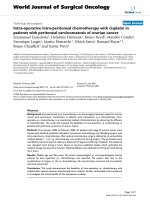
Figure 4 represents and example of ischemic
stroke and hemorrhagic stroke on baseline and
delayed CT with edema developing after 48 hours.
Int. J. Med. Sci. 2008, 5
22
FIGURE 4. A. Ischemic stroke presenting with left
hemiparesis. Normal CT on admission. CRP level: 6.9 m/dL;
NT-PBNP level: 21pmol/L. B. CT scan after 48hours. Large
infarction in the MCA territory showing mass-effect. CRP level:
7.9 mg/dL; NT-PBNP level: 139 pmol/L.
FIGURE 5. A. CT on admission. Hemorrhagic stroke
presenting with headache and left hemiplegia. CRP level: 3.23
mg/dL. NT-PBNP level: 66.3 pmol/L. B. CT scan after 48 hours
in the same patient. See the surrounding hypoatenuation of the
hematoma with increased mass-effect. CRP level: 22.6.
NT-PBNP level: 197 pmol/L.
DISCUSSION
The role of CRP in inflammation and
atherosclerosis has attracted the attention of many
researchers but it is not clear whether CRP is only a
marker or a causal factor
[23]. This protein has been
widely studied in the acute phase of ischemic stroke.
Compared with controls CRP levels in the serum were
higher than healthy controls in all stroke subtypes, in
the acute phase and after 3-month follow-up
[16]. The
CRP levels correlated positively with the size of the
infarct and stroke severity
[17,18]. Furthermore CRP
elevation in ischemic stroke indicates a worse
prognosis, as it has been associated with higher
in-hospital mortality
[9,20], higher mortality at 6
months
[21], and more disability
[22]. From the
Perindopril Protection against Recurrent Stroke a
case-control study showed that NT-PBNP levels but
not CRP predicted stroke recurrence
[9]. In this study
we did not find prognostic significance of either CRP
or NT-PBNP but we found relationship between the
levels of these proteins and the size of the stroke lesion
as edema tends to appear in large lesions. The lack of
prognostic significance may be caused by the small
sample size. Initially, one can speculate that the
baseline CRP levels are related to the degree of carotid
arteriosclerosis but two facts are against this
hypothesis: the CRP levels were higher in hemorrhagic
strokes, and that we found no differences of CRP levels
between cardioembolic and atherosclerotic strokes.
The significance of NT-PBNP elevation in the
acute phase of stroke is mostly unknown. In 86
consecutive patients with ischemic stroke the
NT-PBNP levels were related to unfavorable outcome
included death of any cause
[24]. NT-PBNP levels in
250 patients at day 2 after ischemic stroke onset were
predictive of 6-month mortality
[25]. However another
studies including 174 and 30 patients respectively with
ischemic stroke concluded that NT-PBNP levels did
not influence the clinical outcome
[14,15]. The
relationship between NT-PBNP levels and brain
edema in the acute phase of stroke has been little
investigated. Elevations of NT-PBNP were associated
with hyponatremia and natriuresis in subarachnoid
hemorrhage
[26] as well as to hyponatremia and
delayed vasospasm-related ischemic deficits
[27]. Focal
brain edema was related with elevations of ANP and
BNP in 8 patients with subarachnoid hemorrhage
[28].
In rodent model of subarachnoid hemorrhage the
intraventricular administration of ANP reduced the
sodium and water accumulation, as well as brain
edema
[29]. In rats with provoked-hemorrhagic stroke
the intraperitoneal administration of ANP reduced
water, sodium and edema 24 hours after the injury but
mannitol did not so, which suggests that ANP could be
used to treat brain edema
[30]. The role of ANP in
regulating blood flow in the infarcted area was studied
in stroke patients and a statistically significant increase
in the number of ANP-immunoreactive astrocytes was
found in the white matter surrounding the infarction
area
[31].
We observed elevation of NT-PBNP in relation to
brain edema in ischemic and hemorrhagic strokes as
these levels showed the maximum peak after 48 hours
from stroke onset. It is not likely that these elevations
related with blood pressure elevations as it only
occurred in 3 patients with edema. Our findings and
those of the previous studies suggest that natriuretic
peptides are produced in response to edema so as to
reduce it. Whether or not the treatment with BNP may
constitute an alternative of treatment in
stroke-induced brain edema remains to be elucidated.
The lack of efficacious treatment so far for
stroke-induced brain edema makes our hypothesis
Int. J. Med. Sci. 2008, 5
23
worthy of consideration.
The most important shortcoming of our study
was that edema was evaluated only with CT and,
therefore, we focused only on severe edema. The
practice of Diffusion weighted MRI would have added
important information but unfortunately this
technique was unavailable for us.
In conclusion, our work adds something new to
the understanding of the elevation of CRP and
NT-PBNP in the acute phase of stroke. both CRP and
NT-PBNP are markers of brain edema in stroke at
baseline and after 48 hours respectively. Given that
brain edema may be a life-threatening condition,
anti-edema measures could be anticipated in
hyperacute stroke in patients with high baseline levels
of ultrasensitive C-reactive protein. Owing to the small
sample size of our study these results need to be
confirmed in larger samples of stroke patients.
CONFLICT OF INTEREST
The authors have declared that no conflict of
interest exists.
REFERENCES
1. Edema BE. In: Fauci AS, et al, eds. Harrison’s principles of
Internal Medicine, 14
th
ed. McGraw-Hill: New York. 1998:
210-214.
2. Strunk A, Bhalla V, Clopton P, et al. Impact of the history of
congestive heart failure on the utlity of B-Type natriuretic
peptide in the emergency diagnosis of heart failure: results from
the Breathing Not Properly Multinational Study. Am J Med.
2006; 119: 69e1-69e11.
3. Kragelund C, Gronning B, Kober L, Hildebrandt P, Steffensen R.
N-terminal pro-B-Type Natriuretic Peptide and long-term
mortality in stable coronary heart disease. N Engl J Med. 2005;
352: 666-675.
4. Kistorp C, Raymond I, Pedersen F, Gustafsson F, Faber J,
Hildebrandt P. N-terminal pro-brain natriuretic peptide,
C-reactive protein, and urinary albumin levels as predictors of
mortality and cardiovascular events in older adults. JAMA. 2005;
293: 1609-1616.
5. Berger R, Huelsman M, Strecker K, et al. B-type natriuretic
peptide predicts sudden death in patients with chronic heart
failure. Circulation. 2002; 105: 2392-2397.
6. Wang TJ, Larson MG, Levy D, et al. Plasma natriuretic peptide
levels and the risk of cardiovascular events and death. N Engl J
Med. 2004; 350: 655-663.
7. Campbell DJ, Woodward M, Chalmers JP, et al. Prediction of
heart failure by amino terminal-pro-B-type natriuretic peptide
and C-reactive protein in subjects with cerebrovascular disease.
Hypertension. 2005; 45: 69-74.
8. Campbell DJ, Woodward M, Chalmers JP, et al. Prediction of
myocardial infarction by N-terminal-pro-B-type natriuretic
peptide, C-reactive protein, and renin in subjects with
cerebrovascular disease. Circulation. 2005; 112: 110-116.
9. Campbell DJ, Woodward M, Chalmers JP, et al. Soluble vascular
cell adhesion molecule 1 and N-terminal pro-B-type natriuretic
peptide in predicting ischemic stroke in patients with
cerebrovascular disease. Arch Neurol. 2006; 63: 60-65.
10. Rost NS, Wolf PA, Kase CS, et al. Plasma concentration of
C-reactive protein and risk of ischemic stroke and transient
ischemic attack: the Framingham study. Stroke. 2001; 32:
2575-2579.
11. Estrada V, Tellez MJ, Moya J, Fernandez-Durango R, Egido J,
Fernández-Cruz AF. High plasma levels of endothelin-1 and
atrial natriuretic peptide in patients with acute ischemic stroke.
Am J Hypertens. 1994; 7: 1085-1089.
12. Sato Y, Maruoka H, Honda Y, Hachiya N, Oizumi K. Plasma
concentration of atrial natriuretic peptide in cardioembolic
stroke with atrial fibrillation. Kurume Med J. 1995; 42: 71-77.
13. Makikallio AM, Makikallio TH, Korpelainen JT, et al. Natriuretic
peptides and mortality after stroke. Stroke 2005; 36: 1016-1020.
14. Etgen T, Baum H, Sander K, Sander D. Cardiac troponins and
N-terminal pro-brain natriuretic peptide in acute ischemic stroke
do not relate to clinical prognosis. Stroke. 2005; 36: 270-275.
15. Giannakoulas G, Hatzitolios A, Karvounis H, et al. N-terminal
pro-brain natriuretic peptide levels are elevated in patients with
acute ischemic stroke. Angiology. 2005; 56: 723-730.
16. Ladenvall C, Jood K, Blomstrand C, Nilsson S, Jern C, Ladenvall
P. Serum C-reactive protein concentration and genotype in
relation to ischemic stroke subtype. Stroke. 2006; 37: 2018-2023.
17. Audeberg HJ, Rott MM, Eck T, Haberl RL. Systemic
inflammatory response depends on initial stroke severity but is
attenuated by successful thrombolysis. Stroke. 2004; 35:
2128-2133.
18. Marq
uardt L, Ruf A, Mansmann U, Winter R, Buggle F,
Kallenberg K, Grau AJ. Inflammatory response after acute
ischemic stroke. J Neurol Sci. 2005; 236: 65-71.
19. Masotti L, Ceccarelli E, Forconi S, Cappelli R. Prognostic role of
C-reactive protein in very old patients with acute ischemic
stroke. J Intern Med. 2005; 258: 145-152.
20. Rallidis LS, Vikelis M, Panagiotakos DB, et al. Inflammatory
markers and in-hospital mortality in acute ischemic stroke.
Atherosclerosis. 2005.
21. Kocer A, Canbulat C, Gozke E, Ilhan A. C-reactive protein is an
indicator for fatal outcomes in first-time stroke patients. Med Sci
Monit. 2005; 11: CR540-544.
22. Arevalo-Lorido JC, Carretero-Gomez J, Calvo-Romero JM, et al.
C-reactive protein in the acute phase of ischemic stroke. Med
Clin (Barc). 2005; 125: 775-777.
23. Paffen E, DeMaat MP. C-reactive protein in atherosclerosis: A
causal factor? Cardiovasc Res. 2006; 71: 30-39.
24. Yip HK, Sun CK, Chang LT, Chen MC, Liou CW. Time course
and prognostic value of plasma levels of N-terminal pro-brain
natriuretic peptide in patients after ischemic stroke. Circ J. 2006;
70: 447-452.
25. Jensen JK, Mickley H, Bak S, Korsholm L, Kristensen SR. Serial
measurements of N-terminal pro-brain natriuretic peptide alter
acute ischemic stroke. Cerebrovasc Dis 2006; 22:439-444
26. Tomida M, Muraki M, Uemura K, Yamasaki K. Olasma
concentrations of brain natriuretic peptide in patients with
subarachnoid hemorrhage. Stroke. 1998; 29: 1584-1587.
27. McGirt MJ, Blessing R, Nimjee SM, et al. Correlation of serum
brain natriuretic peptide with hyponatremia and delayed
ischemic neurological deficits after subarachnoid hemorrhage.
Neurosurgery. 2004; 54: 1369-1373.
28. Fukui S, Nawashiro H, Otani N, et al. Focal brain edema and
natriuretic peptides in patients with subarachnoid hemorrhage.
Acta Neurochir (Wien). 2003; 86: 489-491.
29. Doczi TP, Joo F, Balas I. Atrial natriuretic peptide (ANP)
attenuates brain edema accompanying experimental
subarachnoid hemorrhage. Acta Neurochir (Wien). 1995; 132:
87-91.
30. Rosenberg GA, Estrada EY. Atrial natriuretic peptide blocks
hemorrhagic brain edema after 4-hour delay in rats. Stroke. 1995;
26: 874-877.
31. Nogami M, Shiga J, Takatsu A, Endo N, Ishiyama I.
Immunohistochemistry of atrial natriuretic peptide in brain
infarction. Histochem J. 2001; 33: 87-90.