Báo cáo y học: " Tumour necrosis factor-α stimulates dehydroepiandrosterone metabolism in human fibroblast-like synoviocytes: a role for nuclear factor-κB and activator protein-1 in the regulation of expression of cytochrome p450 enzyme 7b" doc
Bạn đang xem bản rút gọn của tài liệu. Xem và tải ngay bản đầy đủ của tài liệu tại đây (760.22 KB, 10 trang )
Open Access
Available online />R1271
Vol 7 No 6
Research article
Tumour necrosis factor-α stimulates dehydroepiandrosterone
metabolism in human fibroblast-like synoviocytes: a role for
nuclear factor-κB and activator protein-1 in the regulation of
expression of cytochrome p450 enzyme 7b
John Dulos
1
, Allard Kaptein
1
, Annemieke Kavelaars
2
, Cobi Heijnen
2
and Annemieke Boots
1
1
Department of Pharmacology, Section Autoimmunity, N.V. Organon, Oss, The Netherlands
2
Laboratory for Psychoneuroimmunology, University Medical Center Utrecht, Utrecht, The Netherlands
Corresponding author: John Dulos,
Received: 17 May 2005 Revisions requested: 27 Jun 2005 Revisions received: 4 Aug 2005 Accepted: 11 Aug 2005 Published: 15 Sep 2005
Arthritis Research & Therapy 2005, 7:R1271-R1280 (DOI 10.1186/ar1819)
This article is online at: />© 2005 Dulos et al.; licensee BioMed Central Ltd.
This is an Open Access article distributed under the terms of the Creative Commons Attribution License ( />2.0), which permits unrestricted use, distribution, and reproduction in any medium, provided the original work is properly cited.
Abstract
Glucocorticoids have successfully been used in the
treatment of rheumatoid arthritis. Data suggest that 7α-
hydroxy-dehydroepiandrosterone (7α-OH-DHEA), an
immunostimulating metabolite of dehydroepiandrosterone, can
block glucocorticoid-induced immune suppression. Formation
of 7α-OH-DHEA is catalyzed by activity of cytochrome p450
enzyme 7b (Cyp7b). Recently, we reported that tumour necrosis
factor (TNF)-α, IL-1α, IL-1β and IL-17 enhance Cyp7b mRNA
expression and induce a concomitant increase in the formation
of 7α-OH-DHEA by fibroblast-like synoviocytes (FLS) from
rheumatoid arthritis patients. The aim of this study was to
elucidate which signal transduction pathway is involved in the
TNF-α-mediated induction of Cyp7b activity in FLS. We studied
the effects of inhibitors of different signal transduction pathways
on Cyp7b activity in FLS by measuring Cyp7b mRNA
expression using reverse transcription PCR and by measuring
the formation of 7α-OH-DHEA. We applied SN50, an inhibitor
of nuclear translocation of transcription factors (i.e. activator
protein-1 [AP-1] and nuclear factor-κB [NF-κB]); PSI, a
proteasome inhibitor that prevents IκB degradation and thereby
NF-κB release; SP600125, a c-Jun N-terminal kinase (JNK)
inhibitor; and the mitogen-activated protein kinase inhibitors
PD98059 (extracellular signal-regulated kinase) and
SB203580 (p38). Cyp7b is constitutively expressed in RA FLS
and can be activated in response to TNF-α. SN50 and PSI
prevented the TNF-α-induced increase in Cyp7b activity,
whereas the mitogen-activated protein kinase inhibitors
PD98059 and SB203580 had no effect. In addition, inhibition
of Cyp7b mRNA expression and activity was observed with
SN50, PSI and SP600125, suggesting that NF-κB and AP-1
induce Cyp7b transcription. These findings suggest that NF-κB
and AP-1 are involved in the TNF-α-enhanced formation of the
dehydroepiandrosterone metabolite 7α-OH-DHEA. Our results
are in accordance with presence of AP-1 and NF-κB binding
sites in the Cyp7b promoter.
Introduction
Rheumatoid arthritis (RA) is a chronic inflammatory disease
characterized by hyperplasia of fibroblast-like synoviocytes
(FLS), which is regarded to be important in cartilage and bone
erosion [1]. Steroids such as dehydroepiandrosterone
(DHEA), glucocorticoids, androgens and oestrogens have
been shown to modulate the disease process in RA [2]. Sev-
eral authors have suggested that the natural, abundantly
present steroid DHEA may have immunostimulating effects
[3,4]. Further data indicate that the 7α-hydroxy-dehydroepian-
drosterone (7α-OH-DHEA) metabolite of DHEA, rather than
DHEA itself, is responsible for these immunostimulating
effects [5,6]. In several studies 7α-OH-DHEA was found to
stimulate the immune system both in vitro and in vivo, and it
AP-1 = activator protein-1; Cyp7b = cytochrome p450 enzyme 7b; DHEA = dehydroepiandrosterone; DMEM = Dulbecco's modified Eagle's medium;
ERK = extracellular signal-regulated kinase; FCS = foetal calf serum; FLS = fibroblast-like synoviocytes; IFN = interferon; IL = interleukin; JNK = c-
Jun N-terminal kinase; MAPK = mitogen-activated protein kinase; MEK = mitogen-activated protein kinase kinase; NFAT = nuclear factor of activated
T cells ; NF-κB = nuclear factor-κB; 7α-OH-DHEA = 7α-hydroxy-dehydroepiandrosterone; PBS = phosphate-buffered saline; PCR = polymerase
chain reaction; PMA = phorbol myristate acetate; RA = rheumatoid arthritis; STAT = signal transducer and activator of transcription; TNF = tumor
necrosis factor.
Arthritis Research & Therapy Vol 7 No 6 Dulos et al.
R1272
has been suggested that 7α-OH-DHEA acts as an antigluco-
corticoid [6,7].
The conversion of DHEA into 7α-OH-DHEA is catalyzed by
cytochrome p450 enzyme 7b (Cyp7b) [8]. Because of the
reported immunostimulating effects of 7α-OH-DHEA, we pre-
viously investigated the relation between Cyp7b activity and
arthritis. We showed that the severity of murine collagen-
induced arthritis was associated with an increase in Cyp7b
activity and Cyp7b mRNA level in synovial biopsies [9].
Recently, we reported that Cyp7b mRNA expression and
Cyp7b activity are present in FLS from patients with RA [10].
In addition, expression of Cyp7b in RA FLS was enhanced
after in vitro treatment of these cells with tumour necrosis fac-
tor (TNF)-α, IL-1α, IL-1β and IL-17 [10]. TNF-α is abundantly
produced in inflamed joints and is known to play a crucial role
in the pathogenesis of RA [11]. Therefore, in the present study
we used TNF-α to investigate which signal transduction path-
way is involved in the TNF-α-mediated increase in Cyp7b
activity in human FLS. Signaling pathways that mediate the
effects of TNF-α include mitogen-activated protein kinases
(MAPKs) and nuclear factor-κB (NF-κB) [12]. Three MAPK
families have been implicated to play a role in RA, including
extracellular signal (mitogenic)-regulated protein kinase
(ERK)1/2; the stress-activated protein kinases, also called c-
Jun NH
2
-terminal kinases (JNKs); and the p38 MAPKs [13].
The JNK pathway is of interest because of its capacity to phos-
phorylate the amino acids serine-63 and -73 on the c-Jun acti-
vation domain, which is a component of activator protein-1
(AP-1). AP-1 transcription factors consist of homodimers and
heterodimers of the Jun and Fos family [14]. Apart from
MAPKs, TNF-α activates nuclear translocation of NF-κB,
which plays a central role in inflammatory diseases such as RA
through induction of transcription of proinflammatory genes
[15]. NF-κB is retained in the cytosol of nonstimulated cells by
a noncovalent interaction with IκB. Upon stimulation by TNF-
α, IκB is degraded and NF-κB is released and translocated to
the nucleus inducing inflammatory gene expression [15].
Previous studies implicated a role for TNF receptor I in the reg-
ulation of Cyp7b activity [10], but these studies were inconclu-
sive regarding the role played by TNF receptor II in regulation
of Cyp7b activity. Thus, in order to study which signaling path-
ways are involved in TNF-α-induced Cyp7b activity, we used
different inhibitors with relevance to TNF receptor signaling.
SN50 was initially described as an inhibitor of nuclear translo-
cation of NF-κB. However, in addition to its effect on NF-κB,
SN50 blocks nuclear translocation of the AP-1 transcription
factor [16,17]. For that purpose, the effect of SP600125 – a
recently described inhibitor of JNK – on Cyp7b mRNA expres-
sion and activity was assessed [16]. The proteasome inhibitor
PSI prevents degradation of IκB and thereby indirectly pre-
vents NF-κB nuclear translocation [18]. To determine a possi-
ble role for MAPKs other than JNK in the TNF-α-induced
Cyp7b activity, the ERK1/2 inhibitor PD98059 and the p38
inhibitor SB203580 were used.
In the present study we report that NF-κB and AP-1, but not
ERK1/2 and p38, are probably involved in TNF-α-stimulated
formation of 7α-OH-DHEA.
Materials and methods
Fibroblast-like synoviocytes
FLS cell lines were developed from synovial biopsies obtained
from RA patients, after informed consent had been granted. All
patients fulfilled the 1987 American College of Rheumatology
criteria [19]. FLS were phenotyped as CD55
+
synovial fibrob-
lasts, as described previously [20]. Briefly, the synovial tissue
was minced and digested for 2 hours with 1 mg/ml colla-
genase A in Dulbecco's modified Eagle's medium (DMEM) at
37°C. The tissue homogenate was filtered through a fine sieve
(200 µm), washed and cultured overnight in synoviocyte
medium (Tebu-Bio, Heerhugowaard, The Netherlands) in 5%
carbon dioxide and 37°C to allow separation of adherent cells
from the nonadherent cell population. Nonadherent cells were
separated and adherent cells were cultured further in synovio-
cyte medium. The cells morphologically presenting as FLS
were used between passages 2 and 17 in the experiments.
Antibodies and reagents
The anti-NF-κB-p65 was from Signal Transductions (Becton &
Dickinson, Woerden, The Netherlands), and the biotinylated
anti-mouse IgG antibody was from Brunschwig Chemie
(Amsterdam, The Netherlands). TNF-α was bought from
Peprotech (Tebu-Bio, Heerhugowaard, The Netherlands). The
p38 MAPK inhibitor SB203580 and the ERK1/2-MAPK
kinase (MEK)-1 inhibitor PD98059 were from Omnilabo
(Breda, The Netherlands), dissolved in dimethylsulfoxide or
methanol and used as controls. The proteasome inhibitor PSI
and the JNK inhibitor SP600125 were purchased at Omnilabo
(Breda, The Netherlands) and dissolved in dimethylsulfoxide.
The SN50 peptide (Biomol, Plymouth, USA) was dissolved in
DMEM/Ham's F-12 medium.
Measurement of TNF-α-induced Cyp7b activity in
fibroblast-like synoviocytes
In order to arrest cell growth, synoviocyte medium was
replaced by DMEM/Ham's F-12 medium with 10% foetal calf
serum (FCS) and the FLS were cultured for another 3 days in
a 24-well plate (Greiner, Alphen a/d Rijn, The Netherlands).
FLS were preincubated in the presence or absence of SN50
for 2 hours, or PSI, SP600125, SB203580, or PD98059 for
1 hour in 2% charcoal-treated (depleted from steroids) FCS.
Charcoal-treated FCS were prepared by suspending charcoal
(Norit A) in Tris buffer. The suspension was then centrifuged
for 10 min at 8.000 N/kg, the supernatant was removed and
FCS added to the residue. This suspension was stirred for 30
min at 45°C and the charcoal was removed by centrifugation
for 10 min at 8.000 N/kg. The supernatant was sterilized by
Available online />R1273
membrane filtration using filters of pore sizes 0.8 and 0.2 µm
successively. Following heat inactivation, FCS was stored at -
20°C until use.
FLS were incubated with or without TNF-α and 1,2,6,7-[
3
H]-
DHEA (1.5 × 10E
-8
mol/l: NEN Life Science Products, Boston,
MA, USA) for 24 hours. Steroid-containing medium (1 ml) was
passed over a C18 Solid Phase Extraction cartridge
(Sopachem, Wageningen, The Netherlands) to determine the
conversion of 1,2,6,7-[
3
H]-DHEA into
3
H-labelled 7α-OH-
DHEA as a measure of Cyp7b activity. Steroids were eluted
from the column with methanol. Next,
3
H-labelled 7α-OH-
DHEA and
3
H-labelled DHEA were measured using high-per-
formance liquid chromatography. The amount of 7α-OH-
DHEA is expressed as the percentage of
3
H-labelled 7α-OH-
DHEA of the total amount of
3
H-label measured. Recoveries
after extraction were in the range 85–95%, and identification
of 7α-OH-DHEA was confirmed by Gas Chromatography-
Mass spectrometry GC-MS (data not shown).
Detection of 7α-OH-DHEA levels by radioimmunoassay
To determine 7α-OH-DHEA levels in FLS, a radioimmu-
noassay was performed using antiserum against 7α-OH-
DHEA. The 7α-OH-DHEA metabolite is formed by the activity
of the enzyme Cyp7b. The radioimmunoassay was performed
at the Institute of Endocrinology at Prague (Czech Republic)
in cooperation with Dr R Hampl [21]. In brief, FLS were prein-
cubated in the presence or absence of SN50 for 2 hours or
PSI, SP600125, SB203580, or PD98059 for 1 hour in 2%
charcoal-treated (depleted from steroids) FCS. Thereafter,
FLS were incubated with or without TNF-α and 1.5 × 10E
-8
mol/l DHEA (Diosynth, Oss, The Netherlands) for 24 hours.
Extraction was carried out using diethyl ether. Diethyl ether
extracts containing 7α-OH-DHEA and 7β-OH-DHEA were
evaporated under nitrogen, and the dry residue was dissolved
in assay buffer and measured using radioimmunoassay as pre-
viously described [21].
Immunohistochemistry of fibroblast-like synoviocytes
FLS were grown on chamber slides (Nalge Nunc International;
Fisher Emergo, Landsmeer, The Netherlands) and preincu-
bated for 2 hours in the presence or absence of SN50 (100
µg/ml or 200 µg/ml) and thereafter stimulated for 30 min with
TNF-α (0.5 ng/ml). After washing with phosphate-buffered
saline (PBS), cells were fixed in methanol for 10 min and dried.
The samples were blocked with buffer containing 2% normal
goat serum, 2% human serum, and 2% serum albumin in PBS/
0.01% Triton X-100 (PBS/T) for 30 min. Cells were then incu-
bated with anti-NF-κB p65 antibody in the same buffer for 1
hour at ambient temperature. After washing with PBS-T, the
FLS were incubated for 45 min with biotinylated anti-mouse
IgG. After washing, cells were incubated for 30 min with avi-
din-biotin-peroxidase (Brunschwig Chemie, Amsterdam, The
Netherlands). Following washing, the substrate was incubated
for 10 min with enhanced diaminobenzidine in stable peroxi-
dase buffer (Pierce; Perbio Science, Etten-Leur, The Nether-
lands). Following extensive washing in milli-Q water and
dehydration, coverslips were placed with Entellan (Merck,
Amsterdam, The Netherlands) mounting medium. Slides were
visually analyzed under a Nikon Alphaphot-2 microscope
(Uvikon, Bunnik, The Netherlands).
Cyp7b mRNA levels in fibroblast-like synoviocytes
FLS were preincubated with 200 µg/ml SN50 and then incu-
bated in the presence or absence of TNF-α (0.5 ng/ml) for 6
hours. Cells were washed with PBS and total RNA was
extracted with RNAzol (Campro, Veenendaal, The Nether-
lands). cDNA synthesis was done according to the manufac-
turer's protocol using random hexamer primers (Pharmacia,
Woerden, The Netherlands) and reverse transcriptase (Phar-
macia). For reverse transcription PCR, human Cyp7b sense
(GTCCTGGAGAAATATTATGTGCAG) and antisense
(CGCACACAGTAGTCCCCGG) primers were used. For
GAPDH we used CCCTTCATTGACCTCAACTACATGG
(sense) and GGTCCACCACCCTGTTGCTGTAGCC (anti-
sense) as primers. Reverse transcription PCR was carried out
using an Applied Biosystems (Nieuwerkerk a/d ijssel, The
Netherlands) thermo cycler with an anneal temperature of
53°C.
Computer analysis of the Cyp7b promoter region
The promoter sequence of the human Cyp7b gene was iden-
tified and exported from the Ensembl database (vs19.34b.2; 9
February 2004) using the MartView export function. As pro-
moter region, -1,000 to +100 nucleotides were selected in
relation to the transcription start site. Promoter analysis for
transcription factor binding sites was performed using the
GEMSLauncher version 3.6 from Genomatrix and MatInspec-
tor professional release 7 [22]. Core and matrix similarity set-
tings were 0.75 and optimized -0.03, respectively. The
transcription factor family matrices V$AP1F, V$NFAT,
V$NFKB and V$STAT were used.
Results
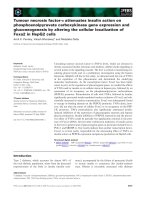
SN50 inhibited TNF-α-stimulated Cyp7b expression and
activity
An FLS cell-line (SCRO.14.SF), obtained from a synovial
biopsy from an RA patient, was used to study the effect of
SN50 on the TNF-α-induced Cyp7b activity. SN50 (200 µg/
ml) significantly reduced basal Cyp7b activity (Fig. 1a). Impor-
tantly, the increase in Cyp7b activity following stimulation of
the cells with TNF-α was dose-dependently inhibited by SN50
(Fig. 1a).
To further substantiate this finding, five other FLS cell lines
generated from RA synovial biopsies obtained from different
RA patients were stimulated with TNF-α with or without the
dose of 200 µg/ml SN50. DHEA was metabolized into 7α-
OH-DHEA in all five untreated FLS cell lines used (Fig. 1b).
TNF-α induced a significant increase in Cyp7b activity in all
Arthritis Research & Therapy Vol 7 No 6 Dulos et al.
R1274
FLS used. When SN50 was applied in combination with TNF-
α, conversion of DHEA into 7α-OH-DHEA was significantly
inhibited in four out of five FLS cell lines.
To investigate whether the effect of SN50 interfered at the
level of Cyp7b activity or expression, we also analyzed the
influence of SN50 on the TNF-α-induced increase in Cyp7b
Figure 1
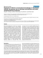
Cyp7b activity and mRNA expression is inhibited by SN50 in fibroblast-like synoviocytesCyp7b activity and mRNA expression is inhibited by SN50 in fibroblast-like synoviocytes. (a) Human fibroblast-like synoviocytes (FLS; SCRO.14.SF,
passages 10–12) were plated at 1 × 10
5
cells/well in a 24-well plate and preincubated in the presence or absence (-) of the SN50 inhibitor for 2
hours. Thereafter, the cells were incubated with (solid bars) or without (open bars) tumour necrosis factor (TNF)-α for another 24 hours with 1.5 ×
10
-8
mol/l
3
H-dehydroepiandrosterone (DHEA). The formation of [
3
H]-7α-hydroxy-dehydroepiandrosterone (7α-OH-DHEA) from [
3
H]-DHEA, repre-
senting cytochrome p450 enzyme 7b (Cyp7b) activity, was determined by high-performance liquid chromatography. The amount of 7α-OH-DHEA is
expressed as the percentage [
3
H]-7α-OH-DHEA of the total amount of [
3
H]-label measured. Results are expressed as the mean ± standard error of
the mean of triplicate samples. The data are representative of two independent experiments. *P < 0.005 (Student's t-test). (b) Human FLS (1 × 10
5
cells/well) were isolated from five different rheumatoid arthritis patient biopsies. Cells (1 × 10
5
/well) were incubated in the presence and absence of
TNF-α and in the presence of SN50 for 2 hours, as described in Materials and methods. Results are representative for one of the two independent
experiments. SCRO.12.SF passage 2, SCRO.11.SF passage 3, SCRO.03.SF passage 8, SCRO.01.SF passage 6 and SCRO.08.SF passage 4
were used. *P < 0.005 versus TNF-α (Student's t-test). (c) FLS (SCRO.14.SF; passages 10–12) were incubated for 6 hours with 0.5 ng/ml TNF-α,
SN50 200 µg/ml plus 0.5 ng/ml TNF-α, or incubated with medium control (-). Reverse transcription PCR was done using GAPDH and Cyp7b spe-
cific primers (35 cycles). The data are representative of two independent experiments. (d) FLS fibroblasts (SCRO.14.SF; passages 10–12) were
grown on chamber slides. Cells were incubated for 2 hours in the presence or absence of 200 µg/ml SN50 before incubation for 30 min in the pres-
ence or absence of TNF-α (0.5 ng/ml). Immunoperoxidase staining was carried out with an antibody against nuclear factor-κB (NF-κB)p65 conju-
gated to peroxidase. Data are representative for three independent experiments.
Available online />R1275
mRNA expression in the SCRO.14.SF cell line. A weak signal
for Cyp7b mRNA was observed in untreated FLS (Fig. 1c).
When stimulated with TNF-α, a marked increase in Cyp7b
mRNA level was observed. Incubation of FLS with SN50
almost completely prevented the TNF-α-induced increase in
Cyp7b mRNA expression (Fig. 1c).
Studies were performed to investigate whether SN50 indeed
inhibits transport of NF-κB to the nucleus. In untreated FLS,
NF-κB is localized in the cytoplasm (Fig. 1d). Incubation of
FLS with TNF-α strongly increased the presence of NF-κB in
the nucleus. This nuclear translocation of NF-κB was inhibited
by SN50 (Fig. 1d).
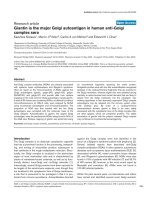
PSI inhibited the TNF-α-induced increase in Cyp7b
activity
In subsequent experiments we examined the effect of PSI, a
proteasome inhibitor that is known to prevent IκB degradation
and thereby activation of NF-κB, on TNF-α-induced Cyp7b
activation in the FLS cell line. PSI (1 × 10E
-6
mol/l) significantly
decreased Cyp7b activity in nonstimulated FLS. Moreover,
PSI prevented the increase in Cyp7b activity following incuba-
tion with TNF-α (Fig. 2). The combined results with SN50 and
PSI imply an involvement of NF-κB in TNF-α-induced Cyp7b
activity.
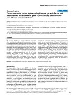
MAPK inhibition did not affect the TNF-α-induced
increase in Cyp7b activity
We further investigated a putative role for MAPKs in the TNF-
α-induced increase in Cyp7b activity by using the MEK1 inhib-
itor PD98059 and the p38 inhibitor SB203580.
The p38 inhibitor (SB203580) did not affect Cyp7b activity in
nonstimulated cells (Fig. 3). Also, following TNF-α stimulation
no effect of SB203580 on the increase in Cyp7b activity was
observed. Similarly, incubation of nonstimulated FLS with the
MEK1/ERK1/2 inhibitor (PD98059) did not affect Cyp7b
activity. Only at a high concentration (1 × 10E
-5
mol/l) did
application of PD98059 result in a small but statistically signif-
icant inhibition of TNF-α-induced increase in Cyp7b activity.
The combination of SB203580 and PD98059 at high concen-
trations, similar to PD98059 alone, also exhibited a small but
significant decrease in TNF-α-induced Cyp7b activity (Fig. 3).
Similar findings were obtained using five additional RA FLS
cell lines; a small inhibitory effect of the p38 inhibitor
SB203580 at high concentration (1 × 10
-5
mol/l) was
observed in one cell line out of five after stimulation with TNF-
α. In none of the five cell lines did we observe any effect on the
TNF-α-induced increase in Cyp7b activity using 1 × 10
-5
mol/
l PD98059 (data not shown). From these results it is con-
cluded that p38 and ERK1/2 do not appear to play a role in
regulating Cyp7b activity.
Regulation of Cyp7b mRNA expression and activity in
fibroblast-like synoviocytes
Previous studies implicated a role for TNF receptor I in regu-
lating Cyp7b activity [10]. Because the TNF receptor I couples
to AP-1 via the JNK pathway, we investigated the effect of the
recently described JNK inhibitor SP600125 [17]. In addition,
we analyzed the effect of NF-κB and MAPK inhibitors on TNF-
α-induced Cyp7b mRNA expression. A weak Cyp7b mRNA
signal was found in untreated FLS (Fig. 4a). Treatment of FLS
with TNF-α resulted in an increase in Cyp7b mRNA expres-
sion. Moreover, SN50 prevented the increase in Cyp7b mRNA
expression following incubation with TNF-α. Furthermore, the
proteasome inhibitor PSI, which is known to prevent IκB deg-
radation, blocked the TNF-α-induced Cyp7b mRNA expres-
sion. In addition, the JNK inhibitor SP600125 prevented the
TNF-α-induced Cyp7b mRNA expression, which further sub-
stantiates a role for AP-1 in TNF-α-induced Cyp7b expression.
Use of the MAPK inhibitors PD98059 and SB203580 did not
result in convincing changes in TNF-α-induced Cyp7b mRNA
expression.
We then determined Cyp7b enzymatic activity in FLS through
the detection of 7α-OH-DHEA. Presence of TNF-α in the cul-
tures resulted in increased Cyp7b activity compared with
baseline (Fig. 4b). We subsequently analyzed the effect on
TNF-α stimulation of the presence or absence of PSI, SN50,
SP600125, PD98059 and or SB203580. TNF-α in combina-
tion with PSI, SN50, or SP600125 significantly decreased the
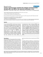
Figure 2
PSI inhibits the TNF-α-induced increase in 7α-OH-DHEAPSI inhibits the TNF-α-induced increase in 7α-OH-DHEA. Human rheu-
matoid arthritis (RA) fibroblast-like synoviocytes (FLS; SCRO.14.SF, 1
× 10
5
cells/well; passages 10–12) were preincubated in the presence
or absence (-) of the PSI inhibitor for 1 hour. Thereafter, the cells were
incubated with (solid bars) or without (open bars) tumour necrosis fac-
tor (TNF)-α for another 24 hours with 1.5 × 10
-8
mol/l
3
H-DHEA, as
described in Materials and methods. Data are expressed as mean ±
standard error of the mean and are representative of four independent
experiments. *P < 0.0005. 7α-OH-DHEA = 7α-hydroxy-dehydroepian-
drosterone.
Arthritis Research & Therapy Vol 7 No 6 Dulos et al.
R1276
Cyp7b activity to basal 7α-OH-DHEA levels (Fig. 4b). In con-
trast, addition of PD98059 or SB203580 did not significantly
affect the TNF-α-induced increase in Cyp7b activity. The
absence of an effect of the MAPK inhibitors PD98059 and
SB203580 on TNF-α-induced Cyp7b activity is in accordance
with our findings at the level of Cyp7b mRNA expression.
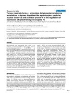
Figure 3
The effect of the MAPK inhibitors PD98059 or SB203580 on TNF-α-induced Cyp7b activityThe effect of the MAPK inhibitors PD98059 or SB203580 on TNF-α-induced Cyp7b activity. (a) Fibroblast-like synoviocytes (FLS; SCRO.14.SF,
passages 8–12) were incubated for 1 hour in the presence or absence (-) of the mitogen-activated protein kinase (MAPK) kinase (MEK)1 inhibitor
PD98059 (PD) or the p38 inhibitor SB203580 (SB). Thereafter, cells were incubated in the presence or absence of 0.5 ng/ml tumour necrosis fac-
tor (TNF)-α plus 1.5 × 10
-8
mol/l [
3
H]-dehydroepiandrosterone (DHEA) for 24 hours and processed using high-performance liquid chromatography.
The amount of 7α-hydroxy-dehydroepiandrosterone (7α-OH-DHEA) is expressed as the percentage [
3
H]-7α-OH-DHEA of the total amount of [
3
H]-
label measured. Results are expressed as the mean ± standard error of the mean of triplicate sample. Data are representative of three independent
experiments. *P < 0.05 versus TNF-α (Student's t-test). (b) The data from panel a (three independent experiments) are combined for the highest
inhibitor concentrations. PD98059 and SB23580 were dissolved in methanol (MeOH) and dimethylsulfoxide (DMSO), respectively, and used as
controls. *P < 0.05 (Student's t-test). Cyp7b = cytochrome p450 enzyme 7b.
Available online />R1277
Presence of NF-κB and AP-1 binding sites within the
Cyp7b promoter
Analysis of the proximal region of the Cyp7b promoter
revealed nucleotide sequences that correspond to putative
binding sites for NF-κB, AP-1, nuclear factor of activated T
cells (NFAT), and signal transducer and activator of transcrip-
tion (STAT)1 (Fig. 5). The presence of putative bindings sites
for NF-κB and AP-1 within the Cyp7b promoter are in accord-
ance with the findings in this report that NF-κB and AP-1 are
involved in the TNF-α-enhanced Cyp7b activity.
Discussion
The findings of the present study suggest involvement of AP-
1 and NF-κB, but not of p38 or ERK1/2, in the TNF-α-
enhanced formation of the immunostimulating 7α-OH-DHEA.
We and others [23]. showed that, upon stimulation of cells
with TNF-α, NF-κB translocates from the cytoplasm to the
nucleus. As expected, translocation of NF-κB to the nucleus
was inhibited by SN50. In addition, SN50 blocks the TNF-α-
induced increases in Cyp7b activity and Cyp7b mRNA level,
which suggests transcriptional involvement of NF-κB and/or
other transcription factors such as AP-1 in TNF-α-induced
Cyp7b activation. Initial reports suggested that SN50 is a spe-
cific inhibitor of NF-κB activation. However, Torgerson and
coworkers [23] reported that SN50 blocks the nuclear trans-
location of the transcription factors AP-1, NFAT and STAT1 in
Jurkat T cells stimulated with IFN-γ or phorbol myristate ace-
tate (PMA) as well.
Figure 4
Regulation of Cyp7b mRNA expression and activity in FLSRegulation of Cyp7b mRNA expression and activity in FLS. (a) Human
fibroblast-like synoviocytes (FLS; STSF.388, passages 8–10) were
incubated for 6 hours with medium control (-), 0.5 ng/ml tumour necro-
sis factor (TNF)-α, TNF-α combined with the proteasome inhibitor (PSI)
1 × 10E
-6
mol/l, SN50 200 µg/ml, SP600125 (SP) 1 × 10E
-5
mol/l,
PD98059 (PD) 1 × 10E
-5
mol/l or SB23580 (SB) 1 × 10E
-5
mol/l, as
described in Materials and methods. RNA was isolated and cDNA was
made and used for reverse transcription with GAPDH and cytochrome
p450 enzyme 7b (Cyp7b) specific primers. The ratio of Cyp7b to
GAPDH mRNA expression was 1.4 (TNF-α alone), 8 × 10
-6
(PSI +
TNF-α), 1.3 × 10
-6
(SN50 + TNF-α), 0.5 × 10
-6
(SP + TNF-α), 0.3 (PD
+ TNF-α) and 0.2 (SB + TNF-α). Data are representative of two inde-
pendent experiments. (b) FLS (STSF.388, passages 8–10) were prein-
cubated in the presence or absence (medium control [-]) of SN50 for 2
hours or SP600125 (SP), PD98059 (PD) or SB23580 (SB) for 1 hour.
Thereafter, FLS were incubated in the presence or absence of 0.5 ng/
ml TNF-α plus 1.5 × 10
-8
mol/l dehydroepiandrosterone (DHEA) for 24
hours and processed for radioimmunoassay detection of 7α-hydroxy-
dehydroepiandrosterone (7α-OH-DHEA). Results are expressed as the
mean ± standard error of the mean of triplicate samples. Data are rep-
resentative for two independent experiments. *P < 0.005 versus TNF-α
(Student's t-test).
Figure 5
NF-κB and AP-1 binding sites within the Cyp7b promoterNF-κB and AP-1 binding sites within the Cyp7b promoter. Putative
binding sites for selected transcription factor family matrices were iden-
tified using the MartView export function. Sequences for putative bind-
ing sites are underlined. *, transcription start side; -, presence of the
transcription binding site on the minus DNA strand; AP-1, activator pro-
tein-1; Cyp7b, cytochrome p450 enzyme 7b; NFAT, nuclear factor of
activated T cells; NF-κB, nuclear factor-κB; STAT, signal transducer
and activator of transcription.
Arthritis Research & Therapy Vol 7 No 6 Dulos et al.
R1278
To determine whether STAT1 could be involved in TNF-α-
induced Cyp7b activity, we analyzed the proximal region of the
Cyp7b promoter for putative binding sites of STAT1, which
revealed such sites in this region. It should be appreciated,
however, that STAT1 is mainly activated by IFN-γ. Also, Cyp7b
is not regulated by IFN-γ, as described previously [10].
Therefore, it is unlikely that STAT1 is involved in Cyp7b activity
regulation.
There is evidence that the dose of SN50 determines the spe-
cificity of the inhibitor [16]. Therefore, it is likely that the doses
of SN50 we used (100–200 µg/ml) can block both transloca-
tion of NF-κB and translocation of AP-1 to the nucleus [24].
Indeed, we observed inhibition of TNF-α-induced NF-κB
nuclear translocation concomitantly with an inhibition of TNF-
α-induced Cyp7b activity by SN50. In order to investigate a
role for AP-1, we used the JNK inhibitor SP600125 [17]. The
results demonstrate an involvement of the AP-1 complex in the
TNF-α-induced Cyp7b expression and activity in FLS from RA
patients. An involvement of NF-κB and AP-1 in the TNF-α-
induced Cyp7b activity is in accordance with the presence of
putative NF-κB and AP-1 binding sites within the Cyp7b
promoter.
Our findings are consistent with data reported by Wu and
coworkers [25] with respect to the presence of putative bind-
ing sites for NF-κB within the Cyp7b promoter. In contrast to
our analysis, those authors [25] did not identify putative AP-1
binding sites, which could be due to the use of the default set-
ting for the matrix score in MartView. However, other
approaches are needed to substantiate further the role played
by NF-κB and AP-1 in the TNF-α-induced increase in Cyp7B
expression. This may be done by analysis of the Cyp7b pro-
moter in a promoter reporter construct, with mutation of the
putative NF-κB and AP-1 response elements. Moreover, the
use of the siRNA technology could contribute to our under-
standing of the importance of NF-κB in the TNF-α-induced
DHEA metabolism in human FLS.
Because the anti-glucocorticoid 7α-OH-DHEA, which is pro-
duced by the activity of the enzyme Cyp7b, might have stimu-
latory effects on the inflammatory process, studies with
administration of 7α-OH-DHEA in animal models with suscep-
tibility for arthritis are needed to elucidate the mechanism by
which 7α-OH-DHEA influences the development of inflamma-
tory processes. In this respect, it would be of interest to inves-
tigate whether inflammation is reduced in Cyp7b knockout
mice, which do not express 7α-OH-DHEA. In addition, intra-
articular delivery of 7α-OH-DHEA and/or Cyp7b expression
systems should add to our understanding of the role played by
Cyp7b in the arthritic process.
The inhibitory effect of PSI on the TNF-α-induced upregulation
of Cyp7b activity is also in accordance with a role for NF-κB in
regulating Cyp7b activity. Although it has not been described
in the original studies of the action of PSI [18], we cannot
exclude the possibility that inhibition of proteasome activity by
PSI may interfere in other signal transduction pathways that
are independent of NF-κB [26].
In this paper we show that inhibitors of the ERK1/2 and p38
signalling pathways did not convincingly affect Cyp7b mRNA
expression and enzymatic activity in RA FLS following stimula-
tion with TNF-α. Barchowsky and coworkers [27] also
reported that there is no role for MAPKs after TNF-α stimula-
tion of collagenase I expression in rabbit synovial fibroblasts.
However, previous studies have reported activation of ERK1/
2 and p38 in several cell lines, including synovial fibroblasts,
after incubation with TNF-α [28]. We observed that, in con-
trast to TNF-α-induced Cyp7b activity, the MEK1/ERK1/2
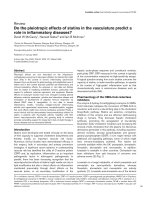
Figure 6
Simplified diagram of the proposed signalling events leading to Cyp7b gene transcription in synovial fibroblastsSimplified diagram of the proposed signalling events leading to Cyp7b
gene transcription in synovial fibroblasts. Using inhibitors of the
mitogen-activated protein kinase (MAPK) kinase (MEK)1/extracellular
signal (mitogenic)-regulated kinase (ERK)1/2 pathway (i.e. PD98059),
the p38 MAPK pathway (i.e. SB203580), the c-Jun-NH
2
-terminal
kinase (JNK) pathway (i.e. SP600125), the IκB/nuclear factor-κB (NF-
κB) pathway (i.e. PSI; dashed line) and the NF-κB/activator protein
(AP)-1 pathway SN50, it was established that the NF-κB and AP-1
pathway is relevant to Cyp7b activity. All experiments were performed
using synovial fibroblasts derived from patients with rheumatoid arthri-
tis. Cyp7b, cytochrome p450 enzyme 7b; IκBα, inhibitor of NF-κB; IKK,
IκB kinase complex (composed of three subunits – IKKα, IKKβ, and
IKKγ [NEMO]; RelA (p65) and NF-κB1 [p50/p105] are subunits of NF-
κB); RIP, receptor interacting protein; TNF, tumour necrosis factor;
TNFR, TNF-α receptor; TRADD, TNF receptor associated death
domain; TRAF, TNF receptor associated factor.
Available online />R1279
inhibitor PD98059 and p38 inhibitor SB203580 reduced the
TNF-α-induced IL-6 production in several RA FLS tested (data
not shown). These results indicate that the inhibitors were
active and can inhibit other effects of TNF-α, but they do not
play a role in regulation of Cyp7b activity by TNF-α. Further-
more, it cannot be excluded that other MAPK isoforms such as
ERK5, ERK7, p38γ and p38δ are regulated by TNF-α as well
in the RA FLS used [29].
Conclusion
Our data suggest that there is a role for both NF-κB and AP-1
in regulating the expression and activity of Cyp7b (Fig. 6),
which strengthens the rationale for specific inhibition of these
pathways in arthritis.
Competing interests
The author(s) declare that they have no competing interests.
Authors' contributions
JD was principle investigator, and designed most of the stud-
ies, carried out most of the assays and wrote the manuscript.
AK (Allard Kaptein) helped in conceiving the study and helped
to draft the manuscript. AK (Annemieke Kavelaars) and CH
were involved in drafting and revising the article. AB helped in
conceiving the study, helped to draft the manuscript and was
the senior scientist responsible for the work. All authors read
and approved the final manuscript.
Acknowledgements
We thank Dr E Bos for critical reading of the manuscript and M Toker
and N Bisseling for photographic reproductions. C Meeuwisse is
acknowledged for performing computer analysis of the Cyp7b promoter.
Dr R Hampl is acknowledged for the performance of the radioimmu-
noassay analysis.
References
1. Zvaifler NJ, Firestein GS: Pannus and pannocytes. Alternative
models of joint destruction in rheumatoid arthritis. Arthritis
Rheum 1994, 37:783-789.
2. Straub RH, Cutolo M: Involvement of the hypothalamic-pitui-
tary-adrenal/gonadal axis and the peripheral nervous system
in rheumatoid arthritis: viewpoint based on a systemic patho-
genetic role. Arthritis Rheum 2001, 44:493-507.
3. Bradlow HL, Murphy J, Byrne JJ: Immunological properties of
dehydroepiandrosterone, its conjugates, and metabolites.
Ann N Y Acad Sci 1999, 876:91-101.
4. Daynes RA, Dudley DJ, Araneo BA: Regulation of murine lym-
phokine production in vivo. II. Dehydroepiandrosterone is a
natural enhancer of interleukin 2 synthesis by helper T cells.
Eur J Immunol 1990, 20:793-802.
5. Morfin R, Courchay G: Pregnenolone and dehydroepiandros-
terone as precursors of native 7-hydroxylated metabolites
which increase the immune response in mice. J Steroid Bio-
chem Mol Biol 1994, 50:91-100.
6. Morfin R: Involvement of steroids and cytochromes P(450)
species in the triggering of immune defenses. J Steroid Bio-
chem Mol Biol 2002, 80:273-290.
7. Lathe R: Steroid and sterol 7-hydroxylation: ancient pathways.
Steroids 2002, 67:967-977.
8. Rose KA, Stapleton G, Dott K, Kieny MP, Best R, Schwarz M, Rus-
sell DW, Bjorkhem I, Seckl J, Lathe R: Cyp7b, a novel brain cyto-
chrome P450, catalyzes the synthesis of neurosteroids
7alpha-hydroxy dehydroepiandrosterone and 7alpha-hydroxy
pregnenolone. Proc Natl Acad Sci U S A 1997, 94:4925-4930.
9. Dulos J, Verbraak E, Bagchus WM, Boots AM, Kaptein A: Severity
of murine collagen-induced arthritis correlates with increased
CYP7B activity: enhancement of dehydroepiandrosterone
metabolism by interleukin-1β. Arthritis Rheum 2004,
50:3346-3353.
10. Dulos J, van der Vleuten MAJ, Kavelaars A, Heijnen CJ, Boots AM:
CYP7B expression and activity in fibroblast-like synoviocytes
from patients with rheumatoid arthritis: regulation by pro-
inflammatory cytokines. Arthritis Rheum 2005, 52:770-778.
11. Elliot MJ, Maini RN, Feldmann M, Long-Fox A, Charles P, Katsikis
P, Brennan FM, Walker J, Bijl H, Ghraveb J, et al.: Treatment of
rheumatoid arthritis with chimeric monoclonal antibodies to
tumor necrosis factor alpha. Arthritis Rheum 1993,
36:1681-1690.
12. Firestein GS, Manning AM: Signal transduction and transcrip-
tion factors in rheumatic disease. Arthritis Rheum 1999,
42:609-621.
13. Chang L, Karin M: Mammalian MAP kinase signalling cascades.
Nature 2001, 410:37-40.
14. Palanki MS: Inhibitors of AP-1 and NF-kappa B mediated tran-
scriptional activation: therapeutic potential in autoimmune
diseases and structural diversity. Curr Med Chem 2002,
9:219-227.
15. Li Q, Verma IM: NF-kappaB regulation in the immune system.
Nat Rev Immunol 2002, 2:725-734.
16. Das J, Chen CH, Yang L, Cohn L, Ray P, Ray A: A critical role for
NF-kappa B in GATA3 expression and TH2 differentiation in
allergic airway inflammation. Nat Immunol 2001, 2:45-50.
17. Hammaker DR, Boyle DL, Chabaud-Riou M, Firestein GS: Regu-
lation of c-Jun N-terminal kinase by MEKK-2 and mitogen-acti-
vated protein kinase kinase kinases in rheumatoid arthritis. J
Immunol 2004, 172:1612-1618.
18. Haas M, Page S, Page M, Neumann FJ, Marx N, Adam M, Zieglar-
Heitbrock HW, Neumeier D, Brand K: Effect of proteasome
inhibitors on monocytic IkappaB-alpha and -beta depletion,
NF-kappaB activation, and cytokine production. J Leukoc Biol
1998, 63:395-404.
19. Saraux A, Berthelot JM, Chales G, Le Henaff C, Thorel JB, Hoang
S, Valls I, Devauchelle V, Martin A, Baron D, et al.: Ability of the
American College of Rheumatology 1987 criteria to predict
rheumatoid arthritis in patients with early arthritis and classifi-
cation of these patients two years later. Arthritis Rheum 2001,
44:2485-2491.
20. Hamann J, Wishaupt JO, van Lier RA, Smeets TJ, Breedveld FC,
Tak PP: Expression of the activation antigen CD97 and its lig-
and CD55 in rheumatoid synovial tissue. Arthritis Rheum 1999,
42:650-658.
21. Lapcik O, Hampl R, Hill M, Starka L: Immunoassay of 7-
hydroxysteroids: 2. Radioimmunoassay of 7alpha-hydroxy-
dehydroepiandrosterone. J Steroid Biochem Mol Biol 1999,
71:231-237.
22. Quandt K, Frech K, Karas H, Wingender E, Werner T: MatInd and
MatInspector: new fast and versatile tools for detection of con-
sensus matches in nucleotide sequence data. Nucleic Acids
Res 1995, 23:4878-4884.
23. Torgerson TR, Colosia AD, Donahue JP, Lin YZ, Hawiger J: Regu-
lation of NF-kappa B, AP-1, NFAT, and STAT1 nuclear import in
T lymphocytes by noninvasive delivery of peptide carrying the
nuclear localization sequence of NF-kappa B p50. J Immunol
1998, 161:6084-6092.
24. Boothby M: Specificity of sn50 for NF-kappa B? Nat Immunol
2001, 2:471-472.
25. Wu Z, Martin KO, Javitt NB, Chiang JY: Structure and functions
of human oxysterol 7alpha-hydroxylase cDNAs and gene
CYP7B1. J Lipid Res 1999, 40:2195-2203.
26. Yoshimura S, Bondeson J, Brennan FM, Foxwell BM, Feldmann M:
Role of NFkappaB in antigen presentation and development of
regulatory T cells elucidated by treatment of dendritic cells
with the proteasome inhibitor PSI. Eur J Immunol 2001,
31:1883-1893.
27. Barchowsky A, Frleta D, Vincenti MP, Okamoto T: Integration of
the NF-kappaB and mitogen-activated protein kinase/AP-1
pathways at the collagenase-1 promoter: divergence of IL-1
and TNF-dependent signal transduction in rabbit primary syn-
ovial fibroblasts. Cytokine 2000, 12:1469-1479.
28. Suzuki M, Tetsuka T, Yoshida S, Watanabe N, Kobayashi M, Mat-
sui N, Okamoto T: The role of p38 mitogen-activated protein
Arthritis Research & Therapy Vol 7 No 6 Dulos et al.
R1280
kinase in IL-6 and IL-8 production from the TNF-alpha- or IL-
1beta-stimulated rheumatoid synovial fibroblasts. FEBS Lett
2000, 465:23-27.
29. Johnson GL, Lapadat R: Mitogen-activated protein kinase path-
ways mediated by ERK, JNK, and p38 protein kinases. Science
2002, 298:1911-1912.