Báo cáo y học: "Identification of citrullinated α-enolase as a candidate autoantigen in rheumatoid arthritis" doc
Bạn đang xem bản rút gọn của tài liệu. Xem và tải ngay bản đầy đủ của tài liệu tại đây (1.19 MB, 9 trang )
Open Access
Available online />R1421
Vol 7 No 6
Research article
Identification of citrullinated α-enolase as a candidate
autoantigen in rheumatoid arthritis
Andrew Kinloch, Verena Tatzer, Robin Wait, David Peston, Karin Lundberg, Phillipe Donatien,
David Moyes, Peter C Taylor and Patrick J Venables
Kennedy Institute of Rheumatology, Imperial College London, Charing Cross Hospital Campus, 1 Aspenlea Road, London W6 8LH, UK
Corresponding author: Patrick J Venables,
Received: 5 May 2005 Revisions requested: 1 Jun 2005 Revisions received: 8 Sep 2005 Accepted: 29 Sep 2005 Published: 19 Oct 2005
Arthritis Research & Therapy 2005, 7:R1421-R1429 (DOI 10.1186/ar1845)
This article is online at: />© 2005 Kinloch et al.; licensee BioMed Central Ltd.
This is an Open Access article distributed under the terms of the Creative Commons Attribution License ( />2.0), which permits unrestricted use, distribution, and reproduction in any medium, provided the original work is properly cited.
Abstract
Antibodies against citrullinated proteins are highly specific for
rheumatoid arthritis (RA), but little is understood about their
citrullinated target antigens. We have detected a candidate
citrullinated protein by immunoblotting lysates of monocytic and
granulocytic HL-60 cells treated with peptidylarginine
deiminase. In an initial screen of serum samples from four
patients with RA and one control, a protein of molecular mass
47 kDa from monocytic HL-60s reacted with sera from the
patients, but not with the serum from the control. Only the
citrullinated form of the protein was recognised. The antigen
was identified by tandem mass spectrometry as α-enolase, and
the positions of nine citrulline residues in the sequence were
determined. Serum samples from 52 patients with RA and 40
healthy controls were tested for presence of antibodies against
citrullinated and non-citrullinated α-enolase by immunoblotting
of the purified antigens. Twenty-four sera from patients with RA
(46%) reacted with citrullinated α-enolase, of which seven
(13%) also recognised the non-citrullinated protein. Six samples
from the controls (15%) reacted with both forms. α-Enolase was
detected in the RA joint, where it co-localised with citrullinated
proteins. The presence of antibody together with expression of
antigen within the joint implicates citrullinated α-enolase as a
candidate autoantigen that could drive the chronic inflammatory
response in RA.
Introduction
Rheumatoid arthritis (RA) is a common and disabling disease
affecting about 1% of the population [1]. Unlike most other
autoimmune rheumatic diseases, the dominant autoantigens
are unknown. Because rheumatoid factors are present in up to
75% of patients with RA, it has been suggested that immu-
noglobulin G is the antigen. However, rheumatoid factors are
also present in patients with other diseases and in up to 5% of
healthy individuals [2]. Other antibodies are also present in
sera from patients with RA, including antiperinuclear factor [3]
and antikeratin antibody [4]. Because both antiperinuclear fac-
tor and antikeratin antibody react with human filaggrin and
related proteins [5] they were collectively designated 'anti-
filaggrin antibodies'. It was subsequently reported that binding
of anti-filaggrin antibody epitopes is dependent on the pres-
ence of citrulline, an amino acid derived from arginine as a
result of a post-translational modification catalysed by the
enzyme peptidylarginine deiminase (PAD) [6,7].
These findings have been exploited in anti-cyclic citrullinated
peptide (anti-CCP) assays, which are more sensitive (80%)
and specific (97%) for RA than rheumatoid factors are [8].
Anti-CCPs may occur early in disease [9], or even before clin-
ical manifestations [10]. Anti-CCP positivity also predicts a
more aggressive form of RA [11,12]. Anti-filaggrin antibodies
have been found at higher concentrations in synovial mem-
brane than in synovial fluid and peripheral blood [13] from
patients with RA. However, filaggrin is notably absent from the
RA joint [8]. This suggested that there might be other citrulli-
nated proteins in the joint driving the immune response. Citrull-
inated fibrin is a candidate because it is present in interstitial
deposits in the synovial membrane [13] and is recognised by
anti-citrullinated-filaggrin antibodies. Endogenous citrullina-
tion of fibrin has also been demonstrated in murine models of
arthritis [14]. However, immunisation of mice with citrullinated
fibrinogen did not induce arthritis [15,16]. Another candidate
is citrullinated vimentin, now known to be identical to the Sa
CCP = cyclic citrullinated peptide; PAD = peptidylarginine deiminase; PBS = phosphate-buffered saline; RA = rheumatoid arthritis.
Arthritis Research & Therapy Vol 7 No 6 Kinloch et al.
R1422
antigen [17,18], the presence of which has been demon-
strated in synovial membrane [19].
It is not known whether citrullinated vimentin and fibrin are just
two of multiple citrullinated autoantigens in RA, or whether
there is a dominant autoantigen that has yet to be described.
The premise of the current study is that, if there were such a
candidate, it is likely to be present in myeloid cells, the domi-
nant cell type in the rheumatoid joint. We therefore studied the
promyelocytic HL-60 cell line, which can readily be differenti-
ated into cells with a monocytic or granulocytic phenotype that
also express PAD [20]. Untreated and citrullinated lysates of
HL-60s were probed with an initial screening panel of serum
from patients with RA, to identify reactive polypeptides. These
were then partly purified and identified by tandem mass spec-
trometry. This approach has enabled us to propose citrulli-
nated α-enolase as a novel candidate autoantigen for RA.
Materials and methods
Patient samples
Serum was obtained with informed consent from 52 patients
with RA attending the Rheumatology Clinic, Charing Cross
Hospital, London. All met the classification criteria for RA [21].
Control serum samples were obtained from healthy volunteers.
Multiple synovial biopsies were taken under direct vision from
each of three predetermined sites within the knee joint during
arthroscopic examination in eight patients with RA and four
with osteoarthritis. Informed consent was obtained from each
patient before arthroscopy. All biopsies taken during a single
examination were fixed for 24 hours in 10% neutral buffered
formalin and then processed into paraffin wax. Ethical approval
was granted by the Riverside Research Ethics Committee and
the Hammersmith NHS Trust Research Ethics Committee.
Isolation of RA synovial cells
Synovial cells were isolated from synovium that had been sur-
gically removed from three patients, undergoing total knee, hip
or elbow replacement. After the removal of fat, synovium was
cut into small pieces in complete medium (RPMI 1640, 10%
fetal calf serum, 1% penicillin and streptomycin) in a plastic tis-
sue culture dish. The tissue was drained in a sieve and
scraped into a beaker containing 20 ml of complete medium,
100 µg of collagenase A and 3 µg of DNase A, mixed thor-
oughly and incubated for up to 90 minutes at 37°C until
'stringy'. The mixture was shaken vigorously and diluted with
complete medium to a final volume of 50 ml. Synovial cells
were pelleted by centrifugation (200 g for 10 minutes at
24°C).
Culture and differentiation of HL-60 cells
HL60 cells were cultured in complete medium and passaged
every third day. For differentiation to PAD-expressing mono-
cytes or granulocytes, 3 × 10
5
cells/ml were incubated for 3
days with either 100 nM 1α,25-dihydroxyvitamin D
3
(Wako
Chemicals, Neuss, Germany) or 1 µM trans-retinoic acid
(Sigma, Poole, UK) [20].
Preparation of whole-cell lysates and subcellular
fractionation
Cells were lysed at 2.5 × 10
6
cells per 150 µl of lysis buffer
(50 mM Tris-HCl, pH 8.0, 150 mM NaCl, 0.1% SDS, 1% Non-
idet P40, 100 mg/ml aprotinin). Protein concentrations were
measured by the Bio-Rad DC Protein assay (Bio-Rad, Her-
cules, CA, USA) and diluted with PBS. Subcellular fractiona-
tion was performed by resuspending PBS-washed cells in
lysis buffer (10 mM Tris-HCl, pH 7.5, 1 mM potassium acetate,
1.5 mM magnesium acetate, 2 mM dithiothreitol, 1 mM phenyl-
methylsulphonyl fluoride, 10 µg/ml aprotinin, 1 µg/ml leupep-
tin, 10 µg/ml pepstatin), incubating on ice for 30 minutes and
disrupting with a Dounce homogeniser. Homogenates were
centrifuged (500 g for 10 minutes) to pellet the nuclear frac-
tion, which was washed, disrupted by sonication and solubi-
lised in 0.5% Nonidet P40. The supernatant from the nuclear
fractionation was centrifuged at 100,000 g, giving a mem-
brane-rich pellet (P100) and a cytosolic supernatant (S100).
Deimination of proteins in vitro
Deimination was performed as described previously [7]. In
brief, whole-cell lysates and subcellular fractions were diluted
to a concentration of 0.86 mg protein/ml in PAD buffer (0.1 M
Tris-HCl, pH 7.4, 10 mM CaCl
2
, 5 mM dithiothreitol, 1 mM
phenylmethylsulphonyl fluoride, 10 µg/ml aprotinin, 1 µg/ml
leupeptin, 10 µg/ml pepstatin) and were deiminated in vitro
with rabbit muscle PAD (7 U/mg of protein; Sigma) for 2 hours
at 50°C. The reaction was stopped by boiling in Laemmli
buffer for 10 minutes. Non-neuronal α-enolase (Hytest, Turku,
Finland) was deiminated in the same buffer at a concentration
of 0.365 mg/ml. All samples were stored at -20°C until use.
Immunoblotting
Whole-cell lysates were separated on 10% NuPAGE Bis-Tris-
Gels (Invitrogen, Paisley, Renfrewshire, UK), transferred to
nitrocellulose membranes, blocked with 5% non-fat milk in
PBS/0.1% Tween, and probed with human serum diluted
100-fold with the blocking solution. Goat anti-α-enolase anti-
body (Santa Cruz Biotechnology, Santa Cruz, CA, USA) was
used at a dilution of 1:100. Membranes were washed three
times for 15 minutes with PBS/0.1% Tween and incubated
with peroxidase-conjugated secondary antibody (Jackson
Immuno Research, West Grove, PA, USA), anti-human (recog-
nising immunoglobulin G, immunoglobulin M and immunoglob-
ulin A) and rabbit anti-goat respectively. After a further wash,
membranes were developed with the use of enhanced chemi-
luminescence (Amersham Biosciences, Little Chalfont, Buck-
inghamshire, UK) in accordance with the manufacturer's
instructions. Deiminated proteins were identified with an anti-
citrulline (modified) detection kit (catalogue no. 07–-390;
Upstate, Lake Placid, NY, USA). The presence of antibodies
against citrullinated and non-citrullinated antigens (1.92 µg
Available online />R1423
per well) was established by blotting with serum at a dilution
of 1:40.
Two-dimensional gel electrophoresis
In vitro deiminated S100 fractions of 1α,25-dihdroxyvitamin
D
3
-differentiated HL-60 cells were desalted with spin desalt-
ing columns (Pierce, Northumberland, UK) and were dissolved
in 2D lysis buffer (9.5 M urea, 1% (w/v) dithiothreitol, 2%
CHAPS and 0.5% carrier ampholyte (Amersham Biosciences)
supplemented with proteinase inhibitors. The samples were
loaded by in-gel rehydration into linear pH 3 to 10 immobilised
pH gradient dry strips 13 cm long (Amersham Biosciences).
Isoelectric focusing was performed with a Multiphor II flatbed
electrophoresis system (Amersham Biosciences) at 300 V for
1 minute, then ramped to 3,500 V for 1.5 hours and main-
tained at 3,500 V for 3.5 hours. Before separation in the sec-
ond dimension, disulphide bonds were reduced by incubation
with 65 mM dithiothreitol (15 minutes in 2% SDS, 6 M Urea,
30% v/v glycerol and 150 mM Tris-HCl, pH 8.8). Free thiol
groups were alkylated by treatment with 260 mM iodoaceta-
mide for 15 minutes. The strips were transferred to a 10%
polyacrylamide gel and run at 8 mA. Gels were fixed and silver
stained with a protocol compatible with mass spectrometry
[22].
Mass spectrometry
In-gel digestion with trypsin was performed with an Investiga-
tor Progest robotic digestion system (Genomic Solutions,
Huntington, UK) as described previously [23]. Tandem elec-
trospray mass spectra were recorded with a Q-Tof hybrid
quadrupole/orthogonal acceleration time-of-flight spectrome-
ter (Micromass, Manchester, UK) interfaced to a Micromass
CapLC chromatograph. Samples were dissolved in 0.1%
aqueous formic acid, and introduced into the spectrometer by
means of a Pepmap C
18
column (300 µm × 0.5 cm; LC Pack-
ings, Amsterdam, The Netherlands), and were eluted with an
acetonitrile/0.1% formic acid gradient (5% to 70% acetonitrile
over 20 minutes).
The capillary voltage was set to 3,500 V, and data-dependent
tandem mass spectrometry acquisitions were performed on
precursors with charge states of 2, 3 or 4 over a survey mass
range of 400 to 1,300. Proteins were identified by correlation
of uninterpreted tandem mass spectra to entries in SwissProt/
TrEMBL, using ProteinLynx Global Server (Version 1.1; Micro-
mass) [24]. The database was created by merging the FASTA
format files of SwissProt, TrEMBL and their associated splice
variants. No taxonomic, mass or pI constraints were applied.
One missed cleavage per peptide was allowed, and the frag-
ment ion mass tolerance window was set to 100 p.p.m. All
matching spectra were reviewed by an expert, and citrullinated
residues were localised by manual interpretation of sequence-
specific fragment ions with the MassLynx program PepSeq
(Micromass).
Slide preparation and immunohistochemistry
Synovial tissue biopsies were processed into paraffin wax by
fixation in 10% neutral buffered formalin for 24 hours. The tis-
sue was then progressively dehydrated by passage through a
series of graded alcohols and xylene. The samples were
mounted on silane-treated slides, which were incubated for 10
minutes with 2% hydrogen peroxide/98% methanol, blocked
for 10 minutes in horse serum and then incubated for 60 min-
utes with anti-α-enolase antibody diluted 1:400. Citrullinated
proteins were detected with the anti-modified citrullinated pro-
tein kit (Upstate). Unmodified sections were used as controls.
The slides were washed in TBS and incubated for 30 minutes
with either biotinylated horse anti-goat (for enolase) or bioti-
nylated pig anti-rabbit (for citrulline) antibodies, at a concentra-
tion of 1:400 and washed again before incubation for 30
minutes with avidin-biotin-HRP (PK6100; Vector Biolabs) at a
1:100 concentration and staining for 5 minutes with diami-
nobenzidine (SK4100; Vector Biolabs). Finally the slides were
counterstained for 1 minute with haematoxylin, washed in tap
water, dehydrated, cleared and mounted.
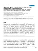
Figure 1
Screen to identify undeiminated and deiminated proteins reacting with RA and non-RA serum samplesScreen to identify undeiminated and deiminated proteins reacting with
RA and non-RA serum samples. Proteins from HL60 lysates incubated
(+) or without (-) peptidylarginine deiminase (PAD) blotted with an anti-
body specific for modified citrulline residues (anti-citrulline) and a
screening panel of rheumatoid arthritis (RA1 to RA4) and non-RA (con-
trol) serum. The rectangular box indicates a citrullinated protein react-
ing strongly with each of the RA serum samples but not the control.
Figure 2
Intracellular expression of immunogenic citrullinated 47 kDa proteinIntracellular expression of immunogenic citrullinated 47 kDa protein.
Presence of citrullinated 47 kDa protein reactive with rheumatoid arthri-
tis serum 1 in different subcellular fractions of undifferentiated HL60s
(U) and HL60 monocytes (M) (S100, cytosolic; P100, membrane; Nuc,
nuclear) showing enrichment in the S100 (cytosolic) fraction.
Arthritis Research & Therapy Vol 7 No 6 Kinloch et al.
R1424
Results
Identification of a 47 kDa citrullinated protein as a target
for antibodies in sera from patients with RA
Each of four serum samples from patients with RA, but not the
control serum, reacted strongly with a band with an apparent
mass of 47 kDa (boxed in Figure 1) in the PAD-treated lysates
of HL-60 cells. No reaction at 47 kDa was observed with non-
deiminated lysates. Reactivity at 47 kDa was strongest with
HL-60 cells that had been differentiated to monocytes,
although a similar polypeptide was also seen in lysates from
cells with the granulocyte phenotype (data not shown). Endog-
enous citrullination in the HL-60 cells was undetectable with
the antibody against modified citrulline, but after treatment
with PAD in vitro, abundant citrullinated polypeptides were
observed. The RA sera, particularly RA1 and RA4, seemed to
be relatively selective for the 47 kDa polypeptide, with only
four to six additional bands identifiable in each blot. Serum
from RA2 and RA3 showed more diffuse reactivity, although a
47 kDa polypeptide predominated. This suggested that,
among the numerous potential antigens generated by citrulli-
nation of proteins in cells of monocytic phenotype, there was
apparent selectivity among our four screening sera for one cit-
rullinated polypeptide migrating at 47 kDa. We therefore per-
formed further experiments to identify this protein.
Nuclear, cytosolic and membrane fractions were prepared
from HL-60 cells by differential centrifugation, and were deim-
inated with PAD as before. Immunoblotting with one of the RA
sera showed that the 47 kDa antigen was enriched in the
cytosolic (S100) fraction (Figure 2).
Identification of the 47 kDa autoantigen as citrullinated
α-enolase
The deiminated S100 fraction was separated by one-dimen-
sional SDS-PAGE and stained with Coomassie blue; the puta-
tive band recognised by sera from patients with RA was
excised, digested with trypsin and analysed by tandem mass
spectrometry. Fourteen peptides were sequenced (Table 1),
all of which mapped onto α-enolase (SwissProt accession
number P06733). In total 242 residues of non-redundant
amino acid sequence were obtained, corresponding to 56%
coverage. To confirm that the stained band co-localised with
the protein recognised by sera from patients with RA, the
deiminated S100 fraction was separated by two-dimensional
electrophoresis, blotted onto nitrocellulose and probed with
serum samples RA1 and RA4. Both recognised a doublet of
spots that matched a feature on the silver-stained gel of appar-
ent molecular mass 47 kDa, and with a pI of 5 (Figure 3).
These spots were excised and identified by mass spectrome-
try as α-enolase. When the membranes were re-probed with
an antibody specific for the carboxy-terminal region of α-eno-
lase, we observed a similar pattern to that obtained with serum
from patients with RA, confirming the identity of the
autoantigen.
Conversion of arginine to citrulline results in a mass increase
of 0.984 Da and a loss of the positive charge from the side
chain, resulting in a significant acidic shift in the two-dimen-
sional electrophoretic migration. Moreover, the pattern of pep-
tides obtained on tryptic digestion will be altered, because the
modified residues are refractory to trypsinolysis and yield pep-
Table 1
Peptides from deiminated α-enolase sequenced by tandem mass spectrometry
m/z (charge) Location Matched sequence
401.24 (2+) 221–227 EGLELLK
480.77 (2+) 81–88 LNVTEQEK
674.34 (2+) 394–405 TGAPC(Cit)SE(Cit)LAK
696.87 (2+) 422–433 FAG(Cit)NF(Cit)NPLAK
817.41 (2+) 343–357 VNQIGSVTESLQACK
837.38 (3+) 285–385 DYPVVSIEDPFDQDDWGAWQK
846.95 (2+) 406–419 YNQLL(Cit)IEEELGSK
980.98 (2+) 202–220 DATNVGDEGGFAPNILENK
878.45 (3+) 5–27 IHA(Cit)EEIFDS(Cit)GNPTVEVDLFTSK
1,177.10 (2+) 372–393 SGETEDTFIADLVVGLCTGQIK
915.14 (3+) 202–227 DATNVGDEGGFAPNILENKEGLELLK
988.15 (3+) 256–280 YDLDFKSPDDPS(Cit)YISPDQLADLYK
1,017.04 (2+) 306–325 FTASAGIQVVGDDLTVTNPK
925.24 (4+) 126–161 GVPLY(Cit)HIADLAGNSEVILPVPAFNVINGGSHAGNK
Cit, citrulline.
Available online />R1425
tides containing internal citrulline, rather than carboxy-terminal
arginine (Tables 1 and 2). Six peptides containing internal cit-
rulline residues were sequenced, which enabled the localisa-
tion of nine sites of modification (Table 1). None of these
peptides were present in tryptic digests of unmodified α-eno-
lase (Table 2). The pI determined by two-dimensional electro-
phoresis was also consistent with this extensive citrullination,
being about 5.0.
Other antigens, recognised more sporadically by sera from
patients with RA (Figure 3), were also characterised by mass
spectrometry. They included elongation factor 1α (SwissProt
accession number P68104) and adenyl cyclase-associated
protein 1 (SwissProt accession number Q01518), both of
which were shown to be citrullinated.
Higher prevalence of antibodies against citrullinated α-
enolase than against native α-enolase in serum from
patients with RA
Twenty-four of the RA serum samples (46%) reacted with the
citrullinated α-enolase, seven of which (13%) also reacted
with the non-citrullinated form of the protein. Six of the controls
(15%) reacted with both (Figure 4). All of the 17 RA samples
Figure 3
Characterisation of the 47 kDa protein by two-dimensional electrophoresisCharacterisation of the 47 kDa protein by two-dimensional electrophoresis. Proteins in the 47 kDa rich monocytic S100 fraction were separated by
two-dimensional electrophoresis according to charge (x-axis) and molecular mass (y-axis). (a) The full complement of proteins was observed by sil-
ver staining. (c,d) Proteins reacting with rheumatoid arthritis serum samples 1 (c) and 4 (d) were highlighted by immunoblotting. (b) The highly reac-
tive 47 kDa protein was confirmed as α-enolase by immunoblotting with the goat anti-α-enolase antibody. CAP1, adenyl cyclase-associated protein
1; EF1α, elongation factor 1α.
Table 2
Peptides from non-citrullinated α-enolase sequenced by
tandem mass spectrometry
m/z (charge) Location Matched sequence
401.24 (2+) 221–227 EGLELLK
403.73 (2+) 406–411 YNQLLR
452.75 (2+) 412–419 IEEELGSK
480.77 (2+) 81–88 LNVTEQEK
572.30 (2+) 183–192 IGAEVYHNLK
703.86 (2+) 15–27 GNPTVEVDLFTSK
713.34 (2+) 269–280 YISPDQADLYK
482.29 (3+) 80–91 KLNVTEQEKIDK
817.41 (2+) 343–357 VNQIGSVTESLQACK
785.09 (3+) 372–393 SGETEDTFIADLVVGLCTGQIK
915.14 (3+) 202–227 DATNVGDEGGFAPNILENKEGLELLK
753.66 (4+) 132–161 HIADLAGNSEVILPVPAFNVINGGSHAGNK
Arthritis Research & Therapy Vol 7 No 6 Kinloch et al.
R1426
that reacted only with the citrullinated form of α-enolase were
positive for anti-CCP2 (data not shown).
α-Enolase is abundant in synovium from patients with
RA
Immunohistochemical analysis of inflamed synovial sections
showed that all eight RA and four osteoarthritis samples exam-
ined expressed α-enolase. Expression in RA sections was
greatest in the more hyperplastic subsynovial layer (Figure 5b).
In the osteoarthritis sections, α-enolase staining was predom-
inantly localised in vascular endothelial cells (Figure 5a). The
antibody against modified citrulline (Figure 6a) indicated that
citrullinated proteins were present in the region that stained
positively for enolase, although the intensity of the staining
indicated that levels of citrullination were relatively low. Immu-
noblotting of synovial cell lysates from three patients with RA
demonstrated that a band co-migrating with purified α-enolase
reacted with the anti-α-enolase antibody (Figure 7).
Discussion
In this study we characterised citrullinated α-enolase as a
dominant antigen in citrullinated lysates of differentiated HL-
60 cells targeted by a screening panel of serum from patients
with RA. The identity of the antigen was established by mass
spectrometry, and the sites of nine citrulline residues within
the protein were determined by tandem mass spectrometry.
Further confirmation was obtained by two-dimensional electro-
phoresis and Western blotting with a specific anti-enolase
antibody. With the use of purified protein, 46% of a larger
panel of sera from patients with RA reacted with citrullinated
α-enolase by immunoblotting. This suggests that citrullinated
α-enolase is at least as immunodominant as citrullinated filag-
grin or citrullinated vimentin, because, by immunoblotting, the
frequency of antibodies against citrullinated filaggrin has been
reported as 41 to 58% [25-28] and against citrullinated
vimentin as 22 to 40% [19,29,30]. Improved sensitivity and
specificity of RA diagnosis may well be obtained by testing RA
sera with peptides derived from citrullinated epitopes of α-
enolase, as has been demonstrated for citrullinated filaggrin in
the first-generation anti-CCP test, in which the sensitivity
increased to more than 70%.
α-Enolase, unlike filaggrin, is abundantly expressed in the syn-
ovial membrane. Several lines of evidence indicate that it is cit-
rullinated in the joint. First, it was detected in the myeloid-like
HL-60s cell line, which expresses PAD and has a similar phe-
notype to that of cells abundant in the joint. Second, it was
detected as a synovial antigen that co-localised with staining
for citrullinated proteins. The staining shown in Figure 6a sug-
gests that only a small proportion of the antigen is citrullinated
in vivo, which might explain why we were unable to demon-
Figure 4
Prevalence of antibodies against deiminated and undeiminated α-eno-lase in RA patients and healthy controlsPrevalence of antibodies against deiminated and undeiminated α-eno-
lase in RA patients and healthy controls. Immunoblotting of in vitro cit-
rullinated and untreated α-enolase with serum samples from patients
with rheumatoid arthritis (RA) and normal controls, showing that about
half of the serum samples from the RA group contain antibodies with
selectivity for citrullinated α-enolase.
Figure 5
Localisation of α-enolase in synovial membranesLocalisation of α-enolase in synovial membranes. Immunohistochemis-
try of biopsy sections from patients with osteoarthritis (a) and rheuma-
toid arthritis (RA) (b) with the goat anti-α-enolase antibody showing
expression of α-enolase (stained brown) in cells in the subsynovium of
the patient with RA and in endothelial cells in the patient with osteoar-
thritis. Cell nuclei are counterstained blue.
Available online />R1427
strate citrullination of α-enolase by Western blotting of immu-
noprecipitates from synovial cells.
Although this is the first report that citrullinated α-enolase is a
common target antigen in RA, native α-enolase has previously
been observed as an infrequent target antigen for several
autoimmune diseases [31-34]. For example, Saulot and
colleagues [34] observed that antibodies against (placental)
α-enolase occurred in 25% of patients with RA and were pre-
dictive of radiological progression. They found that only 8 of
the 36 patients reacting with placental α-enolase also reacted
with the recombinant protein. This contrasted with 19% of
patients with systemic lupus erythematosus and 15% of
patients with systemic sclerosis whose serum samples
reacted with both forms of α-enolase. They hypothesised that
RA sera reacting with placental α-enolase, but not recom-
binant antigen, were recognising a post-translationally modi-
fied form of α-enolase. Although it is tempting to speculate
that the modification they predicted is citrullination, the most
abundant of the triplet of spots identified as α-enolase in their
study migrated in two-dimensional electrophoresis at a pI of
7.0, consistent with native α-enolase. However, it is possible
that the two more acidic α-enolase spots, which they attrib-
uted to phosphorylation, might in fact be citrullinated. The
expression of PAD2 protein in the placenta would account for
a degree of deimination either in vivo or during extraction. It is
also consistent with the identification of the Sa antigen, also of
placental origin, as citrullinated vimentin [17]. The higher fre-
quency of anti-citrullinated α-enolase in our study than that of
Saulot and colleagues might be due to the fact that our cell
lysates were extensively deiminated in vitro. This is
demonstrated by the uniform migration of deiminated enolase
at a pI of 5 and by the replacement of arginine by citrulline in
all the peptides listed in Table 1.
In our study, 15% of the control sera reacted with native α-
enolase and also with citrullinated α-enolase, whereas reactiv-
ity with the citrullinated form alone was restricted to the
patients with RA. This is, again, consistent with the results of
Saulot and colleagues, assuming that the placental form of the
protein was partly deiminated. In turn, this suggests that RA-
specific antibodies might be driven by peptides containing one
or more of the 17 potential citrulline residues in the sequence
of α-enolase. Binding to non-arginine containing regions might
account for the 'background', and hence the apparent loss of
disease specificity seen when immunoblotting with the normal
sera in our study, and the non-RA sera in that of Saulot and
colleagues. One way to test this would be to examine reactivity
to peptides derived from citrullinated epitopes from α-enolase.
Figure 6
Localisation of citrullinated proteins in synovial membranesLocalisation of citrullinated proteins in synovial membranes. (a) Immunostaining of citrullinated proteins by the anti-modified citrulline kit was mainly
confined to the subsynovium. (b) No staining was visible on the control. (c) Staining produced by the anti-α-enolase antibody on an adjacent section
was much stronger, and included the subsynovial cells which also stained for citrullinated antigens. Original magnification × 20 in all cases.
Figure 7
Presence of α-enolase in synovial cells from patients with rheumatoid arthritis (RA)Presence of α-enolase in synovial cells from patients with rheumatoid
arthritis (RA). Immunoblotting of lysates from RA synovial cells with anti-
α-enolase (+PAD, deiminated α-enolase; – PAD, undeiminated α-eno-
lase) showing a 47 kDa protein in synoviocytes, from three patients with
RA, reacting with the goat anti-α-enolase antibody, which co-migrates
with purified α-enolase.
Arthritis Research & Therapy Vol 7 No 6 Kinloch et al.
R1428
Such studies are currently in progress. If both sensitivity and
specificity increases for RA it might provide another assay for
diagnosis of the disease. More importantly it would provide fur-
ther data to support the concept that α-enolase is a driving
autoantigen in RA.
The properties of citrullinated α-enolase make it an attractive
synovial antigen for driving the immune response. α-Enolase is
a highly conserved, multifunctional protein that, in addition to
its role in glycolysis, binds plasminogen. It is known to be
upregulated by hypoxia [35] and by proinflammatory stimuli
[36], both of which are features of the synovial membrane
microenvironment in RA. α-Enolase is expressed during cell
differentiation and is used as marker of differentiation in the
grading of tumours [37]. In myeloid cells, the dominant cell
type in the inflamed synovium, it is co-expressed with PAD2
and PAD4. In the present study we have shown that its distri-
bution in the subsynovium is similar to that of citrullinated pro-
teins and PAD [38], but we have not yet demonstrated its
citrullination in vivo.
There is substantial similarity between human and prokaryotic
α-enolases (47% identity with that from Streptococcus pyo-
genes, for example), and antibodies raised against streptococ-
cal surface α-enolase also recognise the human enzyme [36].
Thus the presence of antibodies against uncitrullinated α-eno-
lase, in serum of individuals without RA, might be attributable
to cross-reaction with bacterial epitopes. Expression of an
enzyme able to citrullinate peptidylarginine has been demon-
strated in the oral organism Porphyromonas gingivalis [39],
which provides a mechanism by which antibacterial antibodies
cross-react with endogenous citrullinated proteins and initiate
loss of tolerance.
Conclusion
We have demonstrated that antibodies against citrullinated α-
enolase are found in 46% of serum samples from patients with
RA, and that native α-enolase is abundantly expressed in rheu-
matoid synovium. It is upregulated by factors such as hypoxia,
which are characteristic of the rheumatoid joint, and its amino-
acid sequence is highly conserved between prokaryotes and
higher eukaryotes, making citrullinated α-enolase a candidate
target antigen in RA; this merits further investigation.
Competing interests
The authors declare that they have no competing interests.
Authors' contributions
AK performed immunoblotting, screened sera for antibodies,
detected α-enolase in synoviocytes, and participated in study
design and drafting of the manuscript. VT performed the initial
Western blotting, cellular fractionation and two-dimensional
electrophoresis experiments. DP, KL and PD performed the
immunohistochemistry. DM participated in the study design
and established the cell culture methodology. PCT performed
the synovial biopsies and participated in study design and
drafting of the manuscript. RW was responsible for mass
spectrometric characterisation of citrullinated α-enolase, par-
ticipated in the study design and helped to draft and edit the
manuscript. PJV conceived of the study, participated in its
design and edited the manuscript. All authors read and
approved the final version.
Acknowledgements
We are grateful to Shajna Begum for assistance with preparation of
samples for mass spectrometry and to Lauren Schewitz for performing
the synovial cell isolation. We thank the Arthritis Research Campaign,
the Medical Research Council and the Wellcome Trust for their support.
References
1. Lee DM, Weinblatt ME: Rheumatoid arthritis. Lancet 2001,
358:903-911.
2. Mageed RA: The RF antigen. In Manual of Biological Markers of
Disease Edited by: van Venrooij WJ, Maini RN. Dordrecht: Kluwer
Academic Publishing; 1996:1-27.
3. Nienhuis RL, Mandema E: A new serum factor in patients with
rheumatoid arthritis; the antiperinuclear factor. Ann Rheum
Dis 1964, 23:302-305.
4. Young BJ, Mallya RK, Leslie RD, Clark CJ, Hamblin TJ: Anti-kera-
tin antibodies in rheumatoid arthritis. Br Med J 1979, 2:97-99.
5. Sebbag M, Simon M, Vincent C, Masson-Bessiere C, Girbal E,
Durieux JJ, Serre G: The antiperinuclear factor and the so-called
antikeratin antibodies are the same rheumatoid arthritis-spe-
cific autoantibodies. J Clin Invest 1995, 95:2672-2679.
6. Schellekens GA, de Jong BA, van den Hoogen FH, van de Putte
LB, van Venrooij WJ: Citrulline is an essential constituent of
antigenic determinants recognized by rheumatoid arthritis-
specific autoantibodies. J Clin Invest 1998, 101:273-281.
7. Girbal-Neuhauser E, Durieux JJ, Arnaud M, Dalbon P, Sebbag M,
Vincent C, Simon M, Senshu T, Masson-Bessiere C, Jolivet-Rey-
naud C, et al.: The epitopes targeted by the rheumatoid arthri-
tis-associated antifilaggrin autoantibodies are
posttranslationally generated on various sites of (pro)filaggrin
by deimination of arginine residues. J Immunol 1999,
162:585-594.
8. Nijenhuis S, Zendman AJ, Vossenaar ER, Pruijn GJ, van Venrooij
WJ: Autoantibodies to citrullinated proteins in rheumatoid
arthritis: clinical performance and biochemical aspects of an
RA-specific marker. Clin Chim Acta 2004, 350:17-34.
9. van Gaalen FA, Linn-Rasker SP, van Venrooij WJ, de Jong BA,
Breedveld FC, Verweij CL, Toes RE, Huizinga TW: Autoantibod-
ies to cyclic citrullinated peptides predict progression to rheu-
matoid arthritis in patients with undifferentiated arthritis: a
prospective cohort study. Arthritis Rheum 2004, 50:709-715.
10. Rantapaa-Dahlqvist S, de Jong BA, Berglin E, Hallmans G, Wadell
G, Stenlund H, Sundin U, van Venrooij WJ: Antibodies against
cyclic citrullinated peptide and IgA rheumatoid factor predict
the development of rheumatoid arthritis. Arthritis Rheum 2003,
48:2741-2749.
11. Vencovsky J, Machacek S, Sedova L, Kafkova J, Gatterova J, Pesa-
kova V, Ruzickova S: Autoantibodies can be prognostic markers
of an erosive disease in early rheumatoid arthritis. Ann Rheum
Dis 2003, 62:427-430.
12. Forslind K, Ahlmen M, Eberhardt K, Hafstrom I, Svensson B, BAR-
FOT Study Group: Prediction of radiological outcome in early
rheumatoid arthritis in clinical practice: role of antibodies to
citrullinated peptides (anti-CCP). Ann Rheum Dis 2004,
63:1090-1095.
13. Masson-Bessiere C, Sebbag M, Durieux JJ, Nogueira L, Vincent C,
Girbal-Neuhauser E, Durroux R, Cantagrel A, Serre G: In the rheu-
matoid pannus, anti-filaggrin autoantibodies are produced by
local plasma cells and constitute a higher proportion of IgG
than in synovial fluid and serum. Clin Exp Immunol 2000,
119:544-552.
14. Vossenaar ER, Nijenhuis S, Helsen MM, van der Heijden A, Sen-
shu T, van den Berg WB, van Venrooij WJ, Joosten LA: Citrullina-
Available online />R1429
tion of synovial proteins in murine models of rheumatoid
arthritis. Arthritis Rheum 2003, 48:2489-2500.
15. Hida S, Miura NN, Adachi Y, Ohno N: Influence of arginine deim-
ination on antigenicity of fibrinogen. J Autoimmun 2004,
23:141-150.
16. Rubin B, Sonderstrup G: Citrullination of self-proteins and
autoimmunity. Scand J Immunol 2004, 60:112-120.
17. Vossenaar ER, Despres N, Lapointe E, van der Heijden A, Lora M,
Senshu T, van Venrooij WJ, Menard HA: Rheumatoid arthritis
specific anti-Sa antibodies target citrullinated vimentin. Arthri-
tis Res Ther 2004, 6:R142-R150.
18. Vossenaar ER, Radstake TR, van der Heijden A, van Mansum MA,
Dieteren C, de Rooij DJ, Barrera P, Zendman AJ, van Venrooij WJ:
Expression and activity of citrullinating peptidylarginine deim-
inase enzymes in monocytes and macrophages. Ann Rheum
Dis 2004, 63:373-381.
19. Despres N, Boire G, Lopez-Longo FJ, Menard HA: The Sa sys-
tem: a novel antigen-antibody system specific for rheumatoid
arthritis. J Rheumatol 1994, 21:1027-1033.
20. Nakashima K, Hagiwara T, Ishigami A, Nagata S, Asaga H,
Kuramoto M, Senshu T, Yamada M: Molecular characterization
of peptidylarginine deiminase in HL-60 cells induced by retin-
oic acid and 1α,25-dihydroxyvitamin D
3
. J Biol Chem 1999,
274:27786-27792.
21. Arnett FC, Edworthy SM, Bloch DA, McShane DJ, Fries JF, Cooper
NS, Healey LA, Kaplan SR, Liang MH, Luthra HS, et al.: The Amer-
ican Rheumatism Association 1987 revised criteria for the
classification of rheumatoid arthritis. Arthritis Rheum 1988,
31:315-324.
22. Shevchenko A, Wilm M, Vorm O, Mann M: Mass spectrometric
sequencing of proteins silver-stained polyacrylamide gels.
Anal Chem 1996, 68:850-858.
23. Wait R, Gianazza E, Eberini I, Sironi L, Dunn MJ, Gemeiner M,
Miller I: Proteins of rat serum, urine, and cerebrospinal fluid: VI.
Further protein identifications and interstrain comparison.
Electrophoresis 2001, 22:3043-3052.
24. Wait R, Miller I, Eberini I, Cairoli F, Veronesi C, Battocchio M,
Gemeiner M, Gianazza E: Strategies for proteomics with incom-
pletely characterized genomes: the proteome of Bos taurus
serum. Electrophoresis 2002, 23:3418-3427.
25. Vittecoq O, Incaurgarat B, Jouen-Beades F, Legoedec J,
Letourneur O, Rolland D, Gervasi G, Menard JF, Gayet A, Fardel-
lone P, et al.: Autoantibodies recognizing citrullinated rat filag-
grin in an ELISA using citrullinated and non-citrullinated
recombinant proteins as antigens are highly diagnostic for
rheumatoid arthritis. Clin Exp Immunol 2004, 135:173-180.
26. Vincent C, Simon M, Sebbag M, Girbal-Neuhauser E, Durieux JJ,
Cantagrel A, Fournie B, Mazieres B, Serre G: Immunoblotting
detection of autoantibodies to human epidermis filaggrin: a
new diagnostic test for rheumatoid arthritis. J Rheumatol
1998, 25:838-846.
27. Palosuo T, Lukka M, Alenius H, Kalkkinen N, Aho K, Kurki P, Heik-
kila R, Nykanen M, von Essen R: Purification of filaggrin from
human epidermis and measurement of antifilaggrin autoanti-
bodies in sera from patients with rheumatoid arthritis by an
enzyme-linked immunosorbent assay. Int Arch Allergy
Immunol 1998, 115:294-302.
28. Paimela L, Palosuo T, Aho K, Lukka M, Kurki P, Leirisalo-Repo M,
von Essen R: Association of autoantibodies to filaggrin with an
active disease in early rheumatoid arthritis. Ann Rheum Dis
2001, 60:32-35.
29. Goldbach-Mansky R, Lee J, McCoy A, Hoxworth J, Yarboro C,
Smolen JS, Steiner G, Rosen A, Zhang C, Menard HA, et al.:
Rheumatoid arthritis associated autoantibodies in patients
with synovitis of recent onset. Arthritis Res 2000, 2:236-243.
30. Hayem G, Chazerain P, Combe B, Elias A, Haim T, Nicaise P,
Benali K, Eliaou J, Kahn MF, Sany J, Meyer O: Anti-Sa antibody is
an accurate diagnostic and prognostic marker in adult rheu-
matoid arthritis. J Rheumatol 1999, 26:7-13.
31. Moodie FD, Leaker B, Cambridge G, Totty NF, Segal AW: Alpha-
enolase: a novel cytosolic autoantigen in ANCA positive
vasculitis. Kidney Int 1993, 43:675-681.
32. Gitlits VM, Sentry JW, Matthew ML, Smith AI, Toh BH: Autoanti-
bodies to evolutionarily conserved epitopes of enolase in a
patient with discoid lupus erythematosus. Immunology 1997,
92:362-368.
33. Pratesi F, Moscato S, Sabbatini A, Chimenti D, Bombardieri S,
Migliorini P: Autoantibodies specific for alpha-enolase in sys-
temic autoimmune disorders. J Rheumatol 2000, 27:109-115.
34. Saulot V, Vittecoq O, Charlionet R, Fardellone P, Lange C, Marvin
L, Machour N, Le Loet X, Gilbert D, Tron F: Presence of autoan-
tibodies to the glycolytic enzyme alpha-enolase in sera from
patients with early rheumatoid arthritis. Arthritis Rheum 2002,
46:1196-1201.
35. Aaronson RM, Graven KK, Tucci M, McDonald RJ, Farber HW:
Non-neuronal enolase is an endothelial hypoxic stress protein.
J Biol Chem 1995, 270:27752-27757.
36. Fontan PA, Pancholi V, Nociari MM, Fischetti VA: Antibodies to
streptococcal surface enolase react with human alpha-eno-
lase: implications in poststreptococcal sequelae. J Infect Dis
2000, 182:1712-1721.
37. Sugio K, Sugaya M, Hanagiri T, Yasumoto K: [Tumor marker in
primary lung cancer]. J UOEH 2004, 26:473-479.
38. Suzuki A, Yamada R, Chang X, Tokuhiro S, Sawada T, Suzuki M,
Nagasaki M, Nakayama-Hamada M, Kawaida R, Ono M, et al.:
Functional haplotypes of PADI4, encoding citrullinating
enzyme peptidylarginine deiminase 4, are associated with
rheumatoid arthritis. Nat Genet 2003, 34:395-402.
39. McGraw WT, Potempa J, Farley D, Travis J: Purification, charac-
terization, and sequence analysis of a potential virulence fac-
tor from Porphyromonas gingivalis, peptidylarginine
deiminase. Infect Immun 1999, 67:3248-3256.