Báo cáo y học: "Polarized subsets of human T-helper cells induce distinct patterns of chemokine production by normal and systemic sclerosis dermal fibroblasts" doc
Bạn đang xem bản rút gọn của tài liệu. Xem và tải ngay bản đầy đủ của tài liệu tại đây (752.31 KB, 12 trang )
Open Access
Available online />Page 1 of 12
(page number not for citation purposes)
Vol 8 No 1
Research article
Polarized subsets of human T-helper cells induce distinct patterns
of chemokine production by normal and systemic sclerosis dermal
fibroblasts
Carlo Chizzolini
1
, Yann Parel
1
, Agneta Scheja
2
and Jean-Michel Dayer
1
1
Immunology and Allergy, Geneva University Hospital, Geneva School of Medicine, Rue Micheli-du-Crest, 24, 1211 Geneva 14, Switzerland
2
Division of Rheumatology, Lund University Hospital, 221 85 Lund, Sweden
Corresponding author: Carlo Chizzolini,
Received: 25 Jun 2005 Revisions requested: 14 Jul 2005 Revisions received: 10 Oct 2005 Accepted: 3 Nov 2005 Published: 30 Nov 2005
Arthritis Research & Therapy 2006, 8:R10 (doi:10.1186/ar1860)
This article is online at: />© 2005 Chizzolini et al.; licensee BioMed Central Ltd.
This is an open access article distributed under the terms of the Creative Commons Attribution License ( />),
which permits unrestricted use, distribution, and reproduction in any medium, provided the original work is properly cited.
Abstract
The role of fibroblasts in inflammatory processes and their
cross-talk with T cells is increasingly being recognized. Our aim
was to explore the capacity of dermal fibroblasts to produce
inflammatory chemokines potentially involved in fibrosis
occurring in response to contact with polarized human T cells.
Our findings indicate that the program of chemokine production
by fibroblasts is differentially regulated depending on the T-
helper (Th) cell subset used to activate them. Thus, Th1 and Th2
cells preferentially induced production of IFN-γ inducible protein
(IP)-10 and IL-8, respectively, whereas monocyte
chemoattractant protein (MCP)-1 was equally induced by both
subsets at mRNA and protein levels. Neutralization experiments
indicated that membrane-associated tumour necrosis factor-α
and IL-1 played a major role in the induction of IL-8 and MCP-1
by Th1 and Th2 cells, whereas membrane-associated IFN-γ
(present only in Th1 cells) was responsible, at least in part, for
the lower IL-8 and higher IP-10 production induced by Th1 cells.
The contributions of tumour necrosis factor-α, IL-1 and IFN-α
were confirmed when fibroblasts were cultured separated in a
semipermeable membrane from living T cells activated by CD3
cross-linking. We observed further differences when we
explored signal transduction pathway usage in fibroblasts.
Pharmacological inhibition of c-Jun N-terminal kinase and
nuclear factor-κB resulted in inhibition of IL-8 mRNA
transcription induced by Th1 cells but not that by Th2 cells,
whereas inhibition of MEK/ERK (mitogen-activated protein
kinase of extracellular signal-regulated kinase/extracellular
signal-regulated kinase) and nuclear factor-κB resulted in
inhibition of MCP-1 mRNA induced by Th2 but not by Th1 cells.
Finally, no distinct differences in chemokine production were
observed when the responses to T cell contact or to prototypic
Th1 and Th2 cytokines were examined in systemic sclerosis
versus normal fibroblasts. These findings indicate that
fibroblasts have the potential to participate in shaping the
inflammatory response through the activation of flexible
programs of chemokine production that depend on the Th
subset eliciting their response.
Introduction
Fibroblasts are cells of mesenchymal origin and are principally
involved in the generation and maintenance of extracellular
matrix. Fibroblast morphology, phenotype and function may
vary depending on the tissue of origin and on whether the tis-
sue is exposed to physiological or pathological conditions.
Thus, cultured fibroblasts derived from skin, breast, lung and
haematopoietic tissue have been shown to express structural,
extracellular matrix and surface proteins differentially, and to
produce different cytokines [1-3]. Chemokine production may
also vary depending on the source of fibroblasts, and differ-
ences in the levels of eotaxin/CC chemokine ligand (CCL)11,
IL-8/CXC chemokine ligand (CXCL)8, monocyte chemoat-
tractant protein (MCP)-1/CCL2, RANTES (regulated upon
activation normal T cell expressed and secreted)/CCL5, and
macrophage inflammatory protein (MIP)-1α/CCL3 have been
CCL = CC chemokine ligand; CCR = CC chemokine receptor; DMEM = Dulbecco's modified Eagle medium; ERK = extracellular signal-regulated
kinase; FCS = foetal calf serum; IFN = interferon; IL = interleukin; IP = IFN-γ inducible protein; JNK = c-Jun N-terminal kinase; mAb = monoclonal
antibody; MCP = monocyte chemoattractant protein; MEK = mitogen-activated protein kinase kinase; MIP = macrophage inflammatory protein; NF-
κB = nuclear factor-κB; PSI = proteasome inhibitor I; RANTES = regulated upon activation normal T cell expressed and secreted; SSc = systemic
sclerosis; TGF = transforming growth factor; Th = T-helper; TNF = tumour necrosis factor.
Arthritis Research & Therapy Vol 8 No 1 Chizzolini et al.
Page 2 of 12
(page number not for citation purposes)
reported [3]. In addition, production by fibroblasts of chemok-
ines may be variably modulated by cytokines, with differences
being related to the origin of the fibroblasts [4-8].
Chemokines are soluble mediators that were originally identi-
fied because of their chemotactic properties in cells express-
ing specific receptors. Indeed, chemokines that influence
chemotaxis regulate leucocyte homeostasis and recruitment
of leucocyte subpopulations at sites of inflammation [9]. How-
ever, their biological functions are broader, comprising rele-
vant roles in virus cell entry, angiogenesis, tumour growth,
metastasis formation and fibrosis [10]. For instance, MCP-1/
CCL2 – a CC chemokine that binds to CC chemokine recep-
tor (CCR)2 – has attracted keen interest in the field of fibrosis
because it appears to play direct roles in collagen and matrix
metalloproteinase-1 induction on fibroblasts [11-13] and is
present at sites undergoing fibrosis. In human systemic scle-
rosis (SSc), MCP-1 mRNA proved to be the most abundant
mRNA when bronchoalveolar lavage cells from SSc lung were
compared with controls using microarray technology and test-
ing a total of 4507 genes [14]. Moreover, it is produced in
large amounts by SSc skin fibroblasts [13,15,16]. Of interest,
IL-4 triggers MCP-1 production by human lung fibroblasts
[17], and MCP-1 may polarize T cells toward a T-helper (Th)2
subset in mouse [18,19]. In a rodent model of fibrotic versus
nonfibrotic pulmonary granulomas, procollagen production
was associated with Th2 cells and MCP-1 production [20].
Furthermore, mice null for CCR2 were resistant to develop-
ment of lung fibrosis induced by transgenic IL-13 [21] and ble-
omycin [22].
Several additional chemokines have been detected by histo-
logical or molecular biological methods at sites undergoing
fibrosis in humans or mouse models, including the CC chem-
okines RANTES [23], MIP-1α [24], PARC (pulmonary and
activation-regulated chemokine)/CCL18 [25] and MCP-3/
CCL7 [26], and CXC chemokines IL-8/CXCL8, GRO (growth
regulated oncogene)-α/CXCL1 [27], ENA-78 (neutrophil-acti-
vating peptide-78)/CXCL5 and MIP-2 [28]. With the excep-
tion of PARC [25], it is not known whether these chemokines
play direct profibrotic or antifibrotic activities apart from
recruiting specific leucocyte subsets [3]. Nonetheless, it has
been suggested that the proangiogenic and antiangiogenic
activities of chemokines play important roles in fibrosis [29]. In
bleomycin-induced lung fibrosis, neutralization of MIP-2 (a
possible murine analogue of human IL-8) attenuates fibrosis
[28], and systemic administration of IFN-γ inducible protein
(IP)-10 or transgenic overexpression of IP-10 reduces fibrosis
[30,31].
SSc is a human disease that is presumably of autoimmune ori-
gin and is characterized by vasculopathy and fibrosis of the
skin and internal organs. In the early stage of the disease,
inflammatory infiltrates rich in T cells dominate in tissues
undergoing fibrosis, and fibroblasts adjacent to T cells exhibit
high metabolic activity (for review, see the report by Chizzolini
[32]). T cells infiltrating the skin or recovered from bronchoal-
veolar lavage fluid from SSc individuals predominantly express
the Th2 phenotype [33-36], which is consistent with a profi-
brotic activity of Th2 cytokines [37-39]. In addition, we
addressed the ability of T cells to regulate extracellular matrix
deposition by cell–cell interaction with fibroblasts [36,40].
However, no data exist on the capacity of T cells to elicit the
production of chemokines by dermal fibroblasts and, in partic-
ular, the capacity of polarized T cells (Th1 and Th2) to modu-
late fibroblast production of chemokines differentially. Thus,
we conducted the present study to assess whether polarized
human T cells could, in a contact-dependent manner, induce
dermal fibroblasts to produce MCP-1, IL-8 and IP-10. We
opted to focus on these chemokines because of their potential
role in the development of fibrosis, particularly in SSc.
Materials and methods
Patients and control individuals
A skin punch biopsy, 3 mm in diameter, was obtained from
areas of affected skin from eight SSc patients at the Division
of Rheumatology in Lund (Sweden) and from eight age-
matched and sex-matched healthy control individuals. All SSc
individuals fulfilled the American College of Rheumatology cri-
teria for SSc [41] and had clinical features of early disease,
and none was undergoing immunosuppressive therapy [42].
Permission to perform this investigation was granted by the
ethics committee. Informed consent was obtained from all indi-
viduals. Peripheral blood from healthy individuals was provided
by the Blood Transfusion Center of the Geneva University
Hospital (Switzerland).
Reagents
Anti-CD3 OKT3 mAb and anti-human IL-4 mAb (clone 25D2)
were from ATCC (Bethesda, MD, USA). Anti-human IFN-γ
mAb was a gift from Dr G Garotta (Serono Biomedical
Research Institute, Geneva, Switzerland). IgG
1
mAb (anti-
TNP, clone G106HN) was a kind gift from S Izui (Department
of Pathology and Immunology, Geneva School of Medicine,
Geneva, Switzerland). Anti-CD40L (mAb 5c8) was a kind gift
from P Lipski (University of Texas, Dallas, TX, USA). Human
recombinant (r)IL-4 was from Schering Plough (Dardilly,
France). Human rIL-2 was from Biogen (Cambridge, MA,
USA). Human rIL-13 was from Sanofi (Montpellier, France).
Human rIFN-γ was from Roussel Uclaf (Paris, France). Human
recombinant transforming growth factor (rTGF)-β was from
R&D (Minneapolis, MN, USA). Anti-TNF (recombinant-methio-
nyl soluble TNF-type I pegylated receptor [sTNF-RI]) [43] and
recombinant human IL-1 receptor antagonist [44] were from
Amgen (Thousand Oaks, CA, USA). Recombinant human
CD40L trimer/leucine zipper fusion protein was from Immunex
(Seattle, WA, USA) [45]. RPMI-1640, Dulbecco's modified
Eagle medium (DMEM), glutamine, penicillin, streptomycin,
trypsin, nonessential amino acids, sodium pyruvate and foetal
calf serum (FCS) were from Gibco (Paisley, Scotland).
Available online />Page 3 of 12
(page number not for citation purposes)
Sucrose, phenyl methyl sulfonyl fluoride, pepstatin, EDTA,
iodoacetamide, NP40 and indomethacin were from Sigma (St.
Louis, MO, USA). Proteasome inhibitor I (PSI; Z-Ile-Glu
[OtBu]-Ala-Leu-CHO), c-Jun NH
2
-terminal kinase (JNK) inhib-
itor SP600125 (anthra [1,9-cd]pyrazol-6(2H)-one), and
mitogen-activated protein kinase of extracellular signal-regu-
lated kinase (MEK)1/2 inhibitor U-0126 (1,4-diamino-2,3-dicy-
ano-1,4-bis [2-aminophenithiol]butadiene) were from
Calbiochem (San Diego, CA, USA).
T cell clones and T cell membrane preparation
Prototypic Th1 and Th2 cell clones were generated from
peripheral blood of normal individuals upon antigen activation
and cloning by limiting dilution in RPMI-1640 medium supple-
mented with IL-2 (20 U/ml), penicillin (50 U/ml), streptomycin
(50 µg/ml), 5% human AB serum, 10% FCS, and irradiated
(3,500 rads) allogeneic peripheral blood mononuclear cells
with phytohaemagglutinin (1 µg/ml) [46]. Growing cells were
further expanded and characterized with respect to their
capacity to produce IFN-γ and IL-4 upon CD3 crosslinking.
High IFN-γ/low IL-4 producers were defined as Th1, whereas
low IFN-γ/high IL-4 producers were defined as Th2. In addi-
tion, we used SSc skin-derived polarized T cell clones (gener-
ation and characterization of which are described elsewhere
[36]). For the preparation of T cell membranes, T cells (8 ×
10
6
) were activated in six-well trays, with plastic-adsorbed
anti-CD3 mAb. Controls consisted of T cells cultured in
medium alone. After six hours of culture the supernatants were
collected and frozen for further cytokine determination, and
cell membranes were prepared as described elsewhere [46].
Skin fibroblast–lymphocyte cocultures
Fibroblasts from skin biopsy were grown in DMEM medium
supplemented with 10% FCS, penicillin, streptomycin, nones-
sential amino acids and sodium pyruvate, and split at conflu-
ence. All of the experiments were performed with fibroblasts at
passages four to nine. In order to study chemokine production,
we plated fibroblasts in 96-well trays at 2 × 10
4
cells/well in
200 µl DMEM supplemented with 10% FCS, which were then
cultured for 72 hours. The culture medium was then replaced
by DMEM supplemented with 1% FCS. To assess the effect
of T cell contact on chemokine production, 5 µl of T cell mem-
branes equivalent to 2 × 10
5
cells/well, unless otherwise
stated, was added to fibroblasts in triplicate wells and cultured
for 48 hours. In no instance were T cells syngeneic with fibrob-
lasts. Supernatant was then harvested and frozen until chem-
okine determination. In blocking experiments anti-TNF (soluble
TNF receptor I; 10
-8
mol/l), anti-IFN-γ (10 µg/ml) [40], IL-1
receptor antagonist (2 µg/ml) [44], anti-IL-4 (10 µg/ml) and
irrelevant monoclonal IgG
1
(10 µg/ml), alone or in combina-
tion, were added to the wells 30 minutes before T cell mem-
branes [47,48]. To evaluate the effect of cytokines on
chemokine production by fibroblasts, TGF-β (10 ng/ml), IL-4
(10 ng/ml), IL-13 (10 ng/ml) and IFN-γ (1000 U/ml) were
added to triplicate wells. When used in combination, TGF-β,
IL-4 and IL-13 were used at 5 ng/ml each. In some experi-
ments 20 × 10
3
fibroblasts were cultured in the upper cham-
ber of a semipermeable polyester membrane transwell and
10
6
living T cells were cultured in the lower chamber, in which
anti-CD3 mAb had been plastic adsorbed (Transwell #347-
clear; Costar, Corning, NY, USA) for 48 hours. The total cul-
ture volume was 1 ml.
RNA extraction and RNase protection assay analysis
Fibroblasts (5 × 10
5
) were plated to confluence in 100 mm
Petri dishes and serum starved overnight. T cell membranes to
the equivalent of 8 × 10
6
cells were then added to the cultures
(typically 200 µl of cell membranes in 3 ml) and fibroblasts
were cultured for four additional hours in 1% FCS medium.
When used, intracellular signalling inhibitors (PSI [40 µmol/l],
JNK-inhibitor [10, 20, and 50 µmol/l], U-0126 [40 µmol/l])
were added 1 hour before adding T cell membranes. Total
fibroblast RNA was isolated with TRIzol™ reagent (Life Tech-
nologies, Invitrogen, Carlsbad, CA, USA). The levels of expres-
sion of IL-8, MCP-1, IP-10 and L-32 mRNAs were assessed
by RiboQuant RPA using the hCK-5 multiprobe template set
from Pharmingen (San Diego, CA, USA), in accordance with
the supplier's instructions. After phosphor imaging (Typhoon
9400, Applied Byosistems, Foster City, CA, USA), signal
intensity was determined by densitometry using ImageQuant
software (Molecular Dynamics, NIH, Bethesda, MD, USA), and
normalized values were used to determine the effect of T cells
and signal inhibitors.
Cytokine determination
Levels of IL-8, MCP-1, IP-10, IL-4, IFN-γ and TNF-α (R&D Sys-
tems, Abingdon, UK), and IL-1α (Immunotech, Marseille,
France) proteins were assessed using commercial enzyme-
linked immunosorbent assay. T cell membranes were solubi-
lized in 1% NP40 to detect cytokine content.
Statistical analysis
Student's t test was used with two-tailed P values or Mann–
Whitney U test using StatView 5.0™ (SAS Institute Inc., Cary,
NC) software on a Macintosh computer. P < 0.05 was consid-
ered statistically significant.
Results
Chemokine production by fibroblasts in response to T
cell contact
We first tested whether human dermal fibroblasts were able to
produce IL-8, MCP-1 and IP-10 when activated in a contact-
dependent manner by polarized T cell clones [36]. To minimize
the effect of soluble mediators, we used T cell membranes as
effectors of contact-dependent activation. Upon T cell con-
tact, dermal fibroblasts produced IL-8, MCP-1 and IP-10, with
the production depending on whether T cells were resting or
activated (CD3 crosslinking) and on the subset of Th cells
(Figure 1a). To induce chemokine production, both Th1 and
Th2 cells had to be activated because chemokine production
Arthritis Research & Therapy Vol 8 No 1 Chizzolini et al.
Page 4 of 12
(page number not for citation purposes)
was marginal in the presence of resting T cells (Figure 1a).
Dose-dependent increases in IL-8, MCP-1 and IP-10 were
observed in the presence of activated T cells. However, Th1
cells were less potent inducers of IL-8 and stronger inducers
of IP-10 than were Th2 cells, although both subsets potently
induced MCP-1 (Figure 1a).
We extended these findings by testing a large panel of polar-
ized T cell clones at a fixed ratio of T cells to fibroblasts of
10:1. Under these settings, Th1 cells induced statistically sig-
nificantly higher amounts of IP-10 than did Th2 cells, and Th2
induced higher amounts of IL-8 than did Th1 cells, with no dif-
ferences being observed in MCP-1 levels (Figure 1b). Of note,
the levels of IP-10 produced by fibroblasts were positively cor-
related (R
2
= 0.813) with the potential for IFN-γ production by
T cell clones. No correlation was observed with the levels of
the other chemokines, or were chemokine levels correlated
with the levels of IL-4.
Finally, we tested whether differences existed between dermal
fibroblasts generated from normal skin and those from SSc
skin. Although no differences were observed in response to T
cell contact between control and SSc fibroblasts, differences
in the levels of cytokines were striking depending on the origin
of T cells, with Th1 cells being more potent inducers of IP-10
and Th2 being stronger inducers of IL-8 (data not shown).
These findings indicate that dermal fibroblasts upregulate their
production of chemokines when activated by T cells and that
the type of T cell dictates which chemokine is preferentially
produced.
Chemokine production by fibroblasts in response to T
cell cytokines and TGF-β
In order to further explore possible differences between SSc
and control fibroblasts, we assessed their capacity to produce
chemokines in response to prototypic Th cytokines or to profi-
brotic TGF-β (Figure 2). None of the cytokines tested induced
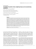
Figure 1
IL-8, MCP-1 and IP-10 production by dermal fibroblasts following contact with T-helper cellsIL-8, MCP-1 and IP-10 production by dermal fibroblasts following contact with T-helper cells. Normal fibroblasts were plated at 2 × 10
4
cells/well;
72 hours later the culture medium was replaced and T cell membranes generated from resting or activated Th1 and Th2 clones were added to the
wells to a final volume of 200 µl. Chemokine levels were determined in the supernatants by enzyme-linked immunosorbent assay after 48 hours of
further culture. (a) The points represent the mean ± standard deviation of triplicate cultures. Similar results were obtained in an additional experi-
ment. (b) T cell membranes corresponding to 2 × 10
5
cells generated from activated Th1 (n = 10) and Th2 (n = 10) clones were added to the wells
in triplicate cultures. The bars represent the mean ± standard error. IL, interleukin; IP, interferon-γ inducible protein; MCP, monocyte chemoattractant
protein; Th, T-helper.
Available online />Page 5 of 12
(page number not for citation purposes)
IL-8, but all of them (IFN-γ, IL-4, IL-13, TGF-β and combina-
tions of IL-4 plus TGF-β and IL-13 plus TGF-β) induced MCP-
1 production. Of interest, IFN-γ induced significantly higher
levels of MCP-1 than did IL-4 or IL-13 (P < 0.005) and IL-4 or
IL-13 higher levels than did TGF-β (P < 0.005). However,
MCP-1 was synergistically induced by TGF-β added together
with IL-4 or IL-13. Predictably, IFN-γ was a potent stimulus
whereas all of the other cytokines inhibited IP-10 production.
It is noteworthy that, although SSc fibroblasts tended to pro-
duce higher amounts of chemokines, the only statistically sig-
nificant difference from control fibroblasts was the higher IP-
10 production in the presence of IFN-γ (Figure 2).
T cell membrane associated TNF-α, IL-1 and IFN-γ play
distinct roles in inducing chemokine production by
dermal fibroblasts
In order to identify some of the mediators present in T cell
membranes that induce chemokine production by fibroblasts,
we used neutralizing reagents to block the biological activity of
several cytokines. IL-8 induction was dependent on IL-1 and
TNF-α, which exhibited additive effects in both Th1 and Th2
cells (Figure 3a,b). Because Th2 cells were more potent
inducers of IL-8 production than were Th1 cells, we tested the
hypothesis that IFN-γ could act as a partial inhibitor. Indeed,
after exogenous addition of IFN-γ to fibroblasts activated by
Th2 cells, IL-8 production was inhibited by more than 50%,
and this inhibition was abrogated by IFN-γ neutralization (Fig-
ure 3c). Similarly, IFN-γ neutralization resulted in enhanced IL-
8 production when fibroblasts were activated by Th1 cells
(Figure 3c). MCP-1 induction by activated Th1 and Th2 cells
was dependent on the synergistic effect of IL-1 and TNF-α.
With Th1 cells, further reduction in MCP-1 production was
observed when IFN-γ was neutralized (Figure 3a). Unsurpris-
ingly, IP-10 induction by Th1 cells was mostly dependent on
IFN-γ and, to a small extent, on TNF-α and IL-1, whereas the
very poor IP-10 production induced by Th2 cells was essen-
tially due to TNF-α with a marginal contribution from IL-1.
We therefore attempted to verify whether IL-1α, TNF-α, IFN-γ
and IL-4 could be detected in the T cell membranes of the
clones used. This was indeed the case. IFN-γ was present in
the membranes of all Th1 and in none but one of the Th2 cell
membranes tested (Th1, n = 6; Th2, n = 6), IL-4 was present
in none of the Th1 and all Th2 cell membranes tested, TNF-α
was detectable in all membranes, and IL-1α was detectable in
three out of six Th1 and in four out of six Th2 cell membranes
tested (Figure 4). Thus, although IFN-γ and IL-4 were clearly
mutually exclusive in polarized T cell membranes, this was not
the case for TNF-α and IL-1α, which were present in both
subsets.
It has been reported that fibroblasts express CD40 and that
they may be activated via interaction with CD40L (CD154) [3].
In our experimental conditions we could not observe any
blocking effect of an anti-CD40L mAb when fibroblasts were
cultured in the presence of T cell membranes whether from
Th1 or Th2 clones, nor did we observe chemokine production
by fibroblasts in response to recombinant human trimeric
CD40L (not shown). We therefore tested whether fibroblasts
would respond to cytokine produced by activated T cells in the
absence of cell–cell contact. To this end, fibroblasts were cul-
tured in the upper chamber of a semipermeable transwell, and
living T cells were put in the lower chamber and activated by
CD3 cross-linking. The results reinforce those observed when
T cell membranes were used to activate fibroblasts (Figure 5).
Fibroblasts exposed to cytokines produced by Th1 cells pro-
duced IL-8 and high levels of MCP-1 and IP-10, whereas
those exposed to cytokines produced by Th2 cells produced
high levels of IL-8 and MCP-1 but marginal amounts of IP-10.
Furthermore, neutralization of TNF-α and IL-1, particularly
Figure 2
IL-8, MCP-1, and IP-10 production by SSc/control fibroblasts in response to TGF-β and T cell cytokinesIL-8, MCP-1, and IP-10 production by SSc/control fibroblasts in response to TGF-β and T cell cytokines. Fibroblasts were plated at 2 × 10
4
cells/
well; 72 hours later the culture medium was replaced and TGF-β (10 ng/ml), IFN-γ (1,000 U/ml), IL-4 (10 ng/ml) and IL-13 (10 ng/ml) were added to
the wells in triplicate cultures. When used in combination TGF-β, IL-4 and IL-13 were used at 5 ng/ml each. Chemokine levels were determined in
the supernatants by enzyme-linked immunosorbent assay upon 48 hours of further culture. The bars represent the mean ± standard error of eight
distinct experiments in which SSc and control fibroblasts, matched for passage, age and sex of the donor, were cultured in parallel. IFN, interferon;
IL, interleukin; IP, interferon-γ inducible protein; ND, not detectable; MCP, monocyte chemoattractant protein; SSc, systemic sclerosis; TGF, trans-
forming growth factor; Th, T-helper.
Arthritis Research & Therapy Vol 8 No 1 Chizzolini et al.
Page 6 of 12
(page number not for citation purposes)
when neutralizing reagents were used jointly, resulted in inhi-
bition of induction of IL-8 and MCP-1 by Th1 and Th2 cells. In
addition, neutralization of IFN-γ greatly enhanced IL-8 and
abrogated IP-10 production induced by Th1 cells, with no
effect on fibroblast responses induced by Th2 cells. Soluble
TNF-α in the lower chamber, used as positive control, induced
substantial amounts of IL-8 and MCP-1 but very little IP-10,
and these effects were abrogated by its neutralization.
All together these findings indicate that TNF-α and IL-1,
whether released in the supernatants or associated with T cell
membranes, play a major role in inducing chemokine produc-
tion by dermal fibroblasts. Differential effects observed with
Th1 and Th2 cells are due to the role played by IFN-γ, which is
produced only by Th1 cells, in that it inhibits IL-8 and stimu-
lates IP-10 production.
Intracellular signalling pathways mediating fibroblast
activation by T cell contact
Consistent with the data obtained when evaluating protein
production, the steady state levels of IL-8, MCP-1 and IP-10
mRNA were upregulated in fibroblasts activated by T cell con-
tact, as compared with resting fibroblasts (Figure 6a). The rel-
ative intensity of the bands we observed was dependent on
Figure 3
Differential effect of T cell membrane-associated cytokines in inducing IL-8, MCP-1, and IP-10Differential effect of T cell membrane-associated cytokines in inducing IL-8, MCP-1, and IP-10. Fibroblasts were plated at 2 × 10
4
cells/well; 72
hours later the culture medium was replaced and T cell membranes corresponding to 2 × 10
5
cells generated from activated Th1 (panel a, n = 2;
panel c, n = 4) and Th2 (panels b and c, n = 2) clones were added to the wells in triplicate cultures. Anti-TNF (soluble TNF receptor I; 10
-8
mol/l), IL-
1 receptor antagonist (2 µg/ml), anti-IFN-γ (10 µg/ml), anti-IL-4 (10 µg/ml), irrelevant monoclonal IgG
1
(10 µg/ml) and IFN-γ (1,000 U/ml), alone or in
combination, were added to the wells 30 minutes before T cell membranes. Chemokine levels were determined in the supernatants by enzyme-linked
immunosorbent assay upon 48 hours of further culture. The bars represent the mean percentage ± standard error of chemokine production in the
presence of T cell membranes without blocking agent. (a) IL-8 was 8.8 ± 1.9 ng/ml, MCP-1 was 4.7 ± 0.2 ng/ml and IP-10 was 7.3 ± 1.1 ng/ml. (b)
IL-8 was 36.5 ± 8.8 ng/ml, MCP-1 was 6.6 ± 0.9 ng/ml and IP-10 was 0.8 ± 0.3 ng/ml. Note that basal IL-8 levels were fourfold lower and IP-10
tenfold higher in the presence of Th1 compared to Th2 cells. (c) With Th1 cells IL-8 was 6.9 ± 0.7 ng/ml; with Th2 cells IL-8 was 12.0 ± 2.2 ng/ml.
*P < 0.05. IFN, interferon; IL, interleukin; IP, interferon-γ inducible protein; MCP, monocyte chemoattractant protein; Th, T-helper.
Available online />Page 7 of 12
(page number not for citation purposes)
the T cell subset used to activate fibroblasts. Thus, IL-8 mRNA
levels were higher and IP-10 mRNA levels lower in the
presence of Th2 cells than in the presence of Th1 cells,
whereas MCP-1 mRNA levels were similar (Figure 6a). To fur-
ther document the capacity of polarized T cells to induce dis-
tinct patterns of chemokine production in dermal fibroblasts,
we used pharmacological inhibitors in a series of five experi-
ments in which we explored some intracellular signalling path-
ways used in response to T cell contact.
IL-8 mRNA levels were differently affected by the inhibitors
tested. The inhibitor of the MEK/ERK pathway U-0126
strongly inhibited IL-8 mRNA induced by both Th1 and Th2
cells (Figure 6a,b). Inhibition of nuclear factor-κB (NF-κB)
pathway by PSI and of JNK by SP600125 resulted in a selec-
tive and dose dependent (JNK inhibitor) decrease in IL-8
mRNA levels induced by Th1 but not by Th2 cells (Figure
6a,b). Of further interest, MCP-1 mRNA induced by Th1 cells
was not significantly affected by any of the inhibitors used, but
the MCP-1 levels induced by Th2 cells were affected strongly
by both U-0126 and PSI and weakly by the JNK inhibitor (Fig-
ure 6a,c). Finally, IP-10 mRNA levels induced by Th1 cells
were decreased by U-0126 and PSI, and dose-dependently
by JNK inhibitor. Overall, these findings indicate that T cell
contact triggers the simultaneous activation on fibroblasts of
several intracellular signalling pathways and that their role in
regulating chemokine transcription may vary according to the
particular chemokine and subset of polarized T cells.
Discussion
Emerging data suggest that chemokines may contribute to the
development of fibrosis in SSc [49], a disease in which Th2-
like responses dominate [33-36], although the presence of
IFN-γ in lesional tissue has also been reported [34,50]. We
specifically focused on the role of Th cell subsets in regulating
the potential of chemokine production by dermal fibroblasts,
because the interplay between these two cell types may be of
particular relevance early in SSc development [32]. Several of
the findings reported here are new. First, the pattern of chem-
okines produced by fibroblasts was shown to depend on the
type of Th cell that stimulates them. This is consistent with the
capacity of fibroblasts to activate flexible patterns of cytokine
production readily. A corollary of this observation is the differ-
ential sensitivity to pharmacological inhibitors of intracellular
signalling when fibroblasts were activated by Th1 and Th2
cells. Second, prototypic Th1 and Th2 cytokines did not
induce on fibroblasts effects of the same quality and magni-
tude as those induced by polarized T cells. For instance, IL-8
was not induced by any of the Th1 and Th2 cytokines, but was
strongly induced by Th2 and to a lesser extent by Th1 cells –
an effect due to cytokines (IL-1 and TNF-α) shared by the two
T cell subsets. This indicates that the biological activities of T
cells do not simply mirror the cytokines that they preferentially
produce and are used for classification purposes.
MCP-1 is highly represented in tissues undergoing fibrosis.
Our findings indicate that fibroblasts are capable of upregulat-
ing MCP-1 production when they are exposed to T cell con-
tact, and comparable amounts of MCP-1 are produced in
response to either Th1 or Th2 cells. The results of blocking
experiments indicate that membrane-bound IL-1 accounted for
50% of the MCP-1-inducing capacity of both subsets syner-
gistically with TNF-α, whereas the effect of membrane-bound
IFN-γ on Th1 cells was marginal. Of interest, IL-4, IL-13 and
TGF-β inhibit MCP-1 production on macrophages and are in
general considered suppressive cytokines [51]. In our experi-
ments, soluble IFN-γ, IL-4 and IL-13, although with differential
efficacy, induced substantial production of MCP-1 by
fibroblasts. In agreement with our data, IL-4 has been shown
to induce MCP-1 in lung fibroblasts [17] and endothelial cells
[52], thus indicating that MCP-1 regulation is strictly depend-
ent on cell type.
IL-8 is present in SSc skin and is produced by SSc fibroblasts
in large amounts [53,54]. Consistent with the notion that IL-8
is induced by inflammatory cytokines, our neutralization exper-
iments revealed that T cell membrane bound IL-1 and TNF-α
were essential and sufficient inducers of massive IL-8 produc-
tion by fibroblasts, whereas soluble Th1 and Th2 cytokines did
not elicit IL-8 production. Of interest, IL-8 levels were signifi-
cantly higher when fibroblasts were activated by Th2 than by
Th1 cells. By assessing the effect of exogenous addition of
IFN-γ and IFN-γ neutralization, we were able to demonstrate
that IFN-γ, at least in part, accounts for the differential capacity
Figure 4
Cytokine content in the membranes of activated Th1 and Th2 cell clonesCytokine content in the membranes of activated Th1 and Th2 cell
clones. Cytokine levels were determined by enzyme-linked immuno-
sorbent assay in Th cell membranes. Th cells were activated by CD3
cross-linking for six hours before membrane preparation. The bars rep-
resent the mean ± standard deviation of six Th1 and six Th2 cell clones,
and indicate the cytokine levels in the cell membranes from 2 × 10
5
cells. When a given cytokine was not detectable, it was assigned the
value 0 for mean determination. *P < 0.05. IFN, interferon; IL, inter-
leukin; ND, not detectable; Th, T-helper; TNF, tumour necrosis factor.
Arthritis Research & Therapy Vol 8 No 1 Chizzolini et al.
Page 8 of 12
(page number not for citation purposes)
of Th1 and Th2 in inducing IL-8. These results are consistent
with the previously reported capacity of IFN-γ to suppress IL-8
production by fibroblast-like synoviocytes activated by inflam-
matory cytokines [55]. In fibroblasts, the transcriptional inhibi-
tion of IL-8 by IFN-γ was mediated by NF-κB [56].
Transcriptional inhibition has also been demonstrated in poly-
morphonuclear phagocytes [57]. However, interferons do not
appear to be uniformly inhibitory to IL-8, because IFN-γ
enhanced IL-8 gene expression by a post-transcriptional
mechanism in monocytic cells [58], and had enhancing or
inhibitory effects according to the timing of exposure on
gingival fibroblasts activated by bacterial lipopolysaccharides
[59]. Thus, the regulatory activity of IFN-γ on IL-8 may depend
on subtle differences within a particular cell type and may vary
depending on cell type.
In our experimental setting IP-10 was massively induced in
fibroblasts by Th1 cells and, as expected, neutralization of
membrane-bound IFN-γ totally inhibited IP-10 production. Of
interest is that antagonists of IL-1 and TNF-α were also able to
reduce IP-10 production substantially in response to Th1 cells.
This is consistent with the reported capacity of these proin-
flammatory cytokines to enhance IP-10 production in cells that
are poor IFN-γ responders [60]. However, the fact that IP-10
production by fibroblasts depends on IFN-γ is highlighted by
the weak IP-10 response induced by Th2 cells. IP-10 has been
detected in SSc serum at higher frequencies than in healthy
individuals [61]. It should be stressed, however, that exoge-
nous IP-10 administration in animal models of fibrotic diseases
results in decreased fibrosis [30,31].
As a general feature, SSc fibroblasts compared with control
fibroblasts exhibit higher spontaneous and stimulated ability to
synthesize proteins, including MCP-1 and IL-8 [54,62]. In our
experimental setting, we only observed that chemokine pro-
duction by SSc fibroblasts tended to be higher, with IP-10
production induced by IFN-γ being significantly higher in SSc
than in control fibroblasts. In this regard, it is important to point
out that we used fibroblasts at early passages (from passages
four to nine) and that, in parallel experiments, we found higher
spontaneous collagen synthesis and enhanced resistance to
inhibition by T cell contact using the same SSc and control
fibroblasts [36].
The aim of our experiments in which pharmacological inhibi-
tors of signal transduction pathways were used was to explore
Figure 5
Living T-helper cells differentially induce IL-8, MCP-1, and IP-10 by fibroblasts cultured in transwell chambersLiving T-helper cells differentially induce IL-8, MCP-1, and IP-10 by fibroblasts cultured in transwell chambers. Fibroblasts were plated at 1.5 × 10
4
cells in the upper transwell chamber; 72 hours later the culture medium was replaced and T cells (1 × 10
6
) were plated in the lower transwell cham-
ber previously coated with anti-CD3 mAb. Chemokine levels were determined by enzyme-linked immunosorbent assay in 48-hour supernatants.
TNF-α (100 ng/ml) was used as a positive control in wells without T cells. Anti-TNF (soluble TNF receptor I; 10
-8
mol/l), IL-1 receptor antagonist (2
µg/ml) and anti-IFN-γ (10 µg/ml), alone or in combination, were added to the lower wells 30 minutes before T cells. The bars represent the mean of
duplicate wells. Note that the scale is different in each panel and for IP-10 it is 100 times smaller for Th2 than for Th1 cells. Similar results were
obtained in an additional experiment. IFN, interferon; IL, interleukin; IP, interferon-γ inducible protein; MCP, monocyte chemoattractant protein; Th, T-
helper; TNF, tumour necrosis factor.
Available online />Page 9 of 12
(page number not for citation purposes)
whether the effector molecules differentially expressed on the
surface of polarized T cells would result in differential
intracellular signalling. This proved to be the case, as is
reflected by the differential efficacy of the inhibitors added in
decreasing the steady state mRNA levels of IL-8, MCP-1 and
IP-10 induced by Th1 and Th2 cells. Of particular interest was
the finding that selective blockade of JNK resulted in major
inhibition of IL-8 mRNA induced by Th1 but not by Th2 cells.
Involvement of JNK in IL-8 induction has been demonstrated in
other cell types [63,64]. In our system Th2 cells induced
higher levels of IL-8 than did Th1 cells, an effect that is in part
due to the inhibitory effect of IFN-γ present in Th1 cell mem-
branes. Thus, it may be hypothesized that signals triggered in
fibroblasts by IFN-γ rendered JNK a limiting signal transducer
for IL-8 transcription. Contrary to the differential effect of JNK
inhibition, blocking the MEK/ERK pathway of the mitogen-acti-
vated protein kinases resulted in IL-8 mRNA reduction on both
Th1 and Th2 cells. This points to commonalities in signals ini-
tiated by membrane-associated molecules of both T cell sub-
sets and involved in IL-8 induction (for example IL-1 and TNF-
α). Furthermore, and consistent with our findings, IL-8 synthe-
sis on fibroblast-like synoviocytes induced by bacterial prod-
Figure 6
IL-8, MCP-1 and IP-10 mRNA in fibroblasts activated by T cell contact and effect of inhibitorsIL-8, MCP-1 and IP-10 mRNA in fibroblasts activated by T cell contact and effect of inhibitors. Fibroblasts were plated to confluence resulting in
about 1 × 10
6
cells per Petri dish. They were serum starved overnight, and T cell membranes equivalent to 8 × 10
6
cells from Th1 and Th2 clones
were then added for 4 hours in 1% FCS medium. Intracellular signalling inhibitors were added one hour before adding T cell membranes. Total
fibroblast RNA was isolated and mRNA levels were assessed by RNase protection assay. (a) A representative analysis from five performed. n = nil;
u = U-0126 (40 µmol/l); p = PSI (40 µmol/l); j = JNK inhibitor (10, 20 and 50 µmol/l). (b) mRNA signal intensity was determined densitometrically
and normalized values, computed by dividing chemokine by housekeeping probe signals, were used to evaluate the effect of T cell contact and intra-
cellular signal inhibitors. The bars represent the percentage of the chemokine signal intensity measured in the presence of T cell membranes with
intracellular signal inhibitors divided by the signal obtained in the presence of T cell membranes without inhibitors. Shown is the mean ± standard
deviation of five distinct experiments. Statistically significant differences versus medium: *P < 0.05,
‡
P < 0.001. ERK, extracellular signal-regulated
kinase; FCS, foetal calf serum; IFN, interferon; IL, interleukin; IP, interferon-γ inducible protein; JNK, c-Jun N-terminal kinase; MCP, monocyte chem-
oattractant protein; PSI, proteasome inhibitor I; Th, T-helper; TNF, tumour necrosis factor.
Arthritis Research & Therapy Vol 8 No 1 Chizzolini et al.
Page 10 of 12
(page number not for citation purposes)
ucts has been shown to depend both on ERK and JNK
mediated signals [64].
As far as MCP-1 mRNA levels are concerned, a significant
decrease was observed exclusively when Th2 cells were used
to activate fibroblasts, whether in the presence of MEK/ERK,
proteasome, or by JNK inhibitors. In different cell systems, NF-
κB dependent signalling (targeted by the proteasome inhibitor
we used) has been implicated in IL-1 and TNF-α induction of
MCP-1 [65,66]. In our experimental system, T cell membrane
associated IL-1 and TNF-α were identified as major inducers
of MCP-1, with a contribution from IFN-γ in Th1 cells. The dif-
ferential effects of signalling inhibitors used thus point to pro-
found differences in the transduction signals triggered by
polarized T cells, although the levels of MCP-1 mRNA and
MCP-1 protein induced by Th1 and Th2 cells were similar.
Finally, proteasome and JNK inhibition resulted in a significant
reduction in IP-10 mRNA levels induced by Th1 cells, whereas
inhibition of MEK/ERK had only a marginal effect. We did not
explore the contribution of STAT (signal transducer and acti-
vator of transcription)-1 signalling, which is the main pathway
used by IFN-γ for IP-10 transcription. In agreement with our
findings, based on the use of a proteasome inhibitor, NF-κB
was previously implicated in IP-10 transcription when TNF-α
cooperates with IFN-γ [67].
Conclusion
We showed that T cells induce dermal fibroblasts to upregu-
late production of selected chemokines. The type of T cells –
Th1 versus Th2 – determines the type of cytokines induced,
and T cell effector molecules may be either membrane associ-
ated or released cytokines. Thus, IP-10 is produced preferen-
tially by Th1 cells and IL-8 mainly by Th2 cells, whereas MCP-
1 is induced equally by both subsets. This illustrates the
capacity of fibroblasts to activate flexible patterns of chemok-
ine production readily in response to the environment in which
they operate, thus favouring different types of inflammatory
responses. Of further interest is that flexibility in potential for
chemokine production is maintained in fibroblasts derived
from SSc skin.
Competing interests
The authors declare that they have no competing interests.
Authors' contributions
CC conceived the study, participated in its design and coordi-
nation, generated most of the T cell clones used, and drafted
the manuscript. YP made substantial contributions to the
acquisition of data (culture, enzyme-linked immunosorbent
assay) and interpretation of data. AS performed skin biopsies
and initiated fibroblast cultures. JMD was involved in revising
the manuscript and provided important intellectual content. All
authors read and approved the final manuscript.
Acknowledgements
We are indebted to Mrs Carmelina de Luca and Mrs Marie Wildt for their
skillful technical support and to Dr Anita Åkesson and Prof. Frank Woll-
heim for their enthusiasm and valuable advice.
This work was supported in part by the Swiss National Science Founda-
tion, grant No 3100-100478 (to CC), by Association des Scléroder-
miques de France (to CC), by the Hans Wilsdorf Foundation (to JMD),
and by the Rheumatikerförbundet and King Gustaf V foundation (to AS).
References
1. Jelaska A, Strehlow D, Korn JH: Fibroblast heterogeneity in
physiological conditions and fibrotic disease. Springer Semin
Immunopathol 1999, 21:385-395.
2. Brouty-Boye D, Doucet C, Clay D, Le Bousse-Kerdiles MC, Lam-
pidis TJ, Azzarone B: Phenotypic diversity in human fibroblasts
from myelometaplasic and non-myelometaplasic hematopoi-
etic tissues. Int J Cancer 1998, 76:767-773.
3. Brouty-Boye D, Pottin-Clemenceau C, Doucet C, Jasmin C, Azzar-
one B: Chemokines and CD40 expression in human
fibroblasts. Eur J Immunol 2000, 30:914-919.
4. Fukuda K, Yamada N, Fujitsu Y, Kumagai N, Nishida T: Inhibition
of eotaxin expression in human corneal fibroblasts by
interferon-gamma. Int Arch Allergy Immunol 2002,
129:138-144.
5. Yu B, Koga T, Urabe K, Moroi Y, Maeda S, Yanagihara Y, Furue M:
Differential regulation of thymus- and activation-regulated
chemokine induced by IL-4, IL-13, TNF-alpha and IFN-gamma
in human keratinocyte and fibroblast. J Dermatol Sci 2002,
30:29-36.
6. Teran LM, Mochizuki M, Bartels J, Valencia EL, Nakajima T, Hirai K,
Schroder JM: Th1- and Th2-type cytokines regulate the expres-
sion and production of eotaxin and RANTES by human lung
fibroblasts. Am J Respir Cell Mol Biol 1999, 20:777-786.
7. Struyf S, Van Collie E, Paemen L, Put W, Lenaerts JP, Proost P,
Opdenakker G, Van Damme J: Synergistic induction of MCP-1
and -2 by IL-1beta and interferons in fibroblasts and epithelial
cells. J Leukoc Biol 1998, 63:364-372.
8. Fukuda K, Fujitsu Y, Seki K, Kumagai N, Nishida T: Differential
expression of thymus- and activation-regulated chemokine
(CCL17) and macrophage-derived chemokine (CCL22) by
human fibroblasts from cornea, skin, and lung. J Allergy Clin
Immunol 2003, 111:520-526.
9. Baggiolini M: Chemokines and leukocyte traffic. Nature 1998,
392:565-568.
10. Gerard C, Rollins BJ: Chemokines and disease. Nat Immunol
2001, 2:108-115.
11. Gharaee-Kermani M, Denholm EM, Phan SH: Costimulation of
fibroblast collagen and transforming growth factor beta1 gene
expression by monocyte chemoattractant protein-1 via spe-
cific receptors. J Biol Chem 1996, 271:17779-17784.
12. Yamamoto T, Eckes B, Mauch C, Hartmann K, Krieg T: Monocyte
chemoattractant protein-1 enhances gene expression and
synthesis of matrix metalloproteinase-1 in human fibroblasts
by an autocrine IL-1 alpha loop. J Immunol 2000,
164:6174-6179.
13. Distler O, Pap T, Kowal-Bielecka O, Meyringer R, Guiducci S,
Landthaler M, Scholmerich J, Michel BA, Gay RE, Matucci-Cerinic
M, et al.: Overexpression of monocyte chemoattractant protein
1 in systemic sclerosis: role of platelet-derived growth factor
and effects on monocyte chemotaxis and collagen synthesis.
Arthritis Rheum 2001, 44:2665-2678.
14. Luzina IG, Atamas SP, Wise R, Wigley FM, Xiao HQ, White B:
Gene expression in bronchoalveolar lavage cells from sclero-
derma patients. Am J Respir Cell Mol Biol 2002, 26:549-557.
15. Galindo M, Santiago B, Rivero M, Rullas J, Alcami J, Pablos JL:
Chemokine expression by systemic sclerosis fibroblasts:
abnormal regulation of monocyte chemoattractant protein 1
expression. Arthritis Rheum 2001, 44:1382-1386.
16. Yamamoto T, Eckes B, Krieg T: High expression and autoinduc-
tion of monocyte chemoattractant protein-1 in scleroderma
fibroblasts. Eur J Immunol 2001, 31:2936-2941.
17. Doucet C, Brouty-Boye D, Pottin-Clemenceau C, Canonica GW,
Jasmin C, Azzarone B: Interleukin (IL) 4 and IL-13 act on human
Available online />Page 11 of 12
(page number not for citation purposes)
lung fibroblasts. Implication in asthma. J Clin Invest 1998,
101:2129-2139.
18. Karpus WJ, Lukacs NW, Kennedy KJ, Smith WS, Hurst SD, Barrett
TA: Differential CC chemokine-induced enhancement of T
helper cell cytokine production. J Immunol 1997,
158:4129-4136.
19. Gu L, Tseng S, Horner RM, Tam C, Loda M, Rollins BJ: Control of
TH2 polarization by the chemokine monocyte chemoattractant
protein-1. Nature 2000, 404:407-411.
20. Hogaboam CM, Bone-Larson CL, Lipinski S, Lukacs NW, Chen-
sue SW, Strieter RM, Kunkel SL: Differential monocyte chem-
oattractant protein-1 and chemokine receptor 2 expression by
murine lung fibroblasts derived from Th1- and Th2-type pul-
monary granuloma models. J Immunol 1999, 163:2193-2201.
21. Zhu Z, Ma B, Zheng T, Homer RJ, Lee CG, Charo IF, Noble P, Elias
JA: IL-13-induced chemokine responses in the lung: role of
CCR2 in the pathogenesis of IL-13-induced inflammation and
remodeling. J Immunol 2002, 168:2953-2962.
22. Gharaee-Kermani M, McCullumsmith RE, Charo IF, Kunkel SL,
Phan SH: CC-chemokine receptor 2 required for bleomycin-
induced pulmonary fibrosis. Cytokine 2003, 24:266-276.
23. Distler O, Rinkes B, Hohenleutner U, Scholmerich J, Landthaler M,
Lang B, Gay S, Muller-Ladner U: Expression of RANTES in biop-
sies of skin and upper gastrointestinal tract from patients with
systemic sclerosis. Rheumatol Int 1999, 19:39-46.
24. Hasegawa M, Sato S, Takehara K: Augmented production of
chemokines (monocyte chemotactic protein-1 (MCP-1), mac-
rophage inflammatory protein-1alpha (MIP-1alpha) and MIP-
1beta) in patients with systemic sclerosis: MCP-1 and MIP-
1alpha may be involved in the development of pulmonary
fibrosis. Clin Exp Immunol 1999, 117:159-165.
25. Atamas SP, Luzina IG, Choi J, Tsymbalyuk N, Carbonetti NH,
Singh IS, Trojanowska M, Jimenez SA, White B: Pulmonary and
activation-regulated chemokine stimulates collagen produc-
tion in lung fibroblasts. Am J Respir Cell Mol Biol 2003,
29:743-749.
26. Ong VH, Evans LA, Shiwen X, Fisher IB, Rajkumar V, Abraham DJ,
Black CM, Denton CP: Monocyte chemoattractant protein 3 as
a mediator of fibrosis: overexpression in systemic sclerosis
and the type 1 tight-skin mouse. Arthritis Rheum 2003,
48:1979-1991.
27. Furuse S, Fujii H, Kaburagi Y, Fujimoto M, Hasegawa M, Takehara
K, Sato S: Serum concentrations of the CXC chemokines inter-
leukin 8 and growth-regulated oncogene-alpha are elevated in
patients with systemic sclerosis. J Rheumatol 2003,
30:1524-1528.
28. Keane MP, Belperio JA, Moore TA, Moore BB, Arenberg DA, Smith
RE, Burdick MD, Kunkel SL, Strieter RM: Neutralization of the
CXC chemokine, macrophage inflammatory protein-2, attenu-
ates bleomycin-induced pulmonary fibrosis. J Immunol 1999,
162:5511-5518.
29. Keane MP, Arenberg DA, Lynch JP, Whyte RI, Iannettoni MD,
Burdick MD, Wilke CA, Morris SB, Glass MC, DiGiovine B, et al.:
The CXC chemokines, IL-8 and IP-10, regulate angiogenic
activity in idiopathic pulmonary fibrosis. J Immunol 1997,
159:1437-1443.
30. Keane MP, Belperio JA, Arenberg DA, Burdick MD, Xu ZJ, Xue YY,
Strieter RM: IFN-gamma-inducible protein-10 attenuates bleo-
mycin-induced pulmonary fibrosis via inhibition of
angiogenesis. J Immunol 1999, 163:5686-5692.
31. Tager AM, Kradin RL, LaCamera P, Bercury SD, Campanella GS,
Leary CP, Polosukhin V, Zhao LH, Sakamoto H, Blackwell TS, Lus-
ter AD: Inhibition of pulmonary fibrosis by the chemokine IP-
10/CXCL10. Am J Respir Cell Mol Biol 2004, 31:395-404.
32. Chizzolini C: T lymphocyte and fibroblast interactions: the case
of skin involvement in systemic sclerosis and other examples.
Springer Semin Immunopathol 1999, 21:431-450.
33. Atamas SP, Yurovsky VV, Wise R, Wigley FM, Goter Robinson CJ,
Henry P, Alms WJ, White B: Production of type 2 cytokines by
CD8
+
lung cells is associated with greater decline in pulmo-
nary function in patients with systemic sclerosis. Arthritis
Rheum 1999, 42:1168-1178.
34. Mavilia C, Scaletti C, Romagnani P, Carossino AM, Pignone A,
Emmi L, Pupilli C, Pizzolo G, Maggi E, Romagnani S: Type 2
helper T cell predominance and high CD30 expression in sys-
temic sclerosis. Am J Pathol 1997, 151:1751-1758.
35. Scaletti C, Vultaggio A, Bonifacio S, Emmi L, Torricelli F, Maggi E,
Romagnani S, Piccinni MP: Th2-oriented profile of male off-
spring T cells present in women with systemic sclerosis and
reactive with maternal major histocompatibility complex
antigens. Arthritis Rheum 2002, 46:445-450.
36. Chizzolini C, Parel Y, De Luca C, Tyndall A, Akesson A, Scheja A,
Dayer JM: Systemic sclerosis Th2 cells inhibit collagen produc-
tion by dermal fibroblasts via membrane-associated tumor
necrosis factor α. Arthritis Rheum 2003, 48:2593-2604.
37. Postlethwaite AE, Holness MA, Katai H, Raghow R: Human
fibroblasts synthesize elevated levels of extracellular matrix
proteins in response to interleukin 4. J Clin Invest 1992,
90:1479-1485.
38. Serpier H, Gillery P, Salmon-Ehr V, Garnotel R, Georges N, Kalis
B, Maquart FX: Antagonistic effects of interferon-gamma and
interleukin-4 on fibroblast cultures. J Invest Dermatol 1997,
109:158-162.
39. Luzina IG, Atamas SP, Wise R, Wigley FM, Choi J, Xiao HQ, White
B: Occurrence of an activated, profibrotic pattern of gene
expression in lung CD8
+
T cells from scleroderma patients.
Arthritis Rheum 2003, 48:2262-2274.
40. Chizzolini C, Rezzonico R, Ribbens C, Burger D, Wollheim FA,
Dayer JM: Inhibition of type I collagen production by dermal
fibroblasts upon contact with activated T cells. Different sensi-
tivity to inhibition of systemic sclerosis and control fibroblasts.
Arthritis Rheum 1998, 41:2039-2047.
41. Subcommittee for Scleroderma Criteria of the American Rheuma-
tism Association Diagnostic and Therapeutic Criteria Committee:
Preliminary criteria for the classification of systemic sclerosis
(scleroderma). Arthritis Rheum 1980, 23:581-590.
42. LeRoy EC, Black C, Fleischmajer R, Jablonska S, Krieg T, Medsger
TA Jr, Rowell N, Wollheim F: Scleroderma (systemic sclerosis):
classification, subsets and pathogenesis. J Rheumatol 1988,
15:202-205.
43. Edwards CK, Martin SW, Seely J, Kinstler O, Buckel S, Bendele
AM, Ellen Cosenza M, Feige U, Kohno T: Design of PEGylated
soluble tumor necrosis factor receptor type I (PEG sTNF-RI)
for chronic inflammatory diseases. Adv Drug Deliv Rev 2003,
55:1315-1336.
44. Burger D, Chicheportiche R, Giri JG, Dayer JM: The inhibitory
activity of human interleukin-1 receptor antagonist is
enhanced by type II interleukin-1 soluble receptor and hin-
dered by type I interleukin-1 soluble receptor. J Clin Invest
1995, 96:38-41.
45. Molnarfi N, Hyka-Nouspikel N, Gruaz L, Dayer JM, Burger D: The
production of IL-1 receptor antagonist in IFN-beta-stimulated
human monocytes depends on the activation of phosphatidyli-
nositol 3-kinase but not of STAT1. J Immunol 2005,
174:2974-2980.
46. Chizzolini C, Chicheportiche R, Burger D, Dayer JM: Human Th1
cells preferentially induce interleukin (IL)-1beta while Th2
cells induce IL-1 receptor antagonist production upon cell/cell
contact with monocytes. Eur J Immunol 1997, 27:171-177.
47. Ferrari-Lacraz S, Nicod LP, Chicheportiche R, Welgus HG, Dayer
JM: Human lung tissue macrophages, but not alveolar macro-
phages, express matrix metalloproteinases after direct con-
tact with activated T lymphocytes. Am J Respir Cell Mol Biol
2001, 24:442-451.
48. Chizzolini C, Rezzonico R, De Luca C, Burger D, Dayer JM: Th2
cells membrane factors in association with IL-4 enhance
matrix metalloproteinase-1 (MMP-1) while decreasing MMP-9
production by granulocyte-macrophage colony-stimulating
factor-differentiated on human monocytes. J Immunol 2000,
164:5952-5960.
49. Atamas SP, White B: The role of chemokines in the pathogen-
esis of scleroderma. Curr Opin Rheumatol 2003, 15:772-777.
50. Gruschwitz MS, Vieth G: Up-regulation of class II major histo-
compatibility complex and intercellular adhesion molecule 1
expression on scleroderma fibroblasts and endothelial cells
by intreferon-gamma and tumor necrosis factor-alpha in the
early disease stage. Arthritis Rheum 1997, 40:540-550.
51. Rollins BJ: MCP-1, MCP-2, MCP-3, MCP-4, and MCP-5. In
Cytokine Reference Edited by: Oppenheim JJ, Feldman M. Lon-
don: Academic Press; 2001:1145-1160.
52. Rollins BJ, Pober JS: Interleukin-4 induces the synthesis and
secretion of MCP-1/JE by human endothelial cells. Am J
Pathol 1991, 138:1315-1319.
Arthritis Research & Therapy Vol 8 No 1 Chizzolini et al.
Page 12 of 12
(page number not for citation purposes)
53. Koch AE, Kronfeld-Harrington LB, Szekanecz Z, Cho MM, Haines
GK, Harlow LA, Strieter RM, Kunkel SL, Massa MC, Barr WG, et
al.: In situ expression of cytokines and cellular adhesion mol-
ecules in the skin of patients with systemic sclerosis. Their
role in early and late disease. Pathobiology 1993, 61:239-246.
54. Kadono T, Kikuchi K, Ihn H, Takehara K, Tamaki K: Increased pro-
duction of interleukin 6 and interleukin 8 in scleroderma
fibroblasts. J Rheumatol 1998, 25:296-301.
55. Seitz M, Loetscher P, Dewald B, Towbin H, Ceska M, Baggiolini M:
Production of interleukin-1 receptor antagonist, inflammatory
chemotactic proteins, and prostaglandin E by rheumatoid and
osteoarthritic synoviocytes: regulation by IFN-gamma and IL-
4. J Immunol 1994, 152:2060-2065.
56. Oliveira IC, Mukaida N, Matsushima K, Vilcek J: Transcriptional
inhibition of the interleukin-8 gene by interferon is mediated
by the NF-kappa B site. Mol Cell Biol 1994, 14:5300-5308.
57. Cassatella MA, Gasperini S, Calzetti F, McDonald PP, Trinchieri G:
Lipopolysaccharide-induced interleukin-8 gene expression in
human granulocytes: transcriptional inhibition by interferon-
gamma. Biochem J 1995, 310:751-755.
58. Bosco MC, Gusella GL, Espinoza-Delgado I, Longo DL, Varesio L:
Interferon-gamma upregulates interleukin-8 gene expression
in human monocytic cells by a posttranscriptional mechanism.
Blood 1994, 83:537-542.
59. Sakuta T, Tokuda M, Tamura M, Jimi E, Ikebe T, Koba T, Nagaoka
S, Takada H: Dual regulatory effects of interferon-alpha, -beta,
and -gamma on interleukin-8 gene expression by human gin-
gival fibroblasts in culture upon stimulation with lipopolysac-
charide from Prevotella intermedia, interleukin-1alpha, or
tumor necrosis factor-alpha. J Dent Res 1998, 77:1597-1605.
60. Sauty A, Dziejman M, Taha RA, Iarossi AS, Neote K, Garcia-
Zepeda EA, Hamid Q, Luster AD: The T cell-specific CXC chem-
okines IP-10, Mig, and I-TAC are expressed by activated
human bronchial epithelial cells. J Immunol 1999,
162:3549-3558.
61. Fujii H, Shimada Y, Hasegawa M, Takehara K, Sato S: Serum lev-
els of a Th1 chemoattractant IP-10 and Th2 chemoattractants,
TARC and MDC, are elevated in patients with systemic
sclerosis. J Dermatol Sci 2004, 35:43-51.
62. Yamamoto T, Eckes B, Krieg T: High expression and autoinduc-
tion of monocyte chemoattractant protein-1 in scleroderma
fibroblasts. Eur J Immunol 2001, 31:2936-2941.
63. Natarajan R, Gupta S, Fisher BJ, Ghosh S, Fowler AA: Nitric oxide
suppresses IL-8 transcription by inhibiting c-Jun N-terminal
kinase-induced AP-1 activation. Exp Cell Res 2001,
266:203-212.
64. Neff L, Zeisel M, Druet V, Takeda K, Klein JP, Sibilia J, Wachsmann
D: ERK 1/2- and JNKs-dependent synthesis of interleukins 6
and 8 by fibroblast-like synoviocytes stimulated with protein I/
II, a modulin from oral streptococci, requires focal adhesion
kinase. J Biol Chem 2003, 278:27721-27728.
65. Ping D, Boekhoudt G, Zhang F, Morris A, Philipsen S, Warren ST,
Boss JM: Sp1 binding is critical for promoter assembly and
activation of the MCP-1 gene by tumor necrosis factor. J Biol
Chem 2000, 275:1708-1714.
66. Parry GC, Martin T, Felts KA, Cobb RR: IL-1beta-induced mono-
cyte chemoattractant protein-1 gene expression in endothelial
cells is blocked by proteasome inhibitors. Arterioscler Thromb
Vasc Biol 1998, 18:934-940.
67. Ohmori Y, Schreiber RD, Hamilton TA: Synergy between inter-
feron-gamma and tumor necrosis factor-alpha in transcrip-
tional activation is mediated by cooperation between signal
transducer and activator of transcription 1 and nuclear factor
kappaB. J Biol Chem 1997, 272:14899-14907.