Báo cáo y học: "CD95-induced osteoarthritic chondrocyte apoptosis and necrosis: dependency on p38 mitogen-activated protein kinase" pps
Bạn đang xem bản rút gọn của tài liệu. Xem và tải ngay bản đầy đủ của tài liệu tại đây (267.62 KB, 7 trang )
Open Access
Available online />Page 1 of 7
(page number not for citation purposes)
Vol 8 No 2
Research article
Association between microscopic brain damage as indicated by
magnetization transfer imaging and anticardiolipin antibodies in
neuropsychiatric lupus
Stefan CA Steens
1
, Gerlof PTh Bosma
1
, Gerda M Steup-Beekman
2
, Saskia le Cessie
3
,
Tom WJ Huizinga
2
and Mark A van Buchem
1
1
Department of Radiology, Leiden University Medical Center, Leiden, The Netherlands
2
Department of Rheumatology, Leiden University Medical Center, Leiden, The Netherlands
3
Department of Medical Statistics and Bio-informatics, Leiden University Medical Center, Leiden, The Netherlands
Corresponding author: Stefan CA Steens,
Received: 1 Aug 2005 Revisions requested: 31 Aug 2005 Revisions received: 18 Dec 2005 Accepted: 20 Dec 2005 Published: 16 Jan 2006
Arthritis Research & Therapy 2006, 8:R38 (doi:10.1186/ar1892)
This article is online at: />© 2006 Steens et al.; licensee BioMed Central Ltd.
This is an open access article distributed under the terms of the Creative Commons Attribution License ( />),
which permits unrestricted use, distribution, and reproduction in any medium, provided the original work is properly cited.
Abstract
The pathogenetic role of anticardiolipin antibodies (aCLs) in
patients with neuropsychiatric systemic lupus erythematosus
(NPSLE) without cerebral infarcts remains elusive.
Magnetization transfer imaging (MTI) has proved to be a
sensitive tool for detecting diffuse microscopic brain damage in
NPSLE patients. In this study we examined the correlation
between grey and white matter magnetization transfer ratio
(MTR) parameters and the presence of IgM and IgG aCLs and
lupus anticoagulant in 18 patients with systemic lupus
erythematosus and a history of NPSLE but without cerebral
infarcts on conventional magnetic resonance imaging. Lower
grey matter mean MTR (P < 0.05), white matter mean MTR (P <
0.05), white matter peak location (P < 0.05) and grey matter
peak location (trend toward statistical significance) were
observed in IgM aCL-positive patients than in IgM aCL-negative
patients. No significant differences were found in MTR
histogram parameters with respect to IgG aCL and lupus
anticoagulant status, nor with respect to anti-dsDNA or anti-
ENA (extractable nuclear antigen) status. This is the first report
of an association between the presence of aCLs and cerebral
damage in grey and white matter in NPSLE. Our findings
suggest that aCLs are associated with diffuse brain involvement
in NPSLE patients.
Introduction
Central nervous system (CNS) involvement causes neuropsy-
chiatric manifestations in up to 75% of patients with systemic
lupus erythematosus (SLE) [1]. If these neuropsychiatric
symptoms are not attributable to secondary factors such as
infections, medication, or metabolic derangements, then they
can often be attributed to the SLE disease directly affecting
the CNS [2,3]. In SLE patients with neuropsychiatric manifes-
tations such as cognitive dysfunction, conventional magnetic
resonance imaging (MRI) may be unremarkable or show only
nonspecific abnormalities [4]. Nevertheless, using magnetiza-
tion transfer imaging (MTI) – a quantitative MRI technique that
is sensitive to macroscopic and microscopic brain tissue
changes [5] – global brain involvement has been detected in
patients with neuropsychiatric systemic lupus erythematosus
(NPSLE) without explanatory abnormalities on conventional
MRI [6-8]. Correlations have been reported between MTI
parameters and measures of neurologic, psychiatric and cog-
nitive function [9], as well as parameters from other quantita-
tive neuroimaging techniques [10].
The pathogenesis of neuropsychiatric symptoms in SLE
patients without explanatory MRI abnormalities remains largely
unknown [3]. Various autoantibodies have been implicated in
the pathogenesis of NPSLE, including anticardiolipin antibod-
ies (aCLs) [11,12]. Because of their prothrombotic tendency,
aCLs may cause cerebral infarctions and as such they are cor-
related with focal neurological syndromes [13-15]. Although
aCL = anticardiolipin antibody; ACR = American College of Rheumatology; CNS = central nervous system; ELISA = enzyme-linked immunosorbent
assay; ENA = extractable nuclear antigen; Lac = lupus anticoagulant; MRI = magnetic resonance imaging; MTI = magnetization transfer imaging;
MTR = magnetization transfer ratio; NPSLE = neuropsychiatric systemic lupus erythematosus; SLE = systemic lupus erythematosus;
Arthritis Research & Therapy Vol 8 No 2 Steens et al.
Page 2 of 7
(page number not for citation purposes)
associations with nonfocal neuropsychiatric manifestations
have been reported [16-20], the role of aCLs in the pathogen-
esis of neuropsychiatric symptoms in patients without cerebral
infarcts is less clear. The aim of the present study was to eval-
uate whether the presence of aCLs in SLE patients with a his-
tory of neuropsychiatric manifestations but without explanatory
abnormalities on conventional MRI is associated with brain
involvement detected by MTI.
Materials and methods
Study design
In this study we examined the relation between brain damage
as indicated by quantitative MTI parameters and the presence
of aCLs, lupus anticoagulant (Lac) and antibodies directed
against DNA and extractable nuclear antigen (ENA).
Participants
Eighteen female patients diagnosed with SLE in accordance
with the 1982 revised American College of Rheumatology
(ACR) criteria [21] and with a history of CNS involvement
were asked to participate (age 23–65 years, mean 34 years).
The mean SLE disease duration was nine years (range 7
months to 29 years); neuropsychiatric symptoms had been
diagnosed one month to 18 years (mean 5 years) before scan-
ning. At the time of the study, no active neuropsychiatric symp-
toms or any concurrent other neurological or psychiatric
diseases were present. Patients with radiological evidence of
cerebral infarctions were not included. Before laboratory and
imaging data were acquired, all patients were classified
according to the 1999 ACR NPSLE case definitions [2] by
one experienced rheumatologist. None of the patients had
clinical symptoms compatible with the antiphosphlipid syn-
drome. The institutional review board approved the research
protocol, and informed consent was obtained.
Laboratory examination
Mean time between the MRI/MTI examination and laboratory
examination was 1.3 days (range 0–13 days). The presence of
IgM and IgG aCLs (phospholipid units/ml) was assessed
using commercial ELISA kits (Pharmacia & Upjohn Diagnos-
tics GmbH, Freiburg, Germany) in a procedure that is stand-
ard in our rheumatology department. The assays used for the
detection of Lac were lupus-aPTT (activated partial thrombo-
plastin time) and LA-screen and LA-confirm (Gradipore Inc,
New York, NY, USA). The presence of antibodies against ENA
(anti-ENA) was assessed using QUANTA Lite™ ENA 6 ELISA
kit (INOVA Diagnostics Inc, San Diego, CA, USA); an immun-
ofluorescent assay (Biomedical Diagnostics, Antwerp, Bel-
gium) was used to detect antibodies against double-stranded
DNA (anti-dsDNA).
Magnetic resonance imaging protocol
MRI was carried out on a Philips Gyroscan Intera ACS-NT 1.5
T MR scanner (Philips Medical Systems, Best, The Nether-
lands). Scans were aligned parallel to the axial plane through
the anterior and posterior commissure and covered the whole
brain in all sequences. Conventional T1-weighted spin-echo,
fluid-attenuated inversion recovery and dual (fast spin-echo
proton density and T2-weighted) images were acquired in all
patients and interpreted by one experienced neuroradiologist
[9]. Subsequently, MTI was performed using a three-dimen-
sional gradient-echo pulse sequence with a TE (echo time) of
6 ms, TR (repetition time) of 106 ms and a flip angle of 12°.
Scan parameters were chosen to minimize T1 and T2 weight-
ing, resulting in proton density contrast in the absence of mag-
netization transfer saturation pulses [22]. A matrix of 128 ×
256 pixels was used for 28 contiguous slices, with 5 mm slice
thickness and a field of view of 220 mm. Two consecutive sets
of axial images were acquired: the first with and the second
without a sinc-shaped radio frequency saturation pulse 1,100
Hz upfield of H
2
O resonance. Scanning time for MTI was 11
min and 21 s [23].
Image processing
All analyses were performed by one observer. Using the soft-
ware platform SNIPER (Software for Neuro-Image Processing
in Experimental Research; Division of Image Processing,
Department of Radiology, Leiden University Medical Center,
Leiden, The Netherlands) on an offline workstation, the mag-
netization transfer ratio (MTR) was calculated per voxel using
the equation MTR = ([M
0
- M
s
])/M
0
) × 100%, with M
0
and M
s
representing the intensity of voxels in a nonsaturated state and
in a saturated state, respectively [5]. Then, MTR histograms
were generated for grey matter and white matter separately
according to a method described previously [23] using Statis-
tical Parametric Mapping '99 (Wellcome Department of Cog-
nitive Neurology, Institute of Neurology, London, UK [24]). In
summary, M
s
images were segmented and probability maps
for grey matter, white matter and cerebrospinal fluid were pro-
duced automatically. All images were inspected visually to
confirm adequate extraction of intracranial contents. Binary
masks were then produced for grey matter and white matter
separately based on conservative thresholds to avoid partial
voluming at tissue interfaces, and these binary masks were
applied to the original MTR maps producing grey matter and
white matter MTR maps. From these MTR maps, grey matter
and white matter MTR histograms were generated and normal-
ized for volume differences. Then, the mean MTR (percent
unit), peak height (arbitrary unit) and peak location (percent
unit) were read from the normalized histogram without any
function fitting [23]. The mean MTR indicates the average
MTR value, the peak height is a measure of the uniformity of
brain tissue in terms of MTR values, and the peak location is
an indicator of the MTR value occurring most often. In NPSLE,
lowering of MTR values probably indicates neuronal and
axonal injury, atrophy, or demyelination or gliosis [10].
Statistical analysis
Average and standard deviation were calculated for the clini-
cal parameters age, duration of SLE and duration of NPSLE,
Available online />Page 3 of 7
(page number not for citation purposes)
Table 1
Patient characteristics, NPSLE manifestations and abnormalities detected on conventional MRI
Age (years) Neuropsychiatric symptoms
a
IgM aCLs IgG aCLs Lac Anti-dsDNA Anti-ENA Radiological abnormalities
23 Acute confusional state - - - + + NDA
24 Primary generalized tonic clonic
seizures
+++- -NDA
25 Primary generalized absence seizures + + + - - PAIS (6 lesions, 4 mm) cerebral
atrophy
26 Primary generalized absence seizures - + - - + NDA
27 Primary generalized tonic clonic
seizures
++++ +NDA
27 Primary generalized tonic clonic
seizures
+- +NDA
29 Cerebrovascular disease
b
+ - + - NA NDA
30 Anxiety disorder - - - - + NDA
30 Cognitive dysfunction - + + - + NDA
30 Aseptic meningitis - - - - + NDA
32 Cerebrovascular disease
b
+ + + NA - PAIS (29 lesions, 6 mm) cerebral
atrophy, cerebellar infarction (9
mm)
36 Cerebrovascular disease
b
; cognitive
dysfunction
+- -NDA
38 Primary generalized tonic clonic
seizures
+ + + - - PAIS (2 lesions, 6 mm) cerebral
atrophy, cerebellar infarction (9
mm)
39 Aseptic meningitis + + + - + NDA
41 Chorea - - + + + PAIS (7 lesions, 4 mm)
41 Mononeuropathy (single); cognitive
dysfunction
- + + + + PAIS (1 lesion, 7 mm)
49 Mood disorder with depressive
features
+ - - - - PAIS (21 lesions, 3 mm)
65 Cerebrovascular disease
b
+-+- +CAIS
a
According to the American College of Rheumatology (ACR) nomenclature and case definitions for neuropsychiatric lupus syndromes [2].
b
Chronic multifocal disease. aCL, anticardiolipin antibody; anti-dsDNA, antibodies directed agains double stranded DNA; anti-ENA, antibodies
directed against extractable nuclear antigen; Lac, lupus anticoagulant; MRI, magnetic resonance imaging; NA, not available; NDA, no detectable
abnormalities on conventional MRI; CAIS/PAIS, confluent and punctate areas of increased signal (number of lesions, mean size of lesions);
NPSLE, neuropsychiatric systemic lupus erythematosus.
and for the grey and white matter MTR histogram parameters
mean MTR, peak height and peak location. Nonparametric
Mann–Whitney tests were performed to compare clinical and
grey and white matter MTR histogram parameters between
patients with and without IgM aCLs, IgG aCLs, Lac, anti-
dsDNA and anti-ENA (SPSS for Windows, Rel. 11, 2002;
SPSS Inc., Chicago. IL, USA).
Results
Table 1 lists the observed NPSLE manifestations according to
1999 ACR case definitions [2], antibody status, and findings
on conventional MRI. Nine patients tested positive for IgM
aCLs, nine for IgG aCLs and 13 for Lac, yielding a comparison
of nine versus nine patients for IgM aCL, nine versus nine
patients for IgG aCL, and 13 versus five patients for Lac. For
anti-dsDNA and anti-ENA, four and 11 patients tested posi-
tive, respectively. In two patients, anti-dsDNA or anti-ENA sta-
tus was unavailable.
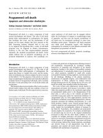
All images showed accurate segmentation of grey and white
matter, an example of which is shown in Figure 1. The stringent
probability thresholds excluded voxels with a partial volume
effect at the interfaces of grey matter, white matter and cere-
brospinal fluid, providing pure grey and white matter MTR
maps (Figure 1). Grey and white matter MTR values showed
considerable overlap, with higher MTR values for the white
matter (Figure 2).
Mann–Whitney tests revealed a lower grey matter mean MTR,
white matter mean MTR and white matter peak location (P <
Arthritis Research & Therapy Vol 8 No 2 Steens et al.
Page 4 of 7
(page number not for citation purposes)
0.05) and grey matter peak location (trend toward signifi-
cance) in IgM aCL-positive as compared with IgM aCL-nega-
tive patients (Table 2, Figure 3). Lower values were also
observed for grey and white matter mean MTR and peak loca-
tion in IgG aCL-positive than in IgG aCL-negative patients (not
significant) and in Lac-positive than in Lac-negative patients
(trend toward significance for grey and white matter mean
MTR). No significant differences were found for the MTR his-
togram parameters with respect to anti-dsDNA or anti-ENA
status (P > 0.2 and P > 0.3 for all MTR parameters, respec-
tively), or for age or SLE or NPSLE disease duration in all com-
parisons.
Discussion
This is the first study to investigate the relation between MTI
parameters of the brain and aCLs in NPSLE patients. MTI
parameters demonstrated brain damage in aCL-positive SLE
patients in the absence of explanatory abnormalities on con-
ventional MRI. Therefore, our results suggest that, apart from
giving rise to macroscopic cerebral infarctions, aCLs may play
a role in the pathogenesis of diffuse microscopic brain dam-
age in NPSLE.
MTI has proved to be a sensitive tool for detecting diffuse brain
involvement in NPSLE patients [4]. In previous work, based on
whole-brain MTR histograms, it was found that SLE patients
with active neuropsychiatric symptoms, past neuropsychiatric
symptoms, and SLE patients without neuropsychiatric symp-
toms could be distinguished, suggesting diagnostic potential
for these parameters [6-8]. The previously observed correla-
tions between whole-brain MTR histogram parameters and
measures of neurological, psychiatric and cognitive function
[9] emphasized the functional relevance of MTI parameters in
such patients. In the present study SLE patients with a history
of neuropsychiatric symptoms were included. Apart from overt
diffuse neuropsychiatric manifestations, some patients suf-
fered from chronic multifocal neuropsychiatric symptoms and
were classified as having cerebrovascular disease, subclassi-
fication chronic multifocal disease [2]. Although two of the four
patients classified as such exhibited nonspecific MRI abnor-
malities, in none of the patients was there evidence of cerebral
infarcts or any other abnormality on conventional MRI to
explain their neuropsychiatric symptoms. Therefore, in all
patients diffuse involvement of the CNS was thought to under-
lie the neuropsychiatric manifestations. We observed lower
values for mean MTR and peak location in grey and white mat-
ter in patients positive for aCLs and Lac.
The pathological conditions underlying the MTR histogram
abnormalities and neuropsychiatric manifestations in SLE
patients remain unclear. Although neuropathological studies in
NPSLE patients are limited, vasculopathy and microinfarcts
have been noted in several studies [3]. A recent MTI study
examining cerebral grey and white matter separately in SLE
patients with a history of diffuse neuropsychiatric manifesta-
tions [23] identified MTR histogram abnormalities specifically
in the grey matter, suggesting that neuronal injury is among the
key factors in diffuse NPSLE. This hypothesis is supported by
increased levels of neuronal and astrocytic degradation prod-
ucts observed in the cerebrospinal fluid of NPSLE patients
[25]. Microscopic brain damage was also suggested given the
data from other quantitative neuroimaging techniques, such as
magnetic resonance spectroscopy [26-31], spin-spin relaxa-
tion time measurements [32] and diffusion-weighted imaging
[33]. A recent study combining these MRI techniques [10]
indicated that the presence of neuronal and axonal injury, atro-
phy, demyelination and gliosis are aspects of the processes
involved in neuropsychiatric involvement in SLE.
Although several studies have reported abnormalities on con-
ventional MRI in patients with antiphospholipid antibodies [34-
36], to our knowledge the only previous MTI study in patients
with a known antiphospholipid antibody status is that by Rov-
aris and coworkers [8]. That study included healthy control
individuals, patients suffering from SLE with and without neu-
ropsychiatric symptoms, and patients suffering from the
Figure 1
Example of segmented axial MTR map (level indicated at the sagittal image)Example of segmented axial MTR map (level indicated at the sagittal
image). Visualized are the compartments grey matter (GM), white mat-
ter (WM) and grey and white matter (GM + WM). Signal intensities rep-
resent MTR values. MTR, magnetization transfer ratio.
Figure 2
Average MTR histograms after volume corrections for patients with and without IgM aCLsAverage MTR histograms after volume corrections for patients with and
without IgM aCLs. Visualized are the average MTR histograms for
patients with IgM aCLs (black lines) and patients without IgM aCLs
(grey lines) for the grey matter (GM; continous lines) and white matter
(WM; dashed lines). aCL, anticardiolipin antibodies; MTR, magnetiza-
tion transfer ratio.
Available online />Page 5 of 7
(page number not for citation purposes)
antiphospholipid antibody syndrome. No significant differ-
ences were observed between the patients with antiphosphol-
ipid antibody syndrome patients and healthy control
individuals, whereas lower mean MTR values were observed in
NPSLE patients than in non-NPSLE patients. These observa-
tions and the findings of our study suggest that the mere pres-
ence of antiphospholipid antibodies, including aCLs, does not
lead to diffuse microscopic brain damage as detected by MTI,
but they implicate that aCLs are involved in the pathogenetic
events that lead to neuropsychiatric manifestations in SLE. A
role for antiphospholipid antibodies in the pathogenesis of
NPSLE has also been suggested by studies using magnetic
resonance spectroscopy. In a study conducted by Sabet and
coworkers [28], a reduced N-acetyl-aspartate to creatine ratio
suggesting neuronal loss or injury was observed in SLE
patients with the antiphospholipid antibody syndrome, as com-
pared with SLE patients without – an effect that was mainly
attributed to the presence of IgG aCLs.
Much in the order of the pathogenetic events that occur in SLE
patients with diffuse neuropsychiatric manifestations remains
unknown, although evidence for involvement of antineuronal
antibodies, complement activation and proinflammatory
cytokines has been found [3]. There are at least three possible
explanations for how aCLs could be involved. First, the throm-
botic tendency of antiphospholipid antibodies, including aCLs,
may cause aggregation of thrombocytes and an increase in
blood viscosity [3,11,37]. This may affect blood flow in small
cerebral blood vessels in particular and cause widespread
hypoperfusion, which subsequently causes ischaemic dam-
age to brain tissue [38]. The trend observed with Lac in the
present study supports this hypothesis. Second, aCLs may
activate endothelial cells and cause a diffuse small-vessel vas-
culopathy – a neuropathological finding that was reported as
long ago as 1968 [3,11,37-39]. The resulting increase in
blood–brain barrier permeability permits entrance to the brain
parenchyma of substances such as circulating antibodies
[3,40]. Third, it has been shown in vitro that IgG aCLs them-
selves may interfere with glutamatergic pathways by a mecha-
nism involving over-activation of the N-methyl-d-aspartate
receptor [41,42].
The present study has several limitiations, and the results are
preliminary. First, patient numbers were small, and control indi-
viduals were not available. Second, aCL status at the time of
active neuropsychiatric manifestations was not available in this
SLE patient cohort with past neuropsychiatric symptoms,
which precludes evaluation of our results in the light of fluctu-
Figure 3
Plot of the mean of the MTR histogram for patients with and without IgM aCLsPlot of the mean of the MTR histogram for patients with and without
IgM aCLs. Visualized are the mean MTRs for patients with IgM aCLs
versus patients without IgM aCLs for the grey matter (GM) and white
matter (WM). aCL, anticardiolipin antibodies; MTR, magnetization
transfer ratio.
Table 2
Descriptive statistics and Mann–Whitney test results
Parameter IgM aCL
+
IgM aCL
-
P IgG aCL
+
IgG aCL
-
P Lac
+
Lac
-
P
Number of patients 9 9 - 9 9 - 13 5 -
Age (years) 36.4 ± 13.4 31.6 ± 6.4 0.67 31.3 ± 6.5 36.7 ± 13.2 0.49 34.9 ± 10.9 31.6 ± 10.2 0.50
Duration of SLE (years) 7.4 ± 5.0 10.6 ± 8.8 0.55 8.4 ± 3.6 9.6 ± 9.8 0.67 8.8 ± 7.1 9.6 ± 8.0 0.85
Duration of NPSLE (years) 4.4 ± 4.0 6.2 ± 5.5 0.49 4.9 ± 3.2 5.7 ± 6.1 0.73 5.7 ± 5.2 4.3 ± 3.7 0.57
Grey matter peak location 33.8 ± 0.7 34.7 ± 1.0 0.077 34.1 ± 0.3 34.3 ± 1.3 0.67 34.0 ± 0.1 34.8 ± 1.3 0.34
Grey matter peak height 131 ± 24 138 ± 19 0.67 135 ± 28 133 ± 14 0.93 133 ± 22 138 ± 23 0.63
Grey matter mean MTR 32.6 ± 0.9 33.8 ± 1.0 0.011 33.0 ± 0.8 33.3 ± 1.4 0.49 32.9 ± 1.1 33.8 ± 1.1 0.12
White matter peak location 37.2 ± 1.0 38.4 ± 1.0 0.019 37.8 ± 0.4 37.9 ± 1.6 0.93 37.6 ± 1.0 38.4 ± 1.6 0.50
White matter peak height 184 ± 31 178 ± 20 0.26 185 ± 32 177 ± 18 0.16 180 ± 27 183 ± 24 0.99
White matter mean MTR 37.2 ± 0.9 38.2 ± 1.0 0.014 37.6 ± 0.3 37.8 ± 1.5 0.44 37.4 ± 0.9 38.4 ± 1.3 0.14
Listed are the mean values ± standard deviation for IgM-positive/IgM-negative and IgG-positive/IgG-negative aCLs as well as Lac, and P values of
Mann–Whitney tests between the groups. aCL, anticardiolipin antibody; Lac, lupus anticoagulant; MTR, magnetization transfer ratio; NPSLE,
neuropsychiatric systemic lupus erythematosus; SLE, systemic lupus erythematosus.
Arthritis Research & Therapy Vol 8 No 2 Steens et al.
Page 6 of 7
(page number not for citation purposes)
ation in aCL levels [19]. Possibly, an even stronger association
could be found between MTI measures of brain damage and
aCL status at the time of active neuropsychiatric symptomatol-
ogy. A prospective study should therefore include a larger
NPSLE patient group with inactive and active neuropsychiatric
symptoms, as well as control groups consisting of non-NPSLE
patients and patients suffering from similar neuropsychiatric
conditions, preferably with measurements of aCLs in serum
and cerebrospinal fluid. Also, the specific role of IgM and IgG
aCLs remains to be identified.
Conclusion
This is the first study to find an association between aCLs and
brain damage as detected by MTI in NPSLE patients. These
results suggest that aCLs, in addition to contributing to overt
brain infarcts, may also contribute to widespread microscopic
damage in the brain.
Competing interests
The authors declare that they have no competing interests.
Authors' contributions
SCAS,GPTB,TWJH and MAvB participated in the design of
the study. SCAS, GPTB and GMS performed a literature
search. SCAS, GPTB and GMS carried out data acquisition.
SCAS, GMS, TWJH and MAvB carried out data analysis.
SCAS and SleC performed the statistical analysis. SCAS,
GPTB, GMS, SleC, TWJH and MAvB drafted the manuscript.
All authors read and approved the final manuscript.
Acknowledgements
The authors thank Dr F Admiraal-Behloul, PhD, and H Olofsen, MSc, of
the Division of Image Processing, Department of Radiology, Leiden Uni-
versity Medical Center, Leiden, The Netherlands for providing post-
processing software and helpful discussions.
References
1. Sibbitt WL, Sibbitt RR, Brooks WM: Neuroimaging in neuropsy-
chiatric systemic lupus erythematosus. Arthritis Rheum 1999,
42:2026-2038.
2. Anonymous: The American College of Rheumatology nomen-
clature and case definitions for neuropsychiatric lupus syn-
dromes. Arthritis Rheum 1999, 42:599-608.
3. Scolding NJ, Joseph FG: The neuropathology and pathogene-
sis of systemic lupus erythematosus. Neuropathol Appl Neuro-
biol 2002, 28:173-189.
4. Huizinga TW, Steens SC, van Buchem MA: Imaging modalities
in central nervous system systemic lupus erythematosus.
Curr Opin Rheumatol 2001, 13:383-388.
5. Tofts PS, Steens SC, van Buchem MA: MT: magnetization trans-
fer. In Quantitative MRI of the Brain: Measuring Changes Caused
by Disease Chichester, UK: Wiley; 2003:257-298.
6. Bosma GP, Rood MJ, Zwinderman AH, Huizinga TW, van Buchem
MA: Evidence of central nervous system damage in patients
with neuropsychiatric systemic lupus erythematosus, demon-
strated by magnetization transfer imaging. Arthritis Rheum
2000, 43:48-54.
7. Bosma GP, Rood MJ, Huizinga TW, de Jong BA, Bollen EL, van
Buchem MA: Detection of cerebral involvement in patients with
active neuropsychiatric systemic lupus erythematosus by the
use of volumetric magnetization transfer imaging. Arthritis
Rheum 2000, 43:2428-2436.
8. Rovaris M, Viti B, Ciboddo G, Gerevini S, Capra R, Iannucci G,
Comi G, Filippi M: Brain involvement in systemic immune medi-
ated diseases: magnetic resonance and magnetisation trans-
fer imaging study. J Neurol Neurosurg Psychiatry 2000,
68:170-177.
9. Bosma GP, Middelkoop HA, Rood MJ, Bollen EL, Huizinga TW,
van Buchem MA: Association of global brain damage and clin-
ical functioning in neuropsychiatric systemic lupus erythema-
tosus. Arthritis Rheum 2002, 46:2665-2672.
10. Bosma GP, Steens SC, Petropoulos H, Admiraal-Behloul F, van
den Haak A, Doornbos J, Huizinga TW, Brooks WM, Harville A,
Sibbitt WL Jr, et al.: Multisequence magnetic resonance imag-
ing study of neuropsychiatric systemic lupus erythematosus.
Arthritis Rheum 2004, 50:3195-3202.
11. Greenwood DL, Gitlits VM, Alderuccio F, Sentry JW, Toh BH:
Autoantibodies in neuropsychiatric lupus. Autoimmunity 2002,
35:79-86.
12. Sherer Y, Gorstein A, Fritzler MJ, Shoenfeld Y: Autoantibody
explosion in systemic lupus erythematosus: More than 100
different antibodies found in SLE patients. Semin Arthritis
Rheum 2004, 34:501-537.
13. Sastre-Garriga J, Montalban X: APS and the brain. Lupus 2003,
12:877-882.
14. Provenzale JM, Barboriak DP, Allen NB, Ortel TL: Patients with
antiphospholipid antibodies: CT and MR findings of the brain.
AJR Am J Roentgenol 1996, 167:1573-1578.
15. Sanna G, Bertolaccini ML, Cuadrado MJ, Laing H, Khamashta MA,
Mathieu A, Hughes GR: Neuropsychiatric manifestations in sys-
temic lupus erythematosus: prevalence and association with
antiphospholipid antibodies. J Rheumatol 2003, 30:985-992.
16. Denburg SD, Denburg JA: Cognitive dysfunction and antiphos-
pholipid antibodies in systemic lupus erythematosus. Lupus
2003, 12:883-890.
17. Afeltra A, Garzia P, Mitterhofer AP, Vadacca M, Galluzzo S, Del
Porto F, Finamore L, Pascucci S, Gasparini M, Lagana B, et al.:
Neuropsychiatric lupus syndromes: relationship with
antiphospholipid antibodies. Neurology 2003, 61:108-110.
18. Hanly JG, Hong C, Smith S, Fisk JD: A prospective analysis of
cognitive function and anticardiolipin antibodies in systemic
lupus erythematosus. Arthritis Rheum 1999, 42:728-734.
19. Menon S, Jameson-Shortall E, Newman SP, Hall-Craggs MR,
Chinn R, Isenberg DA: A longitudinal study of anticardiolipin
antibody levels and cognitive functioning in systemic lupus
erythematosus. Arthritis Rheum 1999, 42:735-741.
20. Omdal R: Some controversies of neuropsychiatric systemic
lupus erythematosus. Scand J Rheumatol 2002, 31:192-197.
21. Tan EM, Cohen AS, Fries JF, Masi AT, McShane DJ, Rothfield NF,
Schaller JG, Talal N, Winchester RJ: The 1982 revised criteria for
the classification of systemic lupus erythematosus. Arthritis
Rheum 1982, 25:1271-1277.
22. Dousset V, Grossman RI, Ramer KN, Schnall MD, Young LH,
Gonzalez-Scarano F, Lavi E, Cohen JA: Experimental allergic
encephalomyelitis and multiple sclerosis: lesion characteriza-
tion with magnetization transfer imaging. Radiology 1992,
182:483-491.
23. Steens SC, Admiraal-Behloul F, Bosma GP, Steup-Beekman GM,
Olofsen H, le Cessie S, Huizinga TW, van Buchem MA: Selective
gray matter damage in neuropsychiatric lupus. Arthritis Rheum
2004, 50:2877-2881.
24. Ashburner J, Friston KJ: Voxel-based morphometry: the meth-
ods. Neuroimage 2000, 11:805-821.
25. Trysberg E, Nylen K, Rosengren LE, Tarkowski A: Neuronal and
astrocytic damage in systemic lupus erythematosus patients
with central nervous system involvement. Arthritis Rheum
2003, 48:2881-2887.
26. Chinn RJ, Wilkinson ID, Hall-Craggs MA, Paley MN, Shortall E,
Carter S, Kendall BE, Isenberg DA, Newman SP, Harrison MJ:
Magnetic resonance imaging of the brain and cerebral proton
spectroscopy in patients with systemic lupus erythematosus.
Arthritis Rheum 1997, 40:36-46.
27. Sibbitt WL, Haseler LJ, Griffey RR, Friedman SD, Brooks WM:
Neurometabolism of active neuropsychiatric lupus deter-
mined with proton MR spectroscopy. AJNR Am J Neuroradiol
1997, 18:1271-1277.
28. Sabet A, Sibbitt WL, Stidley CA, Danska J, Brooks WM: Neu-
rometabolite markers of cerebral injury in the antiphospholi-
Available online />Page 7 of 7
(page number not for citation purposes)
pid antibody syndrome of systemic lupus erythematosus.
Stroke 1998, 29:2254-2260.
29. Peterson PL, Howe FA, Clark CA, Axford JS: Quantitative mag-
netic resonance imaging in neuropsychiatric systemic lupus
erythematosus. Lupus 2003, 12:897-902.
30. Lim MK, Suh CH, Kim HJ, Cho YK, Choi SH, Kang JH, Park W, Lee
JH: Systemic lupus erythematosus: brain MR imaging and sin-
gle-voxel hydrogen 1 MR spectroscopy. Radiology 2000,
217:43-49.
31. Brooks WM, Sabet A, Sibbitt WL, Barker PB, van Zijl PC, Duyn JH,
Moonen CT: Neurochemistry of brain lesions determined by
spectroscopic imaging in systemic lupus erythematosus. J
Rheumatol 1997, 24:2323-2329.
32. Petropoulos H, Sibbitt WL, Brooks WM: Automated T2 quantita-
tion in neuropsychiatric lupus erythematosus: a marker of
active disease. J Magn Reson Imaging 1999, 9:39-43.
33. Bosma GP, Huizinga TW, Mooijaart SP, van Buchem MA: Abnor-
mal brain diffusivity in patients with neuropsychiatric systemic
lupus erythematosus. AJNR Am J Neuroradiol 2003,
24:850-854.
34. Toubi E, Khamashta MA, Panarra A, Hughes GR: Association of
antiphospholipid antibodies with central nervous system dis-
ease in systemic lupus erythematosus. Am J Med 1995,
99:397-401.
35. Ishikawa O, Ohnishi K, Miyachi Y, Ishizaka H: Cerebral lesions in
systemic lupus erythematosus detected by magnetic reso-
nance imaging. Relationship to anticardiolipin antibody. J
Rheumatol 1994, 21:87-90.
36. Csepany T, Bereczki D, Kollar J, Sikula J, Kiss E, Csiba L: MRI find-
ings in central nervous system systemic lupus erythematosus
are associated with immunoserological parameters and
hypertension. J Neurol 2003, 250:1348-1354.
37. Connor P, Hunt BJ: Cerebral haemostasis and antiphospholi-
pid antibodies. Lupus 2003, 12:929-934.
38. Meroni PL, Tincani A, Sepp N, Raschi E, Testoni C, Corsini E,
Cavazzana I, Pellegrini S, Salmaggi A: Endothelium and the brain
in CNS lupus. Lupus 2003, 12:919-928.
39. Johnson RT, Richardson EP: The neurological manifestations of
systemic lupus erythematosus. Medicine (Baltimore) 1968,
47:337-369.
40. Abbott NJ, Mendonca LL, Dolman DE: The blood-brain barrier in
systemic lupus erythematosus. Lupus 2003, 12:908-915.
41. Riccio A, Andreassi C, Eboli ML: Antiphospholipid antibodies
bind to rat cerebellar granule cells: the role of N-methyl-D-
aspartate receptors. Neurosci Lett 1998, 257:116-118.
42. Andreassi C, Zoli A, Riccio A, Scuderi F, Lombardi L, Altomonte L,
Eboli ML: Anticardiolipin antibodies in patients with primary
antiphospholipid syndrome: a correlation between IgG titre
and antibody-induced cell dysfunctions in neuronal cell cul-
tures. Clin Rheumatol 2001, 20:314-318.