Báo cáo y học: "proximal interphalangeal joints in rheumatoid arthritis: a comparison with magnetic resonance imaging, conventional radiography and clinical examination" pdf
Bạn đang xem bản rút gọn của tài liệu. Xem và tải ngay bản đầy đủ của tài liệu tại đây (409.96 KB, 11 trang )
Open Access
Available online />Page 1 of 11
(page number not for citation purposes)
Vol 8 No 2
Research article
Ultrasonography of the metacarpophalangeal and proximal
interphalangeal joints in rheumatoid arthritis: a comparison with
magnetic resonance imaging, conventional radiography and
clinical examination
Marcin Szkudlarek
1
, Mette Klarlund
2
, Eva Narvestad
3
, Michel Court-Payen
3
, Charlotte Strandberg
3
,
Karl E Jensen
3
, Henrik S Thomsen
4
and Mikkel Østergaard
1
1
Department of Rheumatology, University of Copenhagen Hvidovre Hospital, Kettegård Allé 30, 2650 Hvidovre, Denmark
2
Magnetic Resonance Research Centre, University of Copenhagen Hvidovre Hospital, Kettegård Allé 30, 2650 Hvidovre, Denmark
3
Department of Radiology, University of Copenhagen Hvidovre Hospital, Kettegård Allé 30, 2650 Hvidovre, Denmark
4
Department of Radiology, University of Copenhagen Herlev Hospital, Herlev Ringvej 75, 2730 Herlev, Denmark
Corresponding author: Marcin Szkudlarek,
Received: 16 Aug 2005 Revisions requested: 26 Sep 2005 Revisions received: 22 Dec 2005 Accepted: 26 Jan 2006 Published: 6 Mar 2006
Arthritis Research & Therapy 2006, 8:R52 (doi:10.1186/ar1904)
This article is online at: />© 2006 Szkudlarek et al.; licensee BioMed Central Ltd.
This is an open access article distributed under the terms of the Creative Commons Attribution License ( />),
which permits unrestricted use, distribution, and reproduction in any medium, provided the original work is properly cited.
Abstract
Signs of inflammation and destruction in the finger joints are the
principal features of rheumatoid arthritis (RA). There are few
studies assessing the sensitivity and specificity of
ultrasonography in detecting these signs. The objective of the
present study was to investigate whether ultrasonography can
provide information on signs of inflammation and destruction in
RA finger joints that are not available with conventional
radiography and clinical examination, and comparable to the
information provided by magnetic resonance imaging (MRI). The
second to fifth metacarpophalangeal and proximal
interphalangeal joints of 40 RA patients and 20 control persons
were assessed with ultrasonography, clinical examination,
radiography and MRI. With MRI as the reference method, the
sensitivity, specificity and accuracy of ultrasonography in
detecting bone erosions in the finger joints were 0.59, 0.98 and
0.96, respectively; they were 0.42, 0.99 and 0.95 for
radiography. The sensitivity, specificity and accuracy of
ultrasonography, with signs of inflammation on T1-weighted MRI
sequences as the reference method, were 0.70, 0.78 and 0.76,
respectively; they were 0.40, 0.85 and 0.72 for the clinical
examination. With MRI as the reference method,
ultrasonography had higher sensitivity and accuracy in detecting
signs of inflammation and destruction in RA finger joints than did
clinical and radiographic examinations, without loss of
specificity. This study shows that ultrasonography has the
potential to improve assessment of patients with RA.
Introduction
New aggressive and powerful treatments that permit fast and
effective suppression of inflammation in rheumatoid arthritis
(RA) demand sensitive and specific methods for detecting dis-
ease signs and monitoring disease activity. Finger joints are
frequently the first to be involved in RA, and therefore methods
of assessment of these joints are of particular importance at
the onset of disease. The methods currently used, including
clinical examination and conventional radiography, are not sen-
sitive, especially in the evaluation of early stages of RA. In
recent years magnetic resonance imaging (MRI) has been rig-
orously tested in patients with RA, and its value has been con-
firmed both in studies of large joints (for example, knee joints
[1,2]) and in finger joints [3] compared with histological evalu-
ation of biopsy specimens acquired at microarthroscopy. Thus
far, because of the expensive equipment required and the
need for highly qualified personnel, it has not become widely
used as a joint assessment tool in RA. However, its benefits of
high sensitivity and specificity in the evaluation of RA joints [4-
6] make it a worthy surrogate 'gold standard' in settings where
FoV = field of view; Gd-DTPA = gadolinium-diethylenetriamine penta-acetic acid; ICC = intraclass correlation coefficient; MCP = metacarpophalan-
geal; MRI = magnetic resonance imaging; PIP = proximal interphalangeal; RA = rheumatoid arthritis; ST = slice thickness; TE = echo time; TR =
repetition time.
Arthritis Research & Therapy Vol 8 No 2 Szkudlarek et al.
Page 2 of 11
(page number not for citation purposes)
acquiring histological specimens is difficult (for example, fin-
ger joints).
Ultrasonography is an imaging technique that has attracted
much interest in the field of rheumatology in recent years [7,8].
As a result of technological improvements and wide availabil-
ity, ultrasonography has the potential to facilitate diagnosis of
RA and improve the assessment of disease activity, and its use
by rheumatologists may soon become routine. Few studies
have compared ultrasonography with other imaging modalities
with respect to their ability to detect signs of destruction and
inflammation; furthermore, data are seldom gathered from
homogenous populations and studies rarely include control
persons. Despite of appearance in the literature of reports pre-
senting the results of longitudinal studies of ultrasonographic
assessment of RA, the more basic issues of agreement, sensi-
tivity and specificity of ultrasonography in detecting RA pathol-
ogy remain to be addressed.
We therefore planned a systematic study in order to investi-
gate whether ultrasonography can provide information on RA
finger joints that is not available with conventional radiography
and clinical examination and comparable to the information
provided by MRI.
Materials and methods
Patients
We examined a total of 158 second to fifth metacarpophalan-
geal (MCP) joints and 140 second to fifth proximal inter-
phalangeal (PIP) joints of 40 patients with RA (fulfilling
American College of Rheumatology 1987 criteria) and 80 sec-
ond to fifth MCP joints and 80 second to fifth PIP joints of 20
healthy control persons. In the first part of the study we
attempted to evaluate the wrists of RA patients, but after we
had examined the first five patients the evaluation was omitted
because of poor accessibility of most bone surfaces. The
median age of the RA patients was 58 (range 23–79) years
and that of the control persons was 52 (27–79) years. The
female/male ratio was 4:1 both in the RA group and in the con-
trol group. The median disease duration in RA patients was 5
(range 0–20) years.
Twenty patients in the series had a disease duration in excess
of 2 years (established disease). Their median age and dis-
ease duration were 64 (range 23–79) years and 8 (2–20)
years, respectively. A further 20 patients had a disease dura-
tion of under 2 years (early disease). Their median and disease
duration were 53 (range 23–72) years and 1 (0–1) year,
respectively. All patients with established RA and 15 patients
with early RA were being treated with disease-modifying
antirheumatic drugs. The healthy control individuals had nei-
ther history of previous nor any current joint complaints.
The patients were recruited from two outpatient hospital-
based arthritis clinics. The study was conducted in accord-
ance with the Declaration of Helsinki and was approved by the
local ethics committee. Signed informed consent was
obtained from each participant. The inclusion criteria for RA
patients were swelling or tenderness of at least three finger
joints (MCP and/or PIP joints). The exclusion criteria were
severe deformity of MCP or PIP joint and contraindications to
MRI.
Ultrasonographic, clinical, laboratory and MRI examinations of
each patient were conducted on the same day.
Ultrasonography
Ultrasonography was performed using a General Electric
LOGIQ 500 unit (General Electric, Solingen, Germany) using
a 7–13 MHz linear array transducer. Ultrasonography was
conducted in the accessible aspects of the second to fifth
MCP joints and the second to fifth PIP joints of the dominant
hand: the dorsal, radial and palmar aspects of the second
MCP joint; the dorsal and palmar aspects of the third and
fourth MCP joints; the dorsal, ulnar and palmar aspects the
fifth MCP joint; and the dorsal, palmar, radial and ulnar aspects
of all PIP joints. Ultrasonographic examination from the dorsal
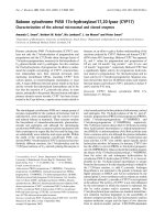
Figure 1
Signs of destruction on ultrasonography in the fourth proximal inter-phalangeal joint: early RASigns of destruction on ultrasonography in the fourth proximal inter-
phalangeal joint: early RA. MRI and conventional radiography revealed
no signs of destruction in the joint. A bone erosion (arrow) is visualized
with ultrasonography in (a) the longitudinal and (b) the transverse
planes. MRI, magnetic resonance imaging; RA, rheumatoid arthritis.
Available online />Page 3 of 11
(page number not for citation purposes)
aspect was performed both in the neutral position and at about
70° of flexion. Each joint was assessed by quadrant for the
presence or absence of bone erosions (Figures 1 and 2) and
each joint was assessed for the presence or absence of signs
of inflammation (joint effusion and synovitis; Figures 2 and 3).
The following definitions of ultrasonographic changes were
employed: bone erosion = break in bone cortex in the area
adjacent to the joint, visualized in two planes; joint effusion =
compressible anechoic intracapsular area; and synovitis =
uncompressible hypoechoic intracapsular area. The ultrasono-
graphic changes were scored according to a semiquantitative
scoring system (grades 0–3) introduced in an earlier report
[9]. In relation to the original system, scoring of synovitis was
widened to include grade 4, defined as a hypoechoic area
bulging out of the joint and stretching over both bone diaphy-
ses of the joint.
Ultrasonographic examinations were performed by two radiol-
ogists with expertise in musculoskeletal ultrasonography and
a rheumatologist with training in the examination of the small
joints of the extremities. Ultrasonography was performed with-
out knowledge of the clinician's assessment or MRI data. The
interobserver variation between one of the radiologists and the
rheumatologist was presented in an earlier report [9].
Clinical examination
Prior to ultrasonography, clinical disease activity (presence or
absence of swelling and/or tenderness) in the MCP and PIP
joints was evaluated in all patients by the consultant rheuma-
tologist on duty. The number and localization of swollen and/
or tender joints was determined.
Conventional radiography
Radiography of the dominant hand was performed using
standard postero-anterior and oblique (Nørgaard's) views
within four weeks of the other examinations. The films were
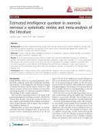
Figure 2
Signs of destruction and inflammation on ultrasonography and MRI in second metacarpophalangeal joint: established RASigns of destruction and inflammation on ultrasonography and MRI in
second metacarpophalangeal joint: established RA. Thin arrows indi-
cate an erosive change; thick arrows indicate synovitis. Ultrasonogra-
phy in the (a) longitudinal and (b) the transverse planes shows both
signs of destruction (grade 2) and inflammation (grade 3). Axial T1-
weighted magnetic resonance images were obtained (c) before and
(d) after contrast administration (grade 3 synovitis). Additionally, a
coronal T1-weighted magnetic resonance image (e) before contrast
administration visualizes the same bone erosion as shown in panels c
and d. The coronal magnetic resonance image of the second metacar-
pophalangeal joint (panel e) is additionally covered by a grid illustrating
division of the assessed joints into quadrants: proximal radial, proximal
ulnar, distal radial and distal ulnar. MRI, magnetic resonance imaging;
RA, rheumatoid arthritis.
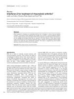
Figure 3
Signs of synovitis on ultrasonography and MRI in fourth proximal inter-phalangeal joint: early RASigns of synovitis on ultrasonography and MRI in fourth proximal inter-
phalangeal joint: early RA. Arrows indicate an area with synovitis. Ultra-
sonography in (a) the longitudinal plane from the dorsal aspect shows
signs of synovitis (grade 4). Axial T1-weighted magnetic resonance
images were obtained (b) before and (c) after contrast administration
(grade 3 synovitis). MRI, magnetic resonance imaging; RA, rheumatoid
arthritis.
Arthritis Research & Therapy Vol 8 No 2 Szkudlarek et al.
Page 4 of 11
(page number not for citation purposes)
Table 1
Number of quadrants with bone erosions in finger joints, stratified by imaging modality and combinations thereof
Joint Quadrants with erosions Quadrants
with no
erosions on
US, MRI or
CR
Agreement Sensitivity Specificity
US + MRI
+ CR
US +
MRI
MRI +
RAD
US +
CR
US
only
MRI
only
CR
only
US versus
MRI (%)
CR versus
MRI (%)
US CR US CR
MCP 2nd 10 10 2 1 2 7 3 205 225(94) 217(90) 0.64 0.41 0.98 0.98
Est. 9 8 2 1 1 2 2 55 72(90) 66(82)
Early 1 2 0 0 1 2 0 74 77(96) 75(94)
Control 0 0 0 0 0 3 1 76 76(95) 76(95)
MCP 3rd 7 7 4 1 4 12 2 230 217(90) 214 (89) 0.47 0.37 0.97 0.98
Est. 6 6 4 1 3 6 2 52 64(80) 62 (78)
Early 1 1 0 0 1 2 0 75 77(96) 76 (95)
Control 0 0 0 0 0 4 0 76 76(95) 76 (95)
MCP 4th 5 2 0 1 1 5 0 222 229 (97) 227 (96) 0.58 0.42 0.99 0.99
Est. 4 1 0 1 1 5 0 64 69 (91) 68 (89)
Early 1 1 0 0 0 0 0 78 80 (100) 79 (99)
Control 0 0 0 0 0 0 0 80 80 (100) 80 (100)
MCP 5th 7 1 0 1 3 2 1 221 229 (97) 228 (97) 0.80 0.70 0.98 0.99
Est. 6 0 0 1 2 0 0 67 73 (96) 73 (96)
Early 1 1 0 0 1 0 1 76 78 (97) 77 (96)
Control 0 0 0 0 0 2 0 78 78 (97) 78 (97)
PIP 2nd 0 0 0 0 6 1 1 212 212 (96) 212 (96) - - 0.97 0.99
Est. 0 0 0 0 5 0 1 54 54 (90) 54 (90)
Early 0 0 0 0 1 0 0 79 79 (99) 79 (99)
Control 0 0 0 0 0 1 0 79 79 (99) 79 (99)
PIP 3rd 1 0 0 1 7 1 0 210 211 (96) 211 (96) 0.50 0.50 0.96 0.99
Est. 1 0 0 1 6 1 0 51 52 (87) 52 (87)
Early 0 0 0 0 1 0 0 79 79 (99) 79 (99)
Control 0 0 0 0 0 0 0 80 80 (100) 80 (100)
PIP 4th 0 0 0 0 2 1 1 216 216 (98) 216(98) - - 0.99 0.99
Est. 0 0 0 0 2 0 1 57 57 (95) 57 (95)
Early 0 0 0 0 0 1 0 79 79 (99) 79 (99)
Control 0 0 0 0 0 0 0 80 80 (100) 80 (100)
PIP 5th 0 0 0 0 1 0 4 215 215 (98) 215(98) - - 0.99 0.98
Est. 0 0 0 0 0 0 2 58 58 (97) 58(97)
Early 0 0 0 0 1 0 2 77 77 (96) 77(96)
Control 0 0 0 0 0 0 0 80 80 (100) 80 (100)
Total 30 20 6 5 26 29 12 1,704 1,754 (96) 1,740 (95) 0.59 0.42 0.98 0.99
Est. 26 15 6 5 20 14 8 458 499 (90) 490 (89)
Early 4 5 0 0 6 5 3 617 626 (98) 621 (97)
Control 0 0 0 0 0 10 1 629 629 (98) 629 (98)
The following numbers of joints were evaluated (1,832 in total): 240 MCP second, 240 MCP third, 236 MCP fourth, 236 MCP sixth, 220 PIP
second, 220 PIP third, 220 PIP fourth, and 220 PIP fifth. All study participants included. CR, conventional radiography; early, early rheumatoid
arthritis; Est., established rheumatoid arthritis; MCP, metacarpophalangeal joint; MRI, magnetic resonance imaging; PIP, proximal interphalangeal
joint; US, ultrasonography.
Available online />Page 5 of 11
(page number not for citation purposes)
assessed by quadrant for the presence or absence of bone
erosions in the second to fifth MCP joints and the second to
fifth PIP joints by an experienced radiologist, who was una-
ware of the findings of the other examinations.
Magnetic resonance imaging
Later in the day on which ultrasonography was performed,
continuous axial and coronal pre-Gd-DTPA (gadolinium-dieth-
ylenetriamine penta-acetic acid) and post-Gd-DTPA T1-
weighted spin-echo magnetic resonance sequences of the
second to fifth MCP and second to fifth PIP joints of the dom-
inant hand were performed. This MRI assessment employed a
1.0 T Siemens Impact MR unit (Siemens, Erlangen, Germany)
equipped with a receive-only, wrap-around flex coil, and was
conducted in the group with established disease, three
patients with early disease and five control persons. The Gd-
DTPA (0.1 mmol/kg body weight) was injected intravenously
between repeated T1-weighted spin-echo magnetic reso-
nance sequences. The patients and control persons were in
the supine position with the hand in the coil along the femur.
The parameters of the applied sequences were as follows for
coronal sequences: repetition time (TR) 600 ms, echo time
(TE) 15 ms, slice thickness (ST) 3 mm, field of view (FoV) 140
mm, and matrix 192 × 256. For axial sequences the parame-
ters were as follows: TR 700 ms, TE 15 ms, ST 3 mm, FoV 120
mm, and matrix 192 × 256.
An extremity coil was used in 17 patients with early RA and 15
control persons. The use of different coils was necessary
because technical problems meant that the wrap-around flex
coil was unavailable for a lengthy period. The persons under-
going MRI were in supine position with the hand stretched
above the head ('Superman' position). The parameters of the
applied sequences for coronal sequences were as follows: TR
600 ms, TE 15 ms, ST 3 mm, FoV 145 mm, and matrix 192 ×
256. For axial sequences the parameters were as follows: TR
600 ms, TE 15 ms, ST 3 mm, FoV 120 mm, and matrix 192 ×
256.
The definitions of the applied MRI RA pathologies were in
accordance with OMERACT recommendations [10].
The examinations were assessed by quadrant for the presence
or absence of bone erosions (Figure 2) and by joint for the
presence or absence of signs of inflammation (joint effusion
and synovitis; Figures 2 and 3). Synovitis was scored accord-
ing to the semiquantitative system (grades 0–4) introduced by
Klarlund and coworkers [11]. The MRI observer was blinded
to clinical and ultrasonographical data.
The numbers of finger joints assessed using ultrasonography/
clinical examination and MRI were different (480 versus 433)
because the MRI data for 47 joints were not available: 20 PIP
joints were not visualized in the five patients in whom MRI of
wrists and MCP joints was performed, and the MRIs of six
MCP and 21 PIP joints were not assessable because the
patients moved between pre- and post-contrast MRI
sequences.
Statistical analysis
The agreement between imaging methods and compared with
clinical examination is reported as the overall agreement,
defined as the proportion of exact agreements to the overall
number of trials (expressed as a percentage). Furthermore,
agreement was expressed as means of sensitivity and specifi-
city. The correlation between ultrasonographic and MRI syno-
vitis scores was estimated using calculations of intraclass
correlation coefficients (ICCs; two-way mixed effects model,
consistency definition).
Results
Signs of bone destruction
A total of 1,832 quadrants of second to fifth MCP joints (952
quadrants) and PIP joints (880 quadrants) from 40 RA
patients and 20 healthy control individuals were examined
using ultrasonography, MRI and radiography (Table 1).
In MCP joints, at least one modality detected bone erosions in
101 of 952 examined quadrants (11%). Agreement between
all modalities on the presence of erosions was found in 29 out
of 101 quadrants (29%), whereas ultrasonography and MRI
agreed in 49 quadrants (49%). In 10 (11%) quadrants only
ultrasonography and in 26 (26%) quadrants only MRI identi-
fied bone erosions. Half of the ultrasonographic erosions in RA
patients that were not visualized by MRI were located in sec-
ond and fifth MCP joints (7 out of 14), whereas MRI quadrants
with erosions in RA patients not visualized with ultrasonogra-
phy were located predominantly in third to fourth MCP joints
(17 out of 23).
In PIP joints, at least one modality detected bone erosions in
27 of 880 quadrants (3%). Of these 27, only one quadrant
(4%) was identified as erosive with all modalities. In 16 (59 %)
quadrants only ultrasonography and in three (11 %) quadrants
only MRI detected bone erosions. Ultrasonographic bone ero-
sions, not visualized with other modalities, were distributed
between all examined PIP joints, but most of them were
located in the second and third PIP joints (15 out of 18). Radi-
ography detected six (22%) quadrants with erosions in PIP
joints that were not detected with other modalities.
Ten of the MRI quadrants with bone erosions in MCP joints
were detected in healthy control persons (10 erosions in 238
MCP joints; frequency 4.2%), which is in contrast to none with
ultrasonography and one with radiography. Ultrasonography
and radiography detected no erosions in PIP joints of the
healthy persons examined; one quadrant with erosions was
found with MRI.
Arthritis Research & Therapy Vol 8 No 2 Szkudlarek et al.
Page 6 of 11
(page number not for citation purposes)
With MRI as the reference method, the sensitivity of ultra-
sonography in detecting bone erosions in the finger joints was
0.59, whereas it was 0.42 for radiography. The specificity of
ultrasonography compared with MRI was 0.98, and for radiog-
raphy compared with MRI it was 0.99. The accuracy of US (for
instance the overall agreement between ultrasonography and
MRI) for bone erosions was 0.96, and the accuracy of radiog-
raphy was 0.95.
Erosive disease (defined as presence of at least one erosion
in the examined finger joints) was found in 13 patients with
radiography, in 20 with MRI, and in 20 with ultrasonography
(15 simultaneously with MRI). Eleven patients with erosions on
radiography were identified as having erosions with MRI and
nine with ultrasonography. In the series of patients with early
RA, ultrasonography visualized erosions in eight, MRI in six
and radiography in three. In 20 control persons, MRI revealed
erosions in seven; in one this was simultaneous with radiogra-
phy.
The lowest grade (grade 1) of ultrasonographic erosive
changes was visualized only in two cases out of 16 identified
with MRI. Most of the definite (grade 2) ultrasonographic bone
erosions were identified with MRI and some were identified
with radiography. Almost all grade 3 ultrasonographic erosive
changes were visualized with both MRI and radiography
(Table 2). All of the most extensive ultrasonographic erosive
changes (for instance grades 2 and 3) that were not detected
with MRI or radiography were localized in PIP joints.
Signs of inflammation
A total of 234 second to fifth MCP joints and 199 second to
fifth PIP joints from 40 RA patients and 20 control persons
were evaluated with ultrasonography, MRI and clinical exami-
nation. Agreement between ultrasonography and MRI regard-
ing the presence or absence of synovitis was achieved in 76%
(331/433) of the examined finger joints (Table 3). Further-
more, ultrasonography revealed signs of synovitis in 59 joints
(14%) that were not detected with MRI, and MRI identified
signs of synovitis in 43 joints (10%) that were not visualized
with ultrasonography. Ultrasonography detected synovitis
more often in patients with early RA than did MRI (88 versus
57 joints – a difference of 36%). The opposite was true in con-
trol persons, in whom MRI revealed synovitis more frequently
than did ultrasonography (20 versus 5 joints – a difference of
75%). MRI did not detect joint effusion in any of the examined
finger joints, whereas ultrasonography revealed joint effusion
in 22 out of the 433 examined finger joints.
Signs of inflammation on ultrasonography (joint effusion and/
or synovitis) were visualized in 194 out of 480 joints, whereas
only 121 joints exhibited signs of inflammation at clinical
assessment (swelling and/or tenderness; Table 4). Ultrasono-
graphic and clinical findings agreed on the presence of signs
of inflammation in 103 joints (21% of the 480 joints) and on
the absence of signs of inflammation in 268 joints (56%). In 91
joints (19%), signs of inflammation (effusion or synovitis) on
ultrasonography were found in clinically uninflamed joints,
whereas no ultrasonographic signs of inflammation were
observed in 18 joints (4%) in which the clinicians described
swelling and/or tenderness. The overall agreement for both
presence and absence of signs of inflammation between ultra-
sonography and clinical assessment was 77% (371 out of
480 examined finger joints).
The number of finger joints assessed with ultrasonography
and MRI differed (480 versus 433) because the magnetic res-
onance data for 47 joints were not available: 20 PIP joints
were not visualized in the five patients in whom MRI of wrists
and MCP joints was performed, and MRIs of six MCP and 21
PIP joints were not assessable because the patients moved
between pre- and post-contrast magnetic resonance
sequences.
The sensitivity of ultrasonography, with signs of inflammation
on T1-weighted MRI sequences as the reference, was 0.70
and the specificity was 0.78. The accuracy (for instance over-
all agreement between ultrasonography and MRI on signs of
inflammation) was 0.76. The sensitivity of clinical examination,
with signs of inflammation on T1-weighted MRI sequences as
the reference, was 0.40 and the specificity was 0.85. The
accuracy (for instance overall agreement between clinical
examination and MRI on signs of inflammation) was 0.72.
Grading of synovitis with ultrasonography and MRI exhibited
moderate-to-good correlations, as expressed by ICCs (two-
way mixed effects model, consistency definition). ICCs for syn-
ovitis in the examined joints were as follows: second MCP
0.71, third MCP 0.61, fourth MCP 0.65, fifth MCP 0.58, sec-
ond PIP 0.58, third PIP 0.58, fourth PIP 0.53, and fifth PIP
0.63. Results for exact agreement between scoring grades on
ultrasonography and MRI are presented in Table 5.
The localization of joint inflammation was investigated,
because signs of inflammation were assessed in all accessible
aspects of the joints and registered separately. In MCP joints,
of which 108 exhibited ultrasonographic signs of inflammation,
these signs were present on both the dorsal and palmar
aspect in 57 joints (52.7%), on the dorsal aspect only in 27
joints (25%), on the palmar aspect only in 19 joints (17.7%),
and on the radial aspect only in five joints (4.6%). In PIP joints,
of which 86 exhibited ultrasonographic signs of inflammation,
these signs were present on both the dorsal and palmar
aspect in 26 joints (30.2%), on the dorsal aspect only in 16
joints (18.6%), on the palmar aspect only in 37 joints (43%),
and on the radial or ulnar aspect only in seven joints (8.1%).
Discussion
In the present study we investigated the agreement between
ultrasonography, radiography and clinical evaluation in the
Available online />Page 7 of 11
(page number not for citation purposes)
assessment of RA and healthy finger joints, with MRI as the
reference method. It showed high agreement between ultra-
sonography and MRI in assessing RA bone erosions in finger
joints. Using MRI as the reference method, ultrasonography
exhibited markedly higher sensitivity in detecting RA bone ero-
sions than did radiography, without loss of specificity.
In agreement with a study of RA MCP joints conducted by
Wakefield and coworkers [12,13], we found that ultrasonog-
raphy of those MCP joints with good accessibility by this
modality (such as second and fifth) exhibited better correla-
tions with MRI than did ultrasonography of joints only accessi-
ble in two planes (third and fourth). We found
ultrasonographic bone erosions in many PIP joints in which
MRI and radiography were unable to detect any destructive
bone changes. This finding is probably explained by the use of
3 mm thick MRI slices, which must be considered suboptimal
for the small PIP joints. In a heterogeneous group of patients
with joint complaints, Backhaus and coworkers [14] did not
find any advantage of ultrasonography over MRI in assessing
bone destruction in PIP joints. This may be due to use of a 7.5
MHz transducer with a distance pad that is inferior to the high-
frequency transducers employed in the present study. Further-
more, Backhaus and coworkers employed thinner MRI slices
than were used in our study (1 mm versus 3 mm), favouring
MRI over ultrasonography. Another possible reason for greater
frequency of detection of erosions in PIP joints with ultra-
sonography may be its higher resolution in relation to MRI.
Preliminary data were reported by Alarcon and coworkers [15]
and Lopez-Ben and colleagues [16] on the detection of bone
erosions with ultrasonography in the second and fifth MCP
joints of RA patients. They reported that ultrasonography had
high accuracy, with MRI as the reference method, in the sec-
ond and fifth MCP joints. In a group of patients with nonerosive
RA on conventional radiography, Magnani and coworkers [17]
visualized significantly more erosions in patients' MCP joints
with ultrasonography than with MRI. Similar to our study, they
used 3 mm thick MRI slices. Optimal technique would proba-
bly have improved the sensitivity of MRI.
Unlike in metatarsophalangeal joints [18], we found no ultra-
sonographic erosive changes in the examined finger joints of
control persons; this is in contrast to MRI, which showed sev-
eral single erosive changes in these joints. Erosive changes in
control persons were detected with MRI with a frequency
twice that reported in another study from our group (4.2% in
the present study versus 2.2% in the study by Ejbjerg and
coworkers [19]), but all except one were small. A possible rea-
son for the MRI finding of erosions in the finger joints is that
Table 2
Detection of bone changes, visualized and scored with
ultrasonography, by other imaging methods
Bone
erosions on
MRI
Bone
erosions on
CR
No bone
erosions on
MRI and
CR
US grades Grade 1
(n = 16)
2014
Grade 2
(n = 55)
32 17 20
Grade 3
(n = 26)
18 18 6
CR, conventional radiography; MRI, magnetic resonance imaging;
US, ultrasonography.
Table 4
Signs of inflammation on ultrasonography versus clinical joint assessment in finger joints
Signs of
inflammation
US + clinical
assessment
US only Clinical
assessment
only
Joints with no signs of
inflammation on US or
clinical assessment
Number of joints
examined
Agreement: US versus
clinical assessment (%)
MCP 2nd 23 12 3 22 60 45 (75)
MCP 3rd 20 10 5 25 60 45 (75)
MCP 4th 8 11 2 39 60 47 (78)
MCP 5th 8 16 2 34 60 42 (70)
PIP 2nd 13 13 1 33 60 46 (77)
PIP 3rd 13 11 3 33 60 46 (77)
PIP 4th 12 7 1 40 60 52 (87)
PIP 5th 6 11 1 42 60 48 (80)
Total 103 91 18 268 480 371 (77)
The number of examined joints is higher than in the other tables because ultrasonography and clinical examination were performed on all finger
joints, whereas MRI data were not available in 47 joints. All study participants included. Ultrasonography detecting signs of synovitis and/or joint
effusion. Clinical joint assessment detecting swelling and/or tenderness. MCP, metacarpophalangeal; PIP, proximal interphalangeal; US,
ultrasonography.
Arthritis Research & Therapy Vol 8 No 2 Szkudlarek et al.
Page 8 of 11
(page number not for citation purposes)
Table 3
Numbers of joints with and without signs of synovitis in finger joints, stratified by imaging modality and combinations thereof
Joint Joints with signs
of synovitis
Joints with no
signs of synovitis
on US or MRI
Number of joints
examined
Agreement: US
versus MRI (%)
US + MRI US only MRI only
MCP 2nd 26 6 7 20 59 46 (78)
Est. 12 3 2 3 20 15 (75)
Early 14 2 0 3 19 17 (89)
Control 0 1 5 14 20 14 (70)
MCP 3rd 21 7 9 22 59 43 (73)
Est. 11 2 2 5 20 16 (80)
Early 10 4 2 3 19 13 (68)
Control 0 1 5 14 20 14 (70)
MCP 4th 11 7 3 37 58 48 (83)
Est. 62291915 (79)
Early 5 4 0 10 19 15 (79)
Control 0 1 1 18 20 18 (90)
MCP 5th 11 12 3 32 58 43 (74)
Est. 65171913 (68)
Early 57071912 (63)
Control 0 0 2 18 20 18 (90)
PIP 2nd 12 9 6 23 50 35 (70)
Est. 6332148 (57)
Early 65151711 (65)
Control 0 1 2 16 19 16 (84)
PIP 3rd 14 4 5 27 50 41 (82)
Est. 61341410 (71)
Early 73071714 (82)
Control 1 0 2 16 19 17 (89)
PIP 4th 10 5 4 31 50 41 (82)
Est. 51351410 (71)
Early 54081713 (76)
Control 0 0 1 18 19 18 (95)
PIP 5th 2 9 6 32 49 34 (69)
Est. 1346147 (50)
Early 1618169 (56)
Control 0 0 1 18 19 18 (95)
Total 107 59 43 224 433 331 (76)
Est. 53 20 20 41 134 94 (70)
Early 53 35 4 51 143 104 (73)
Control 1 4 19 132 156 133 (85)
All study participants included. early, early rheumatoid arthritis; Est., established rheumatoid arthritis; MCP, metacarpophalangeal joint; MRI,
magnetic resonance imaging; PIP, proximal interphalangeal joint; US, ultrasonography.
Available online />Page 9 of 11
(page number not for citation purposes)
the visualized changes were subchondral cysts, which are not
detected with ultrasonography because the employed ultra-
sound frequencies do not penetrate cortical bone. The less
efficient/optimal blinding of the ultrasonographer as compared
with the MRI evaluator might have caused bias toward finding
fewer healthy control joints with erosions and synovitis by
ultrasonography than by MRI.
The rate of detection of ultrasonographic destructive changes
by MRI and radiography increased with the extent of erosion,
as defined by its ultrasonographic grading. Correspondingly,
the gradings of ultrasonographic inflammatory changes corre-
lated with the volume-based MRI scoring of synovitis. Our
results suggest that MRI and ultrasonography both allow
assessment of abnormalities of the bone structures, and that
performance differences are probably caused by technical
aspects such as accessibility for ultrasonographic examina-
tion, high resolution of ultrasonographic assessment, or thick-
ness of the MRI slices, rather than the physical principles of
the examinations.
Ultrasonography had higher sensitivity for detecting signs of
inflammation in the examined finger joints than did clinical
examination, when MRI was considered the reference method,
without considerable loss of specificity. Likewise, regarding
the correlation of detection of synovitis between the methods,
the moderate-to-good ICCs suggest that both ultrasonogra-
phy and MRI were able to detect signs of inflammation. How-
ever, incomplete agreement between the methods suggested
a margin of difference, probably due to ultrasonographic visu-
alization of both 'active' and fibrotic pannus in the joints. The
results are in agreement with those reported by Backhaus and
coworkers [14], who showed greater frequency of detecting
synovitis with ultrasonography than with MRI. A large propor-
tion of 'disagreement', in which ultrasonography alone showed
signs of synovitis, was found in patients with early RA. This
suggests that fibrotic changes, which are probably less fre-
quent in the early stages of the disease, are not the only
changes identified by B-mode ultrasonography and not visual-
ized on MRI. Current knowledge does not allow definite con-
clusions to be drawn regarding the cause of the discrepancy
between ultrasonographic and MRI findings.
The difficulty associated with recognizing both 'active' and
'inactive' synovial tissue may be alleviated by the addition of
Doppler ultrasonography. The growing number of reports
comparing Doppler ultrasonography with MRI [20,21]. and
histology of joints [22,23],. and describing the advantages of
supporting ultrasonography with Doppler evaluation suggests
that it will soon become a routine aspect of the joint assess-
ment. However, many methodological and technical aspects
of the use of Doppler ultrasonography remain to be clarified
[24].
MRI did not permit visualization of joint effusion in RA finger
joints, probably because of the minimal amount of fluid in the
examined joints, whereas ultrasonography detected effusion in
a considerable number of finger joints. This may be explained
by the higher magnification of joints with ultrasonography than
with MRI and the better resolution with ultrasonography. In our
study, magnetic resonance images were read on hard-copy
films. Evaluation on a computer screen, allowing magnification,
would probably increase the sensitivity of MRI in detecting
joint effusions. Additionally, MRI contrast diffusion into the joint
cavity may contribute to making the detection of joint effusions
with MRI more difficult [2,25]. In contrast to MRI, ultrasonog-
raphy is a dynamic, real-time examination method, which per-
mits evaluation of the findings in motion and under
compression. The latter is a distinct feature of joint effusion on
ultrasonography, which may explain the apparent advantage of
this modality over MRI in detecting it.
In the present study the detection of joint effusion on ultra-
sonography did not improve its sensitivity in comparison with
MRI on detecting signs of inflammation because it most often
accompanied synovial thickening. However, in joints in which
accessibility may be problematic, joint effusion could be used
as indirect proof of an ongoing inflammatory process. Other
researchers reported difficulty in differentiating between syno-
vitis and joint effusion [14,26]. Standardization and precise
definitions, as suggested in our earlier study [9], may be help-
ful in this respect.
Localization of signs of inflammation showed the dominance of
the palmar aspect in PIP joints and a slight dominance of the
dorsal aspect in MCP joints. In our opinion, the uneven distri-
bution of signs of inflammation warrants examination of the
joints from all possible aspects in order to avoid losing impor-
tant information on the extent of inflammation [27].
Table 5
Comparison of scoring of synovitis with ultrasonography and
MRI with their respective volume-based scales
MRI grades
01234
US
grades
0 75 128 20 2 0
1138 19 1 1
2142012 16 2
39121331 21
404048
Values in the cells describe the numbers of joints, apart from those
denoting score (first column for US and first row for MRI). Numbers
in bold denote exact agreements between respective identical
scores. MRI, magnetic resonance imaging; US, ultrasonography.
Arthritis Research & Therapy Vol 8 No 2 Szkudlarek et al.
Page 10 of 11
(page number not for citation purposes)
With MRI as the reference method, ultrasonography almost
doubled the sensitivity of assessing RA small joints for signs
of inflammation compared with clinical assessment, without
loss of specificity. The low sensitivity of clinical examination
may account for the deterioration of RA patients despite clini-
cally adequate control of the disease, as reported by Mulherin
and coworkers [28]. Accordingly, a longitudinal study con-
ducted by Backhaus and coworkers [29] showed progression
of erosive changes with both ultrasonography and MRI,
despite limited signs of clinical activity. The present study
strongly suggests that clinical examination is far from optimal
for assessing signs of inflammation in RA finger joints, and that
the use of ultrasonography can considerably improve the
detection of signs of synovial inflammation.
Conclusion
Ultrasonography was shown to permit assessment of destruc-
tive and inflammatory changes in RA finger joints, with high
agreement with MRI. Ultrasonography was more sensitive than
plain film radiography in assessing bone destruction in the
examined joints, and had equal specificity. B-mode ultrasonog-
raphy was more sensitive than clinical examination in assess-
ing signs of inflammation, with only a slight loss of specificity.
The present study strongly encourages further studies of use
of ultrasonography to assess RA finger joints.
Competing interests
The authors declare that they have no competing interests.
Authors' contributions
MS participated in the study development, performed the
ultrasonographic evaluations, conducted the preliminary data
evaluation and statistic analysis, and prepared the draft of the
manuscript. MK took part in the study development, performed
the MRI evaluation, and was involved in patient recruitment.
EN performed the conventional radiographic evaluation. MC-P
and CS performed ultrasonographic evaluations. KEJ took
active part in study development and preliminary data interpre-
tation. HST and MØ took part in the study development and
gave substantial input into data evaluation and manuscript
preparation. All authors read and approved the final manu-
script.
Acknowledgements
The Danish Rheumatism Association is acknowledged for financial sup-
port. We thank Ms Susanne Østergaard for assistance with the medical
images.
References
1. Konig H, Sieper J, Wolf KJ: Rheumatoid arthritis: evaluation of
hypervascular and fibrous pannus with dynamic MR imaging
enhanced with Gd-DTPA. Radiology 1990, 176:473-477.
2. Østergaard M: Magnetic resonance imaging in rheumatoid
arthritis. Quantitative methods for assessment of the inflam-
matory process in peripheral joints. Dan Med Bull 1999,
46:313-344.
3. Ostendorf B, Peters R, Dann P, Becker A, Scherer A, Wedekind F,
Friemann J, Schulitz KP, Modder U, Schneider M: Magnetic reso-
nance imaging and miniarthroscopy of metacarpophalangeal
joints: sensitive detection of morphologic changes in rheuma-
toid arthritis. Arthritis Rheum 2001, 44:2492-2502.
4. McQueen FM, Stewart N, Crabbe J, Robinson E, Yeoman S, Tan
PL, McLean L: Magnetic resonance imaging of the wrist in early
rheumatoid arthritis reveals a high prevalence of erosions at
four months after symptom onset. Ann Rheum Dis 1998,
57:350-356.
5. McGonagle D, Conaghan PG, O'Connor P, Gibbon W, Green M,
Wakefield R, Ridgway J, Emery P: The relationship between syn-
ovitis and bone changes in early untreated rheumatoid arthri-
tis: a controlled magnetic resonance imaging study. Arthritis
Rheum 1999, 42:1706-1711.
6. Klarlund M, Østergaard M, Jensen KE, Madsen JL, Skjødt H, Loren-
zen I: Magnetic resonance imaging, radiography, and scintigra-
phy of the finger joints: one year follow up of patients with
early arthritis. The TIRA Group. Ann Rheum Dis 2000,
59:521-528.
7. Østergaard M, Szkudlarek M: Imaging in rheumatoid arthritis
why MRI and ultrasonography can no longer be ignored.
Scand J Rheumatol 2003, 32:63-73.
8. Wakefield RJ, Brown A, O'Connor P, Grainger A, Karim Z, McG-
onagle D, Conaghan P, Emery P: Rheumatological ultrasound.
Rheumatology (Oxford) 2003, 42:1001
9. Szkudlarek M, Court-Payen M, Jacobsen S, Klarlund M, Thomsen
HS, Østergaard M: Interobserver agreement in ultrasonogra-
phy of the finger and toe joints in rheumatoid arthritis. Arthritis
Rheum 2003, 48:955-962.
10. Østergaard M, Edmonds J, McQueen F, Peterfy C, Lassere M, Ejb-
jerg B, Bird P, Emery P, Genant H, Conaghan P: An introduction
to the EULAR-OMERACT rheumatoid arthritis MRI reference
image atlas. Ann Rheum Dis 2005, 64(Suppl 1):i3-i7.
11. Klarlund M, Østergaard M, Lorenzen I: Finger joint synovitis in
rheumatoid arthritis: quantitative assessment by magnetic
resonance imaging. Rheumatology (Oxford) 1999, 38:66-72.
12. Wakefield RJ, McGonagle D, Green MJ: A comparison of high
resolution sonography with MRI and conventional radiography
for the detection of erosions in early rheumatoid arthritis.
Arthritis Rheum 1997, 40:S511
13. Wakefield RJ, Gibbon WW, Conaghan PG, O'Connor P, McGona-
gle D, Pease C, Green MJ, Veale DJ, Isaacs JD, Emery P: The
value of sonography in the detection of bone erosions in
patients with rheumatoid arthritis: a comparison with conven-
tional radiography. Arthritis Rheum 2000, 43:2762-2770.
14. Backhaus M, Kamradt T, Sandrock D, Loreck D, Fritz J, Wolf KJ,
Raber H, Hamm B, Burmester GR, Bollow M: Arthritis of the fin-
ger joints: a comprehensive approach comparing conventional
radiography, scintigraphy, ultrasound, and contrast-enhanced
magnetic resonance imaging. Arthritis Rheum 1999,
42:1232-1245.
15. Alarcon GS, Lopez-Ben R, Moreland LW: High-resolution ultra-
sound for the study of target joints in rheumatoid arthritis.
Arthritis Rheum 2002, 46:1969-1970.
16. Lopez-Ben R, Bernreuter WK, Moreland LW, Alarcon GS: Ultra-
sound detection of bone erosions in rheumatoid arthritis: a
comparison to routine radiographs of the hands and feet.
Skeletal Radiol 2004, 33:80-84.
17. Magnani M, Salizzoni E, Mule R, Fusconi M, Meliconi R, Galletti S:
Ultrasonography detection of early bone erosions in the met-
acarpophalageal joints of patients with rheumatoid arthritis.
Clin Exp Rheumatol 2004, 22:743-748.
18. Szkudlarek M, Narvestad E, Klarlund M, Court-Payen M, Thomsen
HS, Østergaard M: Ultrasonography of the metatarsophalan-
geal joints in rheumatoid arthritis: comparison with magnetic
resonance imaging, conventional radiography, and clinical
examination. Arthritis Rheum 2004, 50:2103-2112.
19. Ejbjerg B, Narvestad E, Rostrup E, Szkudlarek M, Jacobsen S,
Thomsen HS, Østergaard M: Magnetic resonance imaging of
wrist and finger joints in healthy subjects occasionally shows
changes resembling erosions and synovitis as seen in rheu-
matoid arthritis. Arthritis Rheum 2004, 50:1097-1106.
20. Szkudlarek M, Court-Payen M, Strandberg C, Klarlund M, Klausen
T, Østergaard M: Power Doppler ultrasonography for assess-
ment of synovitis in the metacarpophalangeal joints of
patients with rheumatoid arthritis: a comparison with dynamic
magnetic resonance imaging. Arthritis Rheum 2001,
44:2018-2023.
Available online />Page 11 of 11
(page number not for citation purposes)
21. Terslev L, Torp-Pedersen S, Savnik A, von der Recke P, Qvistgaard
E, Danneskiold-Samsøe B, Bliddal H: Doppler ultrasound and
magnetic resonance imaging of synovial inflammation of the
hand in rheumatoid arthritis: a comparative study. Arthritis
Rheum 2003, 48:2434-2441.
22. Walther M, Harms H, Krenn V, Radke S, Faehndrich TP, Gohlke F:
Correlation of power Doppler sonography with vascularity of
the synovial tissue of the knee joint in patients with osteoar-
thritis and rheumatoid arthritis. Arthritis Rheum 2001,
44:331-338.
23. Walther M, Harms H, Krenn V, Radke S, Kirschner S, Gohlke F:
Synovial tissue of the hip at power Doppler US: correlation
between vascularity and power Doppler US signal. Radiology
2002, 225:225-231.
24. Østergaard M, Wiell C: Ultrasonography in rheumatoid arthri-
tis: a very promising method still needing more validation.
Curr Opin Rheumatol 2004, 16:223-230.
25. Klarlund M: Magnetic Resonance Imaging of Wrist and Finger
Joints in Rheumatoid Arthritis and Early Unclassified Polyar-
thritis. Copenhagen, Denmark: Faculty of Health Sciences, Uni-
versity of Copenhagen;; 2000.
26. Grassi W, Tittarelli E, Pirani O, Avaltroni D, Cervini C: Ultrasound
examination of metacarpophalangeal joints in rheumatoid
arthritis. Scand J Rheumatol 1993, 22:243-247.
27. Østergaard M, Szkudlarek M: Ultrasonography: a valid method
for assessing rheumatoid arthritis? Arthritis Rheum 2005,
52:681-686.
28. Mulherin D, FitzGerald O, Bresnihan B: Clinical improvement and
radiological deterioration in rheumatoid arthritis: evidence
that the pathogenesis of synovial inflammation and articular
erosion may differ. Br J Rheumatol 1996, 35:1263-1268.
29. Backhaus M, Burmester GR, Sandrock D, Loreck D, Hess D,
Scholz A, Blind S, Hamm B, Bollow M: Prospective two year fol-
low up study comparing novel and conventional imaging pro-
cedures in patients with arthritic finger joints. Ann Rheum Dis
2002, 61:895-904.